The Adrenal Cortex


The Adrenal Cortex


A number of endocrine abnormalities involve the adrenal glands, and some of the more common conditions are discussed in this chapter, which should perhaps be read in conjunction with Chapter 7 (on pituitary function) and Chapter 9 (which deals with reproductive endocrinology).

The adrenal glands are divided into two embryologically and functionally distinct parts. The adrenal cortex is part of the hypothalamic-pituitary-adrenal endocrine system. Morphologically, the adult adrenal cortex consists of three layers. The outer thin layer (zona glomerulosa) secretes only aldosterone. The inner two layers (zona fasciculata and zona reticularis) form a functional unit and secrete most of the adrenocortical hormones. In the fetus there is a wider fourth layer, which disappears soon after birth. One of its most important functions during fetal life is, together with the adrenal cortex, to synthesize oestriol, in association with the placenta. The adrenal medulla is part of the sympathetic nervous system.


CHEMISTRY AND BIOSYNTHESIS OF STEROIDS

Steroid hormones are derived from the lipid cholesterol. Figure 8.1 shows the internationally agreed numbering of the 27 carbon atoms of steroid molecules and the lettering of the four rings. The products of cholesterol are also indicated. If the molecule contains 21 carbon atoms, it is referred to as a C21 steroid. The carbon atom at position 21 of the molecule is written as C-21. The side chain on C-17 is the main determinant of the type of hormonal activity (Fig. 8.1), but substitutions in other positions modify activity within a particular group.

The first hormonal product of cholesterol is pregnenolone. Several important synthetic pathways diverge from it (Fig. 8.1). The final product is dependent upon the tissue and its enzymes.

The zona glomerulosa secretes aldosterone, produced by 18-hydroxylation. Synthesis of this steroid is controlled by the renin-angiotensin system and not normally by adrenocorticotrophic hormone (ACTH). Although ACTH is important for maintaining growth of the zona glomerulosa, deficiency does not significantly reduce output.

The zonae fasciculata and reticularis synthesize and secrete two groups of steroid:


  • Cortisol, a glucocorticoid (the most important C21 steroid), is formed by progressive addition of hydroxyl groups at C-17, C-21 and C-11.


  • Androgens (for example androstenedione) are formed after the removal of the side chain to produce C19 steroids.

Adrenocorticotrophic hormone secreted by the anterior pituitary gland stimulates synthesis of these two steroid groups. Its secretion is influenced by negative feedback from changes in plasma cortisol concentrations. Impaired cortisol synthesis due, for example, to an inherited 21-α-hydroxylase or 11-β-hydroxylase deficiency (congenital adrenal hyperplasia) results in increased ACTH stimulation with increased activity of both pathways. The resultant excessive androgen production may cause hirsutism or virilization.


PHYSIOLOGY

The adrenocortical hormones can be classified into groups depending on their predominant physiological effects.


Glucocorticoids

Cortisol and corticosterone are naturally occurring glucocorticoids. They stimulate gluconeogenesis and
the breakdown of protein and fat, that is, they antagonize some of insulin’s action. Glucocorticoids in excess may impair glucose tolerance and alter the distribution of adipose tissue. Cortisol helps maintain the extracellular fluid volume and normal blood pressure.


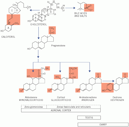
Figure 8.1 Numbering of the steroid carbon atoms of cholesterol and the synthetic pathway of steroid hormones; the chemical groups highlighted determine the biological activity of the steroid.

Circulating cortisol is bound to cortisol-binding globulin (CBG; transcortin) and to albumin. At normal concentrations, only about 5 per cent of the total is unbound and physiologically active. Plasma CBG is almost fully saturated, so that increased cortisol secretion causes a disproportionate rise in the free active fraction. Cortisone is not secreted in significant amounts by the adrenal cortex. It is biologically inactive until it has been converted in vivo to cortisol (hydrocortisone).

Glucocorticoids are conjugated with glucuronate and sulphate in the liver to form inactive metabolites, which, because they are more water soluble than the mainly protein-bound parent hormones, can be excreted in the urine.


Mineralocorticoids

In contrast to other steroids, aldosterone is not transported in plasma bound to specific proteins. It stimulates the exchange of sodium for potassium and hydrogen ions across cell membranes and its renal
action is especially important for sodium and water homeostasis. It is discussed more fully in Chapters 2, 3 and 4. Like the glucocorticoids, it is inactivated by hepatic conjugation and is excreted in the urine.

There is overlap in the actions of C21 steroids. Cortisol, in particular, may have a significant mineralocorticoid effect at high plasma concentrations when the free fraction is significantly increased.


Adrenal androgens

The main adrenal androgens are dehydroepiandrosterone (DHEA), its sulphate (DHEAS) and androstenedione. They promote protein synthesis and are only weakly androgenic at physiological concentrations. Testosterone, the most powerful androgen, is synthesized in the testes or ovaries but not in the adrenal cortex. Most circulating androgens, like cortisol, are protein bound, mainly to sexhormone-binding globulin and albumin.


Figure 8.2 The factors controlling the secretion of cortisol from the adrenal gland, including the site of action of dynamic function tests (shaded). +, stimulates; -, inhibits; ACTH, adrenocorticotrophic hormone; CRH, corticotrophin-releasing hormone.

There is extensive peripheral interconversion of adrenal and gonadal androgens. The end products, androsterone and aetiocholanolone, together with DHEA, are conjugated in the liver and excreted as glucuronides and sulphates in the urine.


THE HYPOTHALAMIC-PITUITARY-ADRENAL AXIS

The hypothalamus, anterior pituitary gland and adrenal cortex form a functional unit – the hypothalamic-pituitary-adrenal axis (see Chapter 7).

Cortisol is synthesized and secreted in response to ACTH from the anterior pituitary gland.

The secretion of ACTH is dependent on corticotrophinreleasing hormone (CRH), released from the hypothalamus. High plasma free cortisol concentrations suppress CRH secretion (negative feedback) and alter the ACTH response to CRH, thus acting on both the hypothalamus and the anterior pituitary gland (Fig. 8.2).


The melanocyte-stimulating effect of high plasma concentrations of ACTH, or related peptides, causes skin pigmentation in two conditions associated with low plasma cortisol concentrations:


  • Addison’s disease,


  • Nelson’s syndrome: after bilateral adrenalectomy for Cushing’s disease (see Causes of Cushing’s syndrome), removal of the cortisol feedback causes a further rise in plasma ACTH concentrations from already high levels.


Inherent rhythms and stress

Adrenocorticotrophic hormone is secreted episodically, each pulse being followed 5-10 min later by cortisol secretion. These episodes are most frequent in the early morning (between the fifth and eighth hour of sleep) and least frequent in the few hours before sleep. Plasma cortisol concentrations are usually highest between about 07.00 and 09.00 h and lowest between 23.00 and 04.00 h.

The secretion of ACTH and cortisol usually varies inversely, and the almost parallel circadian rhythm of the two hormones may be due to cyclical changes in the sensitivity of the hypothalamic feedback centre to cortisol levels. Inappropriately high plasma cortisol concentrations at any time of day suppress ACTH secretion. This effect can be tested by the dexamethasone suppression test. Loss of circadian rhythm is one of the earliest features of Cushing’s syndrome. Stress, either physical or mental, may over-ride the first two mechanisms and cause sustained ACTH secretion. An inadequate stress response may cause acute adrenal insufficiency. Stress caused by insulin-induced hypoglycaemia can be used to test the axis.


FACTORS AFFECTING PLASMA CORTISOL CONCENTRATIONS

Plasma cortisol is usually measured by immunoassay, but the antibody may cross-react with other steroids or drugs. Factors that may affect results include hydrocortisone (cortisol), cortisone (converted to cortisol by metabolism) and prednisolone, which may contribute to the ‘cortisol’ concentration of some immunoassay methods. Thus, it is recommended either that the patient is prescribed dexamethasone (which is less likely to cross-react with cortisol assays) or, if possible, that the prednisolone is gradually reduced and then stopped for about 3 days before sampling depending upon clinical context. Oestrogens and some oral contraceptives increase the plasma CBG concentration, and therefore the proteinbound cortisol concentration.


Adrenocorticotrophic hormone (corticotrophin)

Adrenocorticotrophic hormone is a single-chain polypeptide made up of 39 amino acids with biological activity at the N-terminal end of the peptide. A peptide consisting of this sequence has been synthesized (tetracosactide, Synacthen) and can be used for diagnosis in place of ACTH. The ACTH stimulates cortisol synthesis and secretion by the adrenal cortex. It has much less effect on adrenal androgen production and, at physiological concentrations, virtually no effect on aldosterone production.


DISORDERS OF THE ADRENAL CORTEX

The main disorders of adrenocortical function are shown in Table 8.1.


ADRENOCORTICAL HYPERFUNCTION


Cushing’s syndrome

Cushing’s syndrome is mainly caused by an excess of circulating cortisol but also other steroids such as androgens. Many of the clinical and metabolic disturbances can be explained by glucocorticoid excess. The clinical and metabolic features may include the following:


  • Obesity, typically involving the trunk and face, and a characteristic round, red ‘cushingoid’ face (Fig 8.3).


  • Impaired glucose tolerance and hyperglycaemia. Cortisol has the opposite action to that of insulin, causing increased gluconeogenesis, and some patients may have diabetes mellitus.


    Table 8.1 Disorders of adrenocortical function


    Altered hormone secretion


    Associated clinical disorder


    Hypersecretion


    Cortisol


    Cushing’s syndrome


    Aldosterone


    Primary hyperaldosteronism (Conn’s syndrome)


    Androgens


    Congenital adrenal hyperplasia Adrenocortical carcinoma


    Hyposecretion


    Cortisol and aldosterone


    Primary adrenal disorders, e.g. Addison’s disease or congenital adrenal hyperplasia


    Cortisol and adrenocorticotrophic hormone


    Adrenal insufficiency secondary to pituitary disease


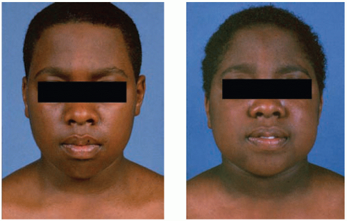
    Figure 8.3 This depicts a patient before and after corticosteroid therapy; note cushingoid appearance. Reproduced with kind permission from Kinirons M and Ellis H. French’s Index of Differential Diagnosis, 15th edition. London: Hodder Arnold, 2011.


  • Increased protein catabolism, which also increases urinary protein loss. Thus, there is a negative nitrogen balance associated with proximal muscle wasting with weakness, thinning of the skin and osteoporosis. The tendency to bruising and the purple striae (most obvious on the abdominal wall) are probably due to this thinning.


  • Hypertension, caused by urinary retention of sodium and therefore of water, which are due to the mineralocorticoid effect of cortisol. Increased urinary potassium loss may cause hypokalaemia.


  • Androgen excess, which may account for the common findings of greasy skin with acne vulgaris and hirsutism, and menstrual disturbances in women.


  • Psychiatric disturbances, such as depression.

Laboratory findings include a hypokalaemic alkalosis, leucocytosis and eosinophilia.


Causes of Cushing’s syndrome

One of the most common causes of Cushing’s syndrome is iatrogenic and related to excessive steroid treatment. Increased endogenous cortisol production may be due to hyperstimulation of the adrenal gland by ACTH, either from the pituitary gland or from an ‘ectopic’ source, or due to largely autonomous secretion by an adrenal tumour such as an adenoma or carcinoma (Fig. 8.4).

The secretion of ACTH is increased in the following conditions.


  • Cushing’s disease It is associated with bilateral adrenal hyperplasia, often secondary to a basophil adenoma of the anterior pituitary gland.


  • Ectopic ACTH secretion In this condition, usually from a small-cell carcinoma of the bronchus, ACTH concentrations may be high enough to cause skin pigmentation. The patient may have weight loss with cachexia. One metabolic complication is a hypokalaemic alkalosis. The clinical features may be indistinguishable from those of Cushing’s disease, although sometimes patients do not have the characteristic cushingoid features as the cortisol rises so quickly.


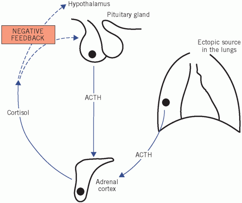
Figure 8.4 Cushing’s disease, indicating excess cortisol production caused either by hyperstimulation of the adrenal gland by adrenocorticotrophic hormone (ACTH), from either the pituitary or an ectopic source, or by autonomous hormone secretion from an adrenal tumour.

The secretion of ACTH is appropriately suppressed in primary cortisol-secreting tumours of the adrenal cortex. The tumours may be benign or malignant and are usually derived from the zona fasciculata/zona reticularis of the adrenal cortex. These glucocorticoidsecreting tumours do not normally secrete aldosterone, which is produced in the zona glomerulosa layer of the adrenal cortex. Benign adenomas occur and also carcinomas. The latter secrete a variety of steroids, including androgens, and thus may cause hirsutism or virilization. In these cases plasma ACTH is suppressed by the excess glucocorticoids.


Basis of investigation of suspected Cushing’s syndrome

The following questions should be asked:


  • Is there abnormal cortisol secretion?


  • If so, does the patient have any other condition that may cause it?


  • If Cushing’s syndrome is confirmed, what is the cause?


Is there abnormal cortisol secretion?

Plasma cortisol concentrations reflect ACTH activity at that moment and, because of the episodic nature of cortisol secretion, such isolated values may be misleading. Indeed, cyclical Cushing’s syndrome may require repeated investigation. The level of cortisol measured in a 24-h urine sample reflects the overall daily secretion.

One of the earliest features of Cushing’s syndrome is the loss of the diurnal variation in cortisol secretion, with high concentrations in the late evening, when secretion is normally at a minimum. However, it is not a diagnostic finding because it can also be caused by, for example, stress and endogenous depression. The assessment of diurnal rhythm is not a practical outpatient procedure.

Out-patient screening tests therefore may include the following.


Estimation of 24-h urinary free cortisol

Only the unbound fraction of cortisol in plasma is filtered at the glomeruli and excreted in the urine (urinary ‘free’ cortisol). In Cushing’s syndrome, because of the loss of circadian rhythm, raised plasma values are present for longer than normal and daily urinary cortisol excretion is further increased (i.e. there is a disproportionately raised free fraction of cortisol).

Plasma and urinary cortisol concentrations are usually much higher when Cushing’s syndrome is due to adrenocortical carcinoma or overt ectopic
ACTH secretion. Determinations of 24-h urinary free cortisol have about a 5 per cent false-negative rate, but if three separate determinations are normal, Cushing’s syndrome is most unlikely.


Low-dose overnight dexamethasone suppression test

A small dose, for example 1 mg, of this synthetic steroid inhibits ACTH, and thereby cortisol secretion by negative feedback. This is usually given at midnight and blood is taken for cortisol assay at 09.00 h the following morning.

The overnight dexamethasone suppression test is a sensitive, but not completely specific, test for evaluating such patients. The false-positive rate is about 12 per cent and the false-negative rate about 2 per cent. A normal fall in plasma cortisol concentrations makes the diagnosis of Cushing’s syndrome very unlikely, but failure to suppress plasma cortisol to less than 50 nmol/L does not confirm it with certainty.

There are some pitfalls, for example certain anticonvulsant drugs, such as phenytoin, may interfere with dexamethasone suppression tests, inducing liver enzymes that increase the rate of metabolism of the drug. Plasma concentrations may therefore be too low to suppress the feedback centre.


Additional tests

In some cases, additional tests are needed to confirm the diagnosis of excess cortisol production. The 48-h lowdose dexamethasone suppression test may be useful as it gives fewer false-positives than the overnight low-dose dexamethasone test: 0.5 mg dexamethasone is given orally at 6-h intervals from 09.00 h on day 1 for eight doses, and then plasma cortisol is measured after 48 h at 09.00 h. Plasma cortisol should normally suppress to less than 50 nmol/L, but not in Cushing’s syndrome.


Table 8.2 Some biochemical test results in patients with Cushing’s syndrome


Pituitary dependent (Cushing’s disease)


Ectopic ACTH


Adrenocortical carcinoma


Adrenocortical adenoma


Plasma cortisol


Morning


Raised or normal


Raised


Raised


Raised or normal


Evening


Raised


Raised


Raised


Raised


After low-dose dexamethasone


No suppression


No suppression


No suppression


No suppression


After high-dose dexamethasone


Suppressed


No suppression


No suppression


No suppression


Urinary free cortisol


Raised


Raised


Raised


Raised


Plasma ACTH


Usually raised


Raised


Low


Low


ACTH, adrenocorticotrophic hormone.

Only gold members can continue reading. Log In or Register to continue

Stay updated, free articles. Join our Telegram channel

Jun 30, 2016 | Posted by in BIOCHEMISTRY | Comments Off on The Adrenal Cortex

Full access? Get Clinical Tree

Get Clinical Tree app for offline access