Chapter 56 Teaching Visual: The Roux-en-Y Limb
Medical Knowledge
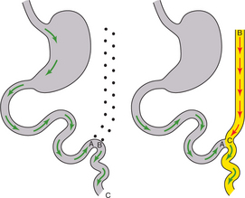
Note points A, B, and C in the drawing on the left in Figure 56-1. To create a Roux-en-Y limb, cut the jejunem at the line between A and B. “Pull up” point B of the mobile jejunem as shown in the figure on the right.
After you master (and can draw) a Roux-en-Y limb, study some of its applications on the following pages.
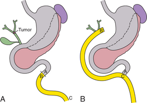
The patient in Figure 56-2A, for example, has a Klatskin tumor, a cholangiocarcinoma at the confluence of the right and left hepatic ducts. Though the bile ducts could be sewn into a loop of jejunem, this would allow the food stream to pass too closely to the biliary tree and runs the risk of ascending cholangitis. A Roux-en-Y hepaticojejunostomy, being isolated from the food stream, solves this problem.
Stay updated, free articles. Join our Telegram channel

Full access? Get Clinical Tree


