Chapter 59 Teaching Visual: Coronary Bypass
Medical Knowledge
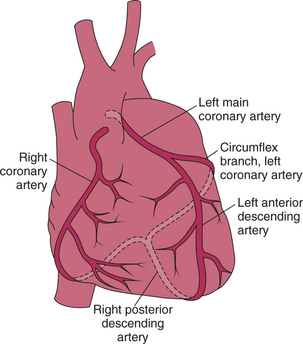
Note the important vessels in Figure 59-1: right, left main, LAD, and left circumflex. The right (∼85%), circumflex (∼10%), or both (∼5%) give off the posterior descending artery.
The gold standard conduit for left anterior descending (LAD) disease is the left IMA. The saphenous vein and the radial artery can also be used as conduits. The right IMA can be used as a graft to the right coronary artery. In poorly controlled or obese diabetic patients, simultaneous use of both IMAs is usually avoided due to a greater risk for sternal wound infections.
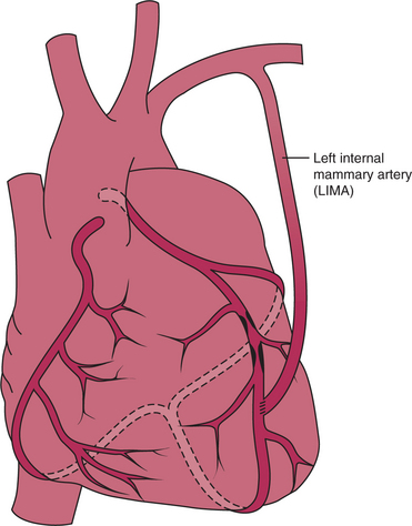
The IMA, which runs along the inside edge of the sternum, is remarkably resistant to atherosclerosis. The IMA is conveniently located near the most important coronary branch, the LAD, allowing transfer of the distal end of the IMA to the LAD after mobilizing the entire length of the IMA (Fig. 59-2).
Stay updated, free articles. Join our Telegram channel

Full access? Get Clinical Tree


