, Kyu Eun Lee1 and June Young Choi1
(1)
Department of Surgery, Seoul National University Hospital, Seoul, Korea, Republic of (South Korea)
Abstract
The thyroid gland is located in front of the neck below the thyroid cartilage. The small, two-or three-in. long gland consists of two lobes connected by isthmus in the middle.
Electronic supplementary material
The online version of this chapter (doi:10.1007/978-3-642-37262-9_1) contains supplementary material, which is available to authorized users.
1.1 The Thyroid Gland
The thyroid gland is located in front of the neck below the thyroid cartilage. The small, two-or three-in. long gland consists of two lobes connected by isthmus in the middle.
Just beneath the skin of the neck, there is a thin platysma muscle, which covers the whole anterior neck. When the platysma muscle is dissected, the strap muscles are exposed. The strap muscles consist of the sternohyoid muscles and sternothyroid muscles. The midline is shown between right and left strap muscles. Dissection along the midline is needed to approach the thyroid gland. To perform thyroidectomy, the strap muscles should be retracted to lateral position or can be transected for better exposure in case of large goiter.
Beneath the thyroid gland, there is a trachea. The trachea is the most important landmark in performing thyroidectomy. The thyroid gland is firmly attached to the trachea from the second to the fourth tracheal ring. In normal adults, the average weight of the thyroid gland is 15–30 g.
The pyramidal lobe can be seen in 20 % of the patients, which ascends from the isthmus or the adjacent part of either lobe up to the hyoid bone.


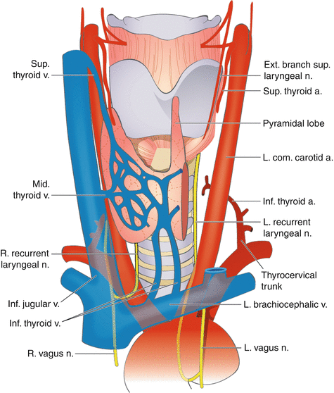
Fig. 1.1
Position and anatomy of the thyroid gland. (a) Normal position of the thyroid gland, (b) anatomy of the thyroid gland

Fig. 1.2
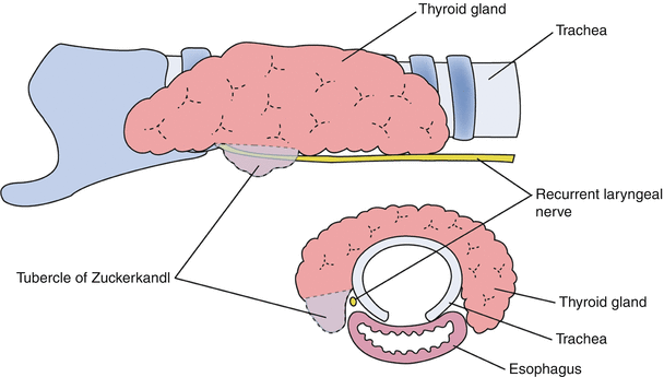
The region of the Tubercle of Zuckerkandl
The relation between the tubercle of Zuckerkandl and the distal course of the recurrent laryngeal nerve (RLN). Tubercle of Zuckerkandl is the most posterior extent of the thyroid lobe as shown in Fig. 1.2.
1.2 Vascular Anatomy of the Thyroid Gland
There are two main arterial blood supplies of the thyroid gland, the superior and inferior thyroid arteries and, to a lesser degree, the thyroid ima artery. The superior thyroid artery is the first branch of the external carotid artery and courses inferiorly to the upper pole of the thyroid gland. It enters the upper pole of the thyroid on its anterosuperior surface. The inferior thyroid artery usually arises from the thyrocervical trunk and passes upward in front of the vertebral artery and Longus colli to the lower pole of the thyroid gland. Before entering the thyroid, the artery usually divides into 2–3 branches. One of the branches supplies the inferior parathyroid.
The thyroid ima artery can be seen in 3–10 % or less of patients. The thyroid ima ascends in front of the trachea to the lower part of the thyroid gland, which it supplies. It varies greatly in size and appears to compensate for deficiency or absence of one of the other thyroid vessels. The thyroid ima artery usually arises from the brachiocephalic trunk (innominate artery). It occasionally arises from the aorta, the right common carotid, the subclavian, or the internal thoracic artery. All of these arteries should be ligated during surgical removal of the thyroid gland.
Venous drainage of the thyroid gland is through a well-developed thyroid venous plexus, which usually drains through the inferior thyroid vein to the left brachiocephalic (innominate) vein. Blood from the thyroid gland also drains to the internal jugular vein via the superior and middle thyroid veins.


