Squamous Papilloma
Elizabeth A. Montgomery, MD
Key Facts
Terminology
Benign tumor composed of hyperplastic squamous epithelium, overlying finger-like cores of lamina propria
Clinical Issues
Recurrences rare
Synchronous or metachronous carcinomas of upper aerodigestive tract reported
May be coincidental, as this has not held up in epidemiologic studies
Squamous papillomas are probably NOT markers for upper aerodigestive malignancy
Papillomas do not themselves undergo malignant transformation
Microscopic Pathology
Bland polypoid squamous mucosa
Fibrovascular cores
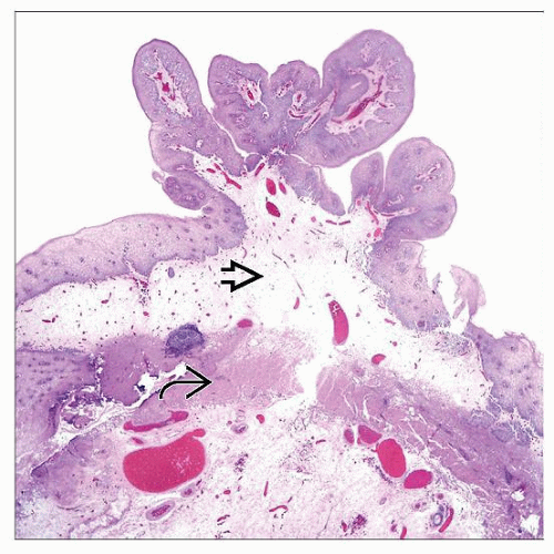 Hematoxylin & eosin low magnification shows a squamous papilloma with fronds and vascular cores after endoscopic removal. Note the edematous lamina propria  and the band of muscularis mucosae and the band of muscularis mucosae  . . |
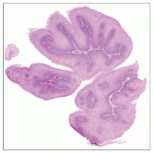 This is a typical appearance of a squamous papilloma on biopsies, as opposed to the appearance of the endoscopic mucosal resection from the previous image. Neither lamina propria nor muscularis mucosae are seen. |
TERMINOLOGY
Definitions
Benign tumor composed of hyperplastic squamous epithelium overlying finger-like cores of lamina propria
Association with human papilloma virus (HPV) is debatable
In most studies, only rare lesions have HPV detected by in situ hybridization or PCR
High prevalence in early studies may reflect faulty PCR technique
In more recent studies, < 10% of lesions are related to HPV
Patients with laryngeal papillomatosis can also have esophageal papillomatosis
In this case, lesions are associated with HPV
ETIOLOGY/PATHOGENESIS
Stay updated, free articles. Join our Telegram channel

Full access? Get Clinical Tree


