INTRODUCTION
The gram-positive spore-forming bacilli are the Bacillus and Clostridium species. These bacilli are ubiquitous, and because they form spores, they can survive in the environment for many years. Bacillus species are aerobes and the Clostridium species are anaerobes (see also Chapter 21).
Of the many species of Bacillus and related genera, most do not cause disease and are not well characterized in medical microbiology. There are a few species, however, that cause important diseases in humans. Anthrax, a classical disease in the history of microbiology, is caused by Bacillus anthracis. Anthrax remains an important disease of animals and occasionally of humans. Because of its potent toxins, B anthracis is a major potential agent of bioterrorism and biologic warfare. Bacillus cereus and Bacillus thuringiensis cause food poisoning and occasionally eye or other localized infections.
The genus Clostridium is extremely heterogeneous and more than 200 species have been described. The list of pathogenic organisms, as well as novel species isolated from human feces whose pathogenic potential remains undetermined, continues to grow. Clostridia cause several important toxin-mediated diseases, including tetanus (Clostridium tetani), botulism (Clostridium botulinum), gas gangrene (Clostridium perfringens), and antibiotic-associated diarrhea and pseudomembranous colitis (Clostridium difficile). Other clostridia are also found in mixed anaerobic infections in humans (see Chapter 21).
BACILLUS SPECIES
The genus Bacillus includes large aerobic, gram-positive rods occurring in chains. The members of this genus are closely related but differ both phenotypically and in terms of pathogenesis. Pathogenic species possess virulence plasmids. Most members of this genus are saprophytic organisms prevalent in soil, water, and air, and on vegetation (eg, Bacillus subtilis). Some are insect pathogens, such as B thuringiensis. This organism is also capable of causing disease in humans. B cereus can grow in foods and cause food poisoning by producing either an enterotoxin (diarrhea) or an emetic toxin (vomiting). Both B cereus and B thuringiensis may occasionally produce disease in immunocompromised humans (eg, meningitis, endocarditis, endophthalmitis, conjunctivitis, or acute gastroenteritis). B anthracis, which causes anthrax, is the principal pathogen of the genus.
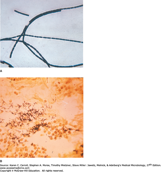
The typical cells, measuring 1 × 3–4 μm, have square ends and are arranged in long chains; spores are located in the center of the bacilli.
Colonies of B anthracis are round and have a “cut glass” appearance in transmitted light. Hemolysis is uncommon with B anthracis but common with B cereus and the saprophytic bacilli. Gelatin is liquefied, and growth in gelatin stabs resembles an inverted fir tree.
The saprophytic bacilli use simple sources of nitrogen and carbon for energy and growth. The spores are resistant to environmental changes, withstand dry heat and certain chemical disinfectants for moderate periods, and persist for years in dry earth. Animal products contaminated with anthrax spores (eg, hides, bristles, hair, wool, bone) can be sterilized by autoclaving.
BACILLUS ANTHRACIS
Anthrax is primarily a disease of herbivores—goats, sheep, cattle, horses, and so on; other animals (eg, rats) are relatively resistant to the infection. Anthrax is endemic among agrarian societies in developing countries in Africa, the Middle East, and Central America. A website maintained by the World Health Organization provides current information on disease in animals and is listed among the references. Humans become infected incidentally by contact with infected animals or their products. In animals, the portal of entry is the mouth and the gastrointestinal tract. Spores from contaminated soil find easy access when ingested with spiny or irritating vegetation. In humans, the infection is usually acquired by the entry of spores through injured skin (cutaneous anthrax) or rarely the mucous membranes (gastrointestinal anthrax) or by inhalation of spores into the lung (inhalation anthrax). A fourth category of the disease, injection anthrax, has caused outbreaks among persons who inject heroin that has been contaminated with anthrax spores. The spores germinate in the tissue at the site of entry, and growth of the vegetative organisms results in formation of a gelatinous edema and congestion. Bacilli spread via lymphatics to the bloodstream, and they multiply freely in the blood and tissues shortly before and after the animal’s death.
B anthracis (Figure 11-1) isolates that do not produce a capsule are not virulent and do not induce anthrax in test animals. The poly-γ-d-glutamic acid capsule is antiphagocytic. The capsule gene is present on a plasmid, pXO2.
Anthrax toxins are made up of three proteins, protective antigen (PA), edema factor (EF), and lethal factor (LF). PA is a protein that binds to specific cell receptors, and after proteolytic activation, it forms a membrane channel that mediates entry of EF and LF into the cell. EF is an adenylate cyclase; with PA, it forms a toxin known as edema toxin. Edema toxin is responsible for cell and tissue edema. LF plus PA form lethal toxin, which is a major virulence factor and cause of death in infected animals and humans. When injected into laboratory animals (eg, rats), the lethal toxin can quickly kill the animals by impairing both innate and adaptive immunity, allowing organism proliferation and cell death. The anthrax toxin genes are encoded on another plasmid, pXO1. In inhalation anthrax (woolsorters’ disease), the spores from the dust of wool, hair, or hides are inhaled; phagocytosed in the lungs; and transported by the lymphatic drainage to the mediastinal lymph nodes, where germination occurs. This is followed by toxin production and the development of hemorrhagic mediastinitis and sepsis, which are usually rapidly fatal. In anthrax sepsis, the number of organisms in the blood exceeds 107/mL just before death. In the Sverdlovsk inhalation anthrax outbreak of 1979 and the U.S. bioterrorism inhalation cases of 2001, the pathogenesis was the same as in inhalation anthrax from animal products.
In susceptible animals and humans, the organisms proliferate at the site of entry. The capsules remain intact, and the organisms are surrounded by a large amount of proteinaceous fluid containing few leukocytes from which they rapidly disseminate and reach the bloodstream.
In resistant animals, the organisms proliferate for a few hours, by which time there is massive accumulation of leukocytes. The capsules gradually disintegrate and disappear. The organisms remain localized.
In humans, approximately 95% of cases are cutaneous anthrax, and 5% are inhalation. Gastrointestinal anthrax is very rare; it has been reported from Africa, Asia, and the United States when people have eaten meat from infected animals.
The bioterrorism events in the fall of 2001 resulted in 22 cases of anthrax—11 inhalation and 11 cutaneous. Five of the patients with inhalation anthrax died. All of the other patients survived.
Cutaneous anthrax generally occurs on exposed surfaces of the arms or hands followed in frequency by the face and neck. A pruritic papule develops 1–7 days after entry of the organisms or spores through a scratch. Initially, it resembles an insect bite. The papule rapidly changes into a vesicle or small ring of vesicles that coalesce, and a necrotic ulcer develops. The lesions typically are 1–3 cm in diameter and have a characteristic central black eschar. Marked edema occurs. Lymphangitis, lymphadenopathy, and systemic signs and symptoms of fever, malaise, and headache may occur. After 7–10 days, the eschar is fully developed. Eventually, it dries, loosens, and separates; healing is by granulation and leaves a scar. It may take many weeks for the lesion to heal and the edema to subside. Antibiotic therapy does not appear to change the natural progression of the disease but prevents dissemination. In as many as 20% of patients, cutaneous anthrax can lead to sepsis, the consequences of systemic infection—including meningitis—and death.
The incubation period in inhalation anthrax may be as long as 6 weeks. The early clinical manifestations are associated with marked hemorrhagic necrosis and edema of the mediastinum. Substernal pain may be prominent, and there is pronounced mediastinal widening visible on chest radiographs. Hemorrhagic pleural effusions follow involvement of the pleura; cough is secondary to the effects on the trachea. Sepsis occurs, and there may be hematogenous spread to the gastrointestinal tract, causing bowel ulceration, or to the meninges, causing hemorrhagic meningitis. The fatality rate in inhalation anthrax is high in the setting of known exposure; it is higher when the diagnosis is not initially suspected.
Animals acquire anthrax through ingestion of spores and spread of the organisms from the intestinal tract. This is rare in humans, and gastrointestinal anthrax is extremely uncommon. Abdominal pain, vomiting, and bloody diarrhea are clinical signs.
Injection anthrax is characterized by extensive, painless, subcutaneous edema and the notable absence of the eschar characteristic of cutaneous anthrax. Patients may progress to hemodynamic instability due to septicemia.
Specimens to be examined are fluid or pus from a local lesion, blood, pleural fluid, and cerebrospinal fluid in inhalational anthrax associated with sepsis and stool or other intestinal contents in the case of gastrointestinal anthrax. Stained smears from the local lesion or of blood from dead animals often show chains of large gram-positive rods. Anthrax can be identified in dried smears by immunofluorescence staining techniques.
When grown on blood agar plates, the organisms produce nonhemolytic gray to white, tenacious colonies with a rough texture and a ground-glass appearance. Comma-shaped outgrowths (Medusa head, “curled hair”) may project from the colony. Demonstration of capsule requires growth on bicarbonate-containing medium in 5–7% carbon dioxide. Gram stain shows large gram-positive rods. Carbohydrate fermentation is not useful. In semisolid medium, anthrax bacilli are always nonmotile, but related organisms (eg, B cereus) exhibit motility by “swarming.” Clinical laboratories that recover large gram-positive rods from blood, cerebrospinal fluid, or suspicious skin lesions, which phenotypically match the description of B anthracis as mentioned, should immediately contact their public health laboratory and send the organism for confirmation. Definitive identification requires lysis by a specific anthrax γ-bacteriophage, detection of the capsule by fluorescent antibody, or identification of toxin genes by polymerase chain reaction (PCR). These tests are available in most public health laboratories.
A rapid enzyme-linked immunoassay (ELISA) that measures total antibody to PA has been approved by the U.S. Food and Drug Administration (FDA), but the test result is not positive early in disease.
Immunization to prevent anthrax is based on the classic experiments of Louis Pasteur. In 1881, he proved that cultures grown in broth at 42–52°C for several months lost much of their virulence and could be injected live into sheep and cattle without causing disease; subsequently, such animals proved to be immune. Active immunity to anthrax can be induced in susceptible animals by vaccination with live attenuated bacilli, with spore suspensions, or with PA from culture filtrates. Animals that graze in known anthrax districts should be immunized for anthrax annually.
In the United States, the current FDA-approved vaccine (AVA BioThrax®, Emergent BioSolutions, Inc, Rockville, MD) is made from the supernatant of a cell-free culture of an unencapsulated but toxigenic strain of B anthracis that contains PA adsorbed to aluminum hydroxide. The dose schedule is 0.5 mL administered intramuscularly at 0 and 4 weeks and then at 6, 12, and 18 months followed by annual boosters. The vaccine is available only to the U.S. Department of Defense and to persons at risk for repeated exposure to B anthracis. Because the current anthrax vaccines provide short-lived immunity and hence require repeated vaccinations, a number of new recombinant PA vaccines (rPA) have been developed. These novel vaccines have been shown to be very well tolerated and highly immunogenic (see discussion under Treatment).
Many antibiotics are effective against anthrax in humans, but treatment must be started early. Ciprofloxacin is recommended for treatment; other agents with activity include penicillin G, doxycycline, erythromycin, and vancomycin. In the setting of potential exposure to B anthracis as an agent of biologic warfare, prophylaxis with ciprofloxacin or doxycycline should be given for 60 days and three doses of vaccine (AVA BioThrax) should be administered.
Raxibacumab (Abthrax®, GlaxoSmithKline, London, UK), a recombinant human monoclonal antibody, was FDA approved for treatment of and prophylaxis against inhalational anthrax in late 2012. The mechanism of action is prevention of binding of PA to its receptors in host cells. The drug is used in combination with appropriate antimicrobial agents.
Anthrax immune globulin intravenous (AIGIV, Cangene Corp. Winnipeg, Manitoba, CA) is not FDA approved but could be made available through the Centers for Disease Control and Prevention. AIGIV is a human polyclonal antiserum that also inhibits binding of PA to its receptors. Like Raxibacumab, it is used as an adjunct to antimicrobial agents for the treatment of severe forms of anthrax.
Soil is contaminated with anthrax spores from the carcasses of dead animals. These spores remain viable for decades. Perhaps, spores can germinate in soil at a pH of 6.5 at proper temperature. Grazing animals infected through injured mucous membranes serve to perpetuate the chain of infection. Contact with infected animals or with their hides, hair, and bristles is the source of infection in humans. Control measures include (1) disposal of animal carcasses by burning or by deep burial in lime pits, (2) decontamination (usually by autoclaving) of animal products, (3) protective clothing and gloves for handling potentially infected materials, and (4) active immunization of domestic animals with live attenuated vaccines. Persons with high occupational risk should be immunized.
BACILLUS CEREUS
Food poisoning caused by B cereus has two distinct forms, the emetic type, which is associated with fried rice, milk, and pasta, and the diarrheal type, which is associated with meat dishes and sauces. B cereus produces toxins that cause disease that is more of intoxication than a food-borne infection. The emetic form is manifested by nausea, vomiting, abdominal cramps, and occasionally diarrhea and is self-limiting, with recovery occurring within 24 hours. It begins 1–5 hours after ingestion of a plasmid-encoded preformed cyclic peptide (emetic toxin) in the contaminated food products. B cereus is a soil organism that commonly contaminates rice. When large amounts of rice are cooked and allowed to cool slowly, the B cereus spores germinate, and the vegetative cells produce the toxin during log-phase growth or during sporulation. The diarrheal form has an incubation period of 1–24 hours and is manifested by profuse diarrhea with abdominal pain and cramps; fever and vomiting are uncommon. In this syndrome, ingested spores that develop into vegetative cells of B cereus secrete one of three possible enterotoxins which induce fluid accumulation and other physiological responses in the small intestine. The presence of B cereus in a patient’s stool is not sufficient to make a diagnosis of B cereus disease because the bacteria may be present in normal stool specimens; a concentration of 105 bacteria or more per gram of food is considered diagnostic.


