Specimen Handling, Whipple
Laura Webb Lamps, MD
Grace E. Kim, MD
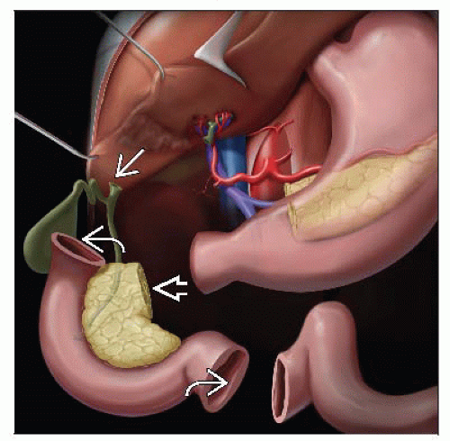
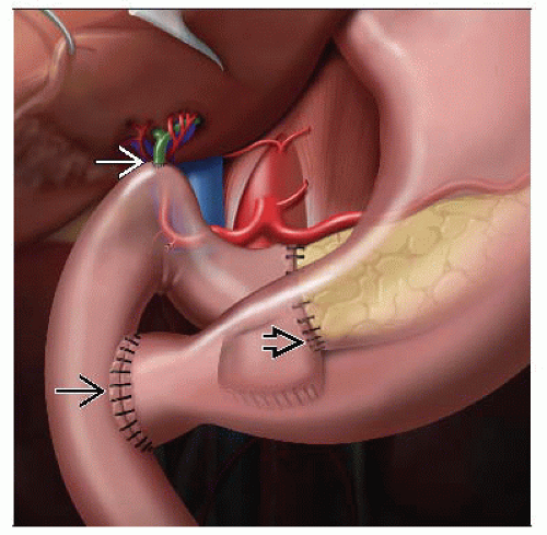
WHIPPLE (PANCREATICODUODENECTOMY)
Major Components
Duodenum
May or may not include pylorus depending on whether it was a pylorus-sparing procedure
Ampulla of Vater
Common bile duct
Pancreas
Anatomic Orientation
Duodenum
Free proximal end usually shorter than free distal segment
Small portion of stomach usually attached to proximal end
Distal end may be either duodenum or jejunum
Common bile duct
Sometimes greenish in color
Posterior and superior to pancreas
May be easier to identify from ampulla than from transected end
If gallbladder is present, can identify insertion of cystic duct and follow to common bile duct
Ampulla of Vater
Usually obvious within duodenum if not obscured by tumor
Some patients have an accessory ampulla that drains accessory duct of Santorini
Pancreas
General anatomic features
Retroperitoneal organ located in C-groove of 2nd part of duodenum
Anterior to pancreas is free space (omental bursa/lesser sac) and then posterior aspect of stomach
Anatomic divisions of pancreas
Head: To right of superior mesenteric vein/portal vein confluence; includes uncinate process
Neck: Constricted region to left of head
Body: Between superior mesenteric vein/portal vein confluence and aorta
Tail: Between aorta and splenic hilum
Pancreatic duct
Usually main pancreatic duct drains bulk of gland into duodenum at major duodenal papilla (ampulla) along with common bile duct
Normal diameter is < 1 cm
Specimen Handing
Identify proximal end of intestines
Usually shorter than distal end
Head of pancreas sits in duodenal C-loop
Neck margin can be identified as oval-cut pancreatic surface with central duct
Determine anterior vs. posterior pancreatic surface
Anterior pancreatic surface bulges
Posterior pancreatic surface is flat
Common bile duct is superior to pancreas near 1st part of duodenum
Adsay trapezoid method of orientation
Useful method to identify essential margins/surfaces
Place proximal intestinal margin to left, distal intestinal margin to right, and medial aspect of pancreas facing toward you
Visualize a trapezoid
Left nonparallel side represents pancreatic neck margin
Right nonparallel side is uncinate margin
Space between sides is vascular groove
Anterior surface is base, and posterior surface is parallel opposite side
Hand method of orientation
Curled left hand resembles pancreas enveloping superior mesenteric artery and portal vein
Thumb is uncinate process; flat fingers are neck, body, tail
Surgical Margins
Common bile duct (shave margin)
Pancreatic resection (shave margin to include duct)
CAP calls this the distal margin
AJCC calls this the pancreatic neck
Uncinate/retroperitoneal (perpendicular margin)
CAP: Uncinate
AJCC: Retroperitoneal
Should be inked, sectioned perpendicularly, and entire area submitted
Additional lymph nodes often found if this method is used
Stay updated, free articles. Join our Telegram channel

Full access? Get Clinical Tree


