Soft Tissue Lesions with Clear or Granular Cells


Soft Tissue Lesions with Clear or Granular Cells


INTRODUCTION

Clear cell change can be seen in some examples of smooth muscle tumors (see Chapter 6), myoepithelial tumors (see Chapter 7), atypical fibroxanthoma (see Chapter 13), Ewing sarcoma (see Chapter 14), rhabdomyosarcoma (see Chapter 14), and gastrointestinal stromal tumor (see Chapter 5). Tumors in which clear cell change can be predominant include glomus tumor (see Chapter 7), alveolar soft part sarcoma, clear cell sarcoma, perivascular epithelioid cell tumor (PEComa), and paraganglioma as well as metastases from other organs including carcinomas. Lesions with lipoblastic and pseudolipoblastic differentiation can also enter the differential diagnosis and are discussed in Chapter 15. Granular cell or oncocytic change is seen in granular cell tumor and in some examples of hibernoma (see Chapter 15), myoepithelial tumor (see Chapter 7), smooth muscle tumors (see Chapter 6), nerve sheath tumors (see Chapter 9), dermatofibroma (see Chapter 4), dermatofibrosarcoma protuberans (see Chapter 4), atypical fibroxanthoma (see Chapter 13), and benign and malignant PEComas. Finally, reactive and neoplastic histiocytic proliferations can present in soft tissue as clear or granular cell tumors. The differential diagnoses are summarized in Tables 12.1 and 12.2.


CLEAR CELL SARCOMA OF SOFT TISSUE


Clinical Features

This is a tumor of young adults (more often in females) that arises predominantly in the extremities, especially the foot and rarely in trunk or head and neck sites as a slowly growing firm mass. The usual location is deep subcutaneous or subfascial tissue, often with involvement of tendon sheath or aponeurosis. This is an aggressive neoplasm that metastasizes to lymph node, lung, and bone, with survival rates of 67%, 33%, and 10% after 5, 10, and 15 years, respectively.1,2 A genetically distinct, clear cell sarcoma-like tumor arises in the gastrointestinal tract (CCSLTGIT),3 where it presents with obstruction, bleeding, or mass effect. Neither subtype is related to clear cell sarcoma of kidney.


TABLE 12.1 Differential Diagnosis of Clear Cell and Granular Cell Tumors


Typical Clinical Features


Microscopic Features


Ancillary Investigations


Clear cell cutaneous fibrous histiocytoma


Raised firm nodule on extremities, young adults


Epidermal hyperplasia, areas of typical dermatofibroma, peripheral collagen bundles


CK−, SMA−, desmin−, S100 protein−


Clear cell atypical fibroxanthoma


Sun-exposed areas, elderly patients


Sheets of clear cells with pleomorphic nuclei, and prominent cell membranes


CD10+ in some cases, S100 protein−, HMB45−, melan-A−, desmin−, CK−


Ossifying fibromyxoid tumor


Subcutaneous well-demarcated nodule, any age


Partial shell of bone in many, thick fibrous capsule, cords of rounded glomus-like or occasionally spindled cells, fibromyxoid stroma


Atypical variant can have increased cellularity, mitoses, intralesional osteoid


S100 protein+, GFAP+, desmin/SMA±, CK+ rarely, t(6;12)(p21;q24.3), EP400-PHF1 fusion


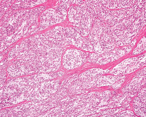
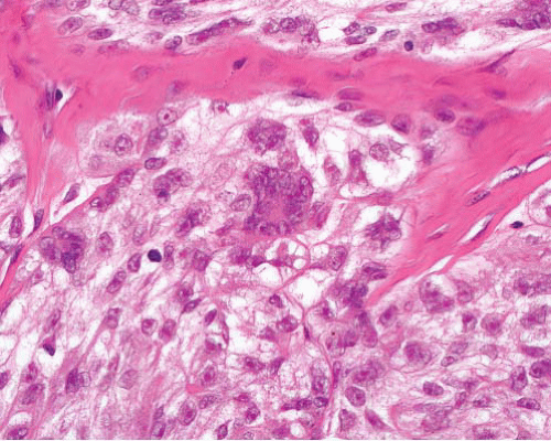
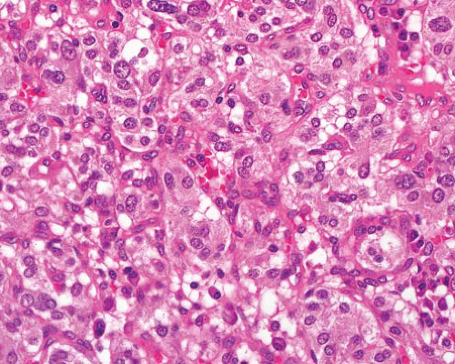
Clear cell sarcoma (soft tissue)


Extremities especially lower limb, young adults


In subcutis or deeper tissue, it involves tendons


Subset in gastrointestinal tract


Nested pattern, round nuclei with central nucleolus, clear or granular cytoplasm, sometimes spindling, multinucleated cells, melanin pigment


No junctional activity


S100 protein+, HMB45+ and melan-A+ (except in GI tract), other markers negative t(12;22)(q13;q12), EWSR1-ATF1 fusion or t(2;22)(q33;q12), EWSR1-CREB1 fusion (more often in GI cases)


Granular cell tumor


Subcutaneous in adults, especially in head and neck sites


Also in various organs


Infiltrative sheets and cords of large polygonal cells, coarsely granular cytoplasm


Clear cell areas often present also


S100 protein+, CD68+, CEA+


Granular cell dermatofibrosarcoma


Like typical dermatofibrosarcoma


Sheets of granular cells involving dermis and superficial subcutis


Areas of typical dermatofibrosarcoma usually present, with transitional forms


CD34+, S100 protein−


Granular cell leiomyoma


Rare focal change in smooth muscle tumors of soft tissue or gynecologic origin


Granular cell areas within epithelioid or spindle cell smooth muscle tumor


SMA+, desmin+, h-caldesmon+, S100 protein−


Paraganglioma


In locations of paraganglia— carotid body, base of skull, mediastinum, retroperitoneum


Some are functional— hypertension, vagal symptoms


Nests of ovoid cells with clear or granular cytoplasm and variable nuclear pleomorphism


Prominent or sinusoidal thin-walled blood vessels, sometimes fibrosis


CD56+, NF+, SYN+, CG+. S100 protein+ in sustentacular cells


Paraganglioma-like dermal melanocytic tumor


Adult females, extremities, dermal nodule


Circumscribed or infiltrative, can extend into subcutis


Nests of cells within slender fibrous septa, uniform rounded nuclei, small nucleoli, abundant clear or amphophilic cytoplasm


S100 protein+, HMB45+, melan-A±


Alveolar soft part sarcoma


Childhood or adult


Single mass or multicentric


Mesenteric, retroperitoneal, other sites


Nests of large polygonal cells with uniform vesicular nuclei and clear or finely granular cytoplasm. A solid pattern of closely packed nests is sometimes seen especially in childhood cases


TFE3+, desmin+ der(17) t(X;17)(p11;q25), ASPSCR1-TFE3 fusion


Rhabdomyoma


Adult or fetal (median: 4 y) type in head and neck, or (mostly female) genital tract locations


Also cardiac rhabdomyoma (adult type)


Sheets of large polygonal cells with abundant eosinophilic cytoplasm


Fetal rhabdomyoma has long spindle cells in myxoid stroma (immature type) or spindled and round cells with variable skeletal muscle differentiation (intermediate type)


Desmin+, myogenin+ (in nuclei), MyoD1+ (in nuclei)


Perivascular epithelioid cell tumor (other than organ-specific subtypes)


Intra-abdominal, soft tissue or gynecologic locations


Nests of ovoid or spindled cells with clear or granular cytoplasm, delicate fibrous septa


Malignant variants often epithelioid with large polygonal cells, pleomorphism, mitoses, necrosis


SMA+, HMB45+, melan-A+, desmin±, CD117±, S100 protein+ rarely, TFE3±, cathepsin K


TFE3 gene rearrangement ±


Chordoma


Mainly in sacrum/coccyx, or skull base, rarely other vertebrae


Can extend into adjacent tissues as tumor mass


Cords and nests of polygonal cells with eosinophilic cytoplasm, resembling adenocarcinoma


Larger vacuolated (physaliferous) cells


Can have areas of dedifferentiation


S100 protein+, CK+, EMA+, brachyury+


Epithelioid leiomyosarcoma


F > M


Gynecologic sites


Retroperitoneum, bowel wall or wall of vessel including inferior vena cava, renal vein


Sheets of polygonal cells with clear or finely granular cytoplasm


Not usually pleomorphic


Typical spindle cell areas can coexist


SMA+, desmin+, h-caldesmon+, CD117−, HMB45±


Epithelioid gastrointestinal stromal tumor


Related to wall of any part of alimentary tract (most commonly stomach, small intestine)


Also, in retroperitoneum, omentum


Sheets of clear or epithelioid cells often with a focal spindle component


Organoid pattern, plasmacytoid or rhabdoid change can be seen


CD117+, DOG1+, CD34+, h-caldesmon+. SMA variable, desmin+ rarely, S100 protein+ rarely. CK± after therapy. KIT or PDGFRA or KIT mutations


Epithelioid pleomorphic liposarcoma


Deep soft tissue, extremities, rarely retroperitoneum, rarely subcutis


Sheets of clear or granular cells, pleomorphic lipoblasts


Usually focal, more typical pleomorphic liposarcoma elsewhere


S100 protein+, AE1/3+, melan-A+, SMA+


Renal cell carcinoma


Origin in kidney, can present as metastasis


Clear or granular cells in sheets, or adenopapillary formations


Variable nuclear grade


Can have sarcomatoid change with divergent differentiation


CK+, EMA+, RCC+, CD117+ (oncocytoma), vimentin+ (chromophobe carcinoma)


Adrenal cortical carcinoma


Origin in adrenal gland


Can be functioning or nonfunctioning


Sheets of clear or granular cells with small nuclei and occasional pleomorphism


Lacks sinusoidal vascular pattern


Inhibin+, melan-A+, CD56+, CK−


TABLE 12.2 Differential Diagnosis of Histiocytic Tumors in Soft Tissue


Typical Clinical Features


Microscopic Features


Ancillary Investigations


Juvenile xanthogranuloma, cutaneous


Infants, head and neck, trunk, proximal limbs


Yellow-brown plaque or nodule


Infiltrative dermal lesion


Sheets of histiocytes


Older lesions have more foamy cells and more Touton giant cells


CD68+, CD163+, S100 protein−, CD1a−


Juvenile xanthogranuloma, deep


Infants or young children, trunk, intramuscular


Monotonous sheets of macrophages, occasional foamy or multinucleated cells


CD68+, S100 protein, CD1a−


Xanthoma


Skin or subcutis, rarely associated with tendon sheath or joint synovium


Hyperlipidemia or normolipidemia


Circumscribed or diffuse, sheets of foamy lipid-laden macrophages, variable admixture of siderophages, cholesterol clefts with giant cells, fibrosis


Reticulohistiocytoma


Solitary cutaneous or multicentric reticulohistiocytosis affecting skin and synovium of joints


Multinucleated giant cells predominate, with plump histiocytes and mixed inflammatory cells


PAS+ (diastase resistant) CD68+, CD163+, MiTF± (nuclear)


Epithelioid benign cutaneous fibrous histiocytoma


Dermal nodule, extremities or trunk, young adults


Overlying epidermal hyperplasia, bland plump cells, sometimes spindle component, peripheral collagen bundles, lymphocytic infiltrate


SMA+, CD34−, S100 protein−, desmin−


Rosai-Dorfman disease


Children or young adults


Lymph node or soft tissue mass


Large cells with abundant clear cytoplasm, phagocytosis of lymphocytes, mild or moderate focal nuclear enlargement and hyperchromasia


Admixed plasma cells, lymphocytes


S100 protein+, CD68+, CD1a−


Langerhans cell histiocytosis


Bone, lung, lymph node, spleen, pituitary, or other affected


Occasionally seen in a biopsy for suspected soft tissue tumor


Sheets of ovoid cells with folded or grooved nuclei, no nucleoli admixed eosinophils


S100 protein+, CD1a+, langerin+, CD68+ in some


Birbeck granules on EM


Silicone granuloma


History of injection or implant, with recent change


Lumpy swelling


Foamy lipoblast-like cells, foreign body giant cells, birefringent material, fibrosis


CK−, S100 protein−


Malakoplakia


History of urinary tract infection


Can form perinephric mass


Sheets of discohesive polygonal cells with eosinophilic cytoplasm and dot-like inclusions (Michaelis-Gutmann)


Von Kossa stain+, CD68+


Granular histiocytic reaction


Related to surgery, often hip joint replacement


Nodules and sheets of histiocytes with granular, eosinophilic cytoplasm infiltrate fat


Sometimes adjacent amorphous foreign material


CD68+, S100 protein−, epithelial markers negative


Polyvinylpyrrolidone storage disease


Following intravenous plasma expander, exposure to some cosmetics or drugs containing polyvinylpyrrolidone


Sheets of macrophages containing bubbly mucin-like material


Mucicarmine+, CD68+, CK−, EMA−, S100 protein−


Crystal-storing histiocytosis


Associated with B-cell lymphoma or myeloma, rarely other causes


Cells expanded by cytoplasmic rod-like crystals


CD68+, S100 protein−


Mycobacterial spindle cell pseudotumor


HIV-positive patients, in lymph nodes, skin, spleen, lung, bone marrow, brain


Mycobacteria often atypical species


Sheets of spindle cells with storiform pattern and plump epithelioid macrophages, foamy and multinucleated cells


Ziehl-Neelsen stain for acid-fast bacilli is positive


Pathologic Features

This is a firm white tumor that can appear lobulated or multinodular (e-Fig. 12.1) and can be focally necrotic. Melanin pigmentation is rarely seen on gross examination. Histologically, the tumor is typically composed of uniform nests, separated by delicate connective tissue septa, of round cells with clear or finely granular eosinophilic cytoplasm, and centrally located rounded nuclei each with a single basophilic nucleolus (Figs. 12.1 and 12.2, e-Figs. 12.2 to 12.9). Cells can be spindled with a fascicular architecture in some cases (e-Fig. 12.5), but pleomorphism is rare and mitotic activity often low. CCSLTGIT can appear similar but can also display sheets of undifferentiated polygonal cells without clear cell features (e-Figs. 12.7 to 12.9). Multinucleated giant cells are seen in some examples (e-Fig. 12.4), and CCSLTGIT can have osteoclast-like giant cells. Rarely, there is myxoid change (e-Fig. 12.6). Melanin pigment is seen in tumor cells in about half of the soft tissue cases.


Ancillary Investigations

Reticulin staining shows a pattern of small nests (e-Fig. 12.10). Histochemical stains for melanin pigment (e.g., Fontana Masson) can sometime reveal it when unapparent on hematoxylin and eosin, and melanosomes can be found with electron microscopy in many cases. Immunohistochemistry is diffusely positive for S100 protein (e-Fig. 12.11) and usually for HMB45 (e-Fig. 12.12) and melan-A. The latter two, however, are negative in CCSLTGIT. Neural markers and rarely cytokeratin have been reported in occasional cases. CD117 is usually negative in contrast to some cases
of melanoma, but because many of the latter are also negative, this is of limited value for diagnosis.


FIGURE 12.1 Clear cell sarcoma. Uniform tumor cells form variably sized nests separated by fibrous septa of varying thickness.


FIGURE 12.2 Clear cell sarcoma. The cells have round nuclei, distinct nucleoli, and moderate amounts of amphophilic cytoplasm. The multinucleated cell shown here is a frequent feature of these tumors in soft tissue.

Genetically, clear cell sarcoma of soft parts is characterized by t(12;22)(q13;q12) with fusion of EWS and ATF1 genes as well as additional aberrations.4 Many examples of the gastrointestinal clear cell sarcoma-like tumor, although morphologically similar, bear a different translocation, t(2;22)(q33;q12), which results in the EWS-CREB1 gene fusion.3 Thus, molecular analysis can exclude metastatic melanoma. The subtypes are not wholly genetically distinct since both fusion genes have been identified in examples of both.5 These two translocations are identical to the genetic rearrangements in some examples of angiomatoid fibrous histiocytoma, a morphologically and immunophenotypically different entity (see Chapter 17)6,7; EWSR1-CREB1 fusion is also found in primary pulmonary myxoid sarcoma.8


PARAGANGLIOMA


Clinical Features

Tumors of paraganglia arise in a variety of locations, of which the principal ones are carotid, mediastinal, retroperitoneal (from aorticosympathetic paraganglia or organ of Zuckerkandl), jugulotympanic, and vagal. Rare sites are larynx, orbit, nasopharynx, heart, and cauda equina. Although the chromaffin reaction has fallen into disuse, the term extra-adrenal paraganglioma is sometimes used for chromaffin-positive tumors, which
are presumed to be similar to pheochromocytoma of adrenal medulla, and can cause raised amounts of urinary catecholamines, whereas those of chemoreceptor (branchiomeric) type are chromaffin-negative and usually nonfunctional.

Paragangliomas usually present with symptoms due to a mass and are sometimes multifocal, especially in familial examples. Some, such as glomus jugulare tumors, can have functional effects, including on blood pressure, and vagal paragangliomas can affect cranial nerves at the base of the skull. Mediastinal tumors, arising from aortic arch or pulmonary artery paraganglia (anterior mediastinum) or sympathetic paraganglia (posterior mediastinum), can be part of Carney triad. Paragangliomas have also been associated with neurofibromatosis type 1, multiple endocrine neoplasia syndromes, and von Hippel-Lindau disease. Most paragangliomas are benign, but those in retroperitoneum or mediastinum tend to be more aggressive. Germline mutations in SDHB are associated with familial paraganglioma. SDHB-deficient extra-adrenal paragangliomas are more likely to be malignant.9 Malignant examples can metastasize to lymph nodes and lung or bone.10


Pathologic Features

These tumors are usually encapsulated and are composed of round or polygonal cells with central nucleoli and finely granular, clear or sometimes vacuolated cytoplasm, arranged in nests or cords within a vascular stroma (Fig. 12.3, e-Figs. 12.13 to 12.16). The cells are larger than normal,
and nuclei can show pleomorphism. The nests are surrounded to a variable extent by sustentacular cells with elongated cytoplasmic processes. A more syncytial pattern can be found in retroperitoneal paragangliomas. Nuclear variation and hyperchromasia are not uncommon (e-Fig. 12.15), and the cells can display clear cytoplasm (e-Fig. 12.16). The stroma can be focally hyalinized11 (e-Fig. 12.17), and the vessels can be hemangiopericytomatous (e-Fig. 12.18), with both features coexisting in some examples. Features suggestive of malignancy include marked nuclear pleomorphism, mitotic activity, necrosis, and vascular invasion. However, none is reliably predictive of behavior, and tumors without these features have metastasized.


FIGURE 12.3 Paraganglioma. Cells in nested pattern. The cells are arranged in discrete nests, separated by delicate fibrovascular septa. The cells have rounded nuclei with small nucleoli. The variation in nuclear size and staining is typical and does not indicate malignant potential.

Jun 18, 2016 | Posted by in PATHOLOGY & LABORATORY MEDICINE | Comments Off on Soft Tissue Lesions with Clear or Granular Cells

Full access? Get Clinical Tree

Get Clinical Tree app for offline access