Rickettsia and Related Organisms
Rickettsia and related organisms are obligate intracellular gram-negative organisms that belong to the subdivision of α-proteobacteria, with a lifecycle that includes both vertebrate and invertebrate (arthropods) hosts. Rickettsiae cause systemic disease and the lungs are affected in human infections.
Part 1 Rickettsia
Juan P. Olano
Rickettsiae are obligate intracellular bacteria that have evolved in association with arthropod vectors. The genus can be subdivided into typhus group and spotted fever group rickettsioses. These infections are present throughout the world. In the United States, the most common rickettsioses are Rocky Mountain spotted fever, caused by Rickettsia rickettsii and transmitted by tick vectors; and endemic typhus, caused by R. typhi and transmitted by fleas. Isolated outbreaks of rickettsial pox, caused by R. akari and transmitted by mites, have been described in New York. A spotted fever rickettsiosis caused by R. parkeri has been documented in the United States and Latin America since 2002.
All rickettsial infections target the endothelium lining the microvasculature; therefore, clinical manifestations are systemic, and the morbidity and mortality of the disease are due to cerebral and pulmonary edema resulting from increased microvascular permeability. In the lungs, the clinical picture is that of a noncardiogenic pulmonary edema and/or adult respiratory distress syndrome.
Histologic Features
Rickettsiae are small (0.3 × 1.0 μm), gram-negative bacilli.
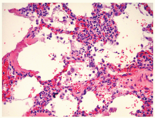
Lungs show alveolar edema and/or hyaline membranes.
Lymphohistiocytic infiltration of alveolar septa (interstitial pneumonitis) and perivascular spaces (pulmonary perivasculitis/vasculitis).
Rickettsiae can be demonstrated in the endothelial cells of the microcirculation by immunoperoxidase or immunofluorescent techniques, using polyclonal antibodies or monoclonal antibodies specific for either typhus group or spotted fever group LPS.
 Figure 98.1 Abundant mononuclear cells in the interstitial compartment and hyaline membranes lining one alveolar septum, from a fatal case of Rocky Mountain spotted fever.
Stay updated, free articles. Join our Telegram channel
Full access? Get Clinical Tree
 Get Clinical Tree app for offline access

|