11 Respiratory Disorders
Acute breathlessness
Case history (1)
You are called by the nurses to see a 63-year-old man who has become breathless. As you walk over to the ward you consider what the causes might be on this rather scanty history.
Remember
Respiratory diseases can cause breathlessness within minutes or hours, or slowly over days, weeks or months.
The systems involved would most probably be cardiac or respiratory. You go through the list in Box 11.1 (p. 346) as you walk to the ward.
Initial assessment
Then make a full cardiac and respiratory examination. Is there any evidence of DVT?
This vague history shows the vast diagnostic possibilities, shown in the table but a shorter differential diagnosis can be made in context of the age of the patient, the past medical history and the current clinical situation. This man turned out to have heart failure.
Upper airways obstruction
Case history (2)
A 29-year-old male had been at a party where he had consumed a lot of alcohol. He managed to get home with the help of his friend but he then vomited and fell to the floor. He was holding his throat and had great difficulty in breathing. The friend realised that he was in great distress and called an ambulance.
Diagnosis: Upper airways obstruction
Fortunately, the emergency crew recognised the problem and tried to clear the airway. There was no improvement so the Heimlich manoeuvre was performed and a large bone was expelled with immediate relief of the man’s respiratory distress. He was taken to A&E for assessment but discharged after 2 hours.
Remember
The Heimlich manoeuvre is used to expel an inhaled foreign body:
• Encircle the upper part of the abdomen, just below the patient’s rib cage, with your arms
• Give a sharp, forceful squeeze, forcing the diaphragm sharply into the thorax.
This should expel sufficient air from the lungs to force the foreign body out of the trachea. If this fails, urgent assessment by experienced ENT or cardiothoracic clinician is required. Fibre optic or rigid bronchoscopy will be required if obstruction beyond vocal cords.
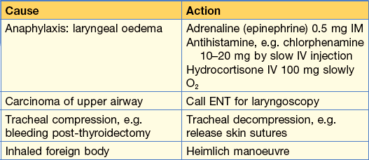
Other causes of upper airway obstruction are shown in Table 11.1, all of which require emergency management, which is also shown.
Cough
Cough is common and when persistent can cause considerable fear and distress. Apart from the immediate discomfort of coughing, paroxysmal cough can interrupt sleep, provoke retching or vomiting and, if severe, result in rib fractures or syncope. Always enquire about sputum and its colour.
Case history
A 67-year-old man is admitted to hospital with a myocardial infarct. Following successful coronary intervention he is discharged on anti-platelet therapy, atorvastatin and enalapril. When seen again 2 weeks later, he was very well but complained of a persistent hacking cough. This had been keeping him awake at night.
In this case, as the patient has only a cough and is not breathless, the cause is very likely to be the ACE inhibitor (enalapril). This should be stopped and an ACE receptor antagonist, e.g. vasartan started. Unlike ACE inhibitors, angiotensin receptor antagonists do not affect bradykinin metabolism and do not produce a cough.
Cough is provoked by stimulation of mucosal and stretch receptors of the lung. Accordingly it has a number of causes:
Acute cough
• Inhalation of direct irritants, e.g. smoke, chlorine gas, ozone and other air pollutants.
• In the asthmatic, inhalation of specific allergen, e.g. pollen or non-specific low-concentration irritants, e.g. perfume, tobacco fumes.
• Upper and lower respiratory tract infections (yellow/green sputum).
Chronic cough
• Large airway obstruction, e.g. carcinoma of bronchus or inhaled foreign body (Note: peanuts and other inhaled food will not be radio-dense).
• Persistent bronchial inflammation, e.g. asthma, bronchiectasis, COPD, smoking.
• Persistent infection, e.g. tuberculosis, lung abscess.
• Interstitial lung disease, e.g. pulmonary fibrosis, asbestosis.
• Raised left atrial pressure, e.g. mitral stenosis, left ventricular failure.
• Gastro-oesophageal reflux (GORD) and bulbar dysfunction.
Enquire about associated features
Investigations
All patients with persistent cough should have a CXR
Other investigations will depend on the likely cause, e.g. serial peak flow in asthma, contrast studies if gastro-oesophageal reflux or aspiration is suspected and sputum culture, including request for Mycobacterium tuberculosis isolation if the cough is productive.
Breathlessness and wheeze
Asthma causes breathlessness, cough and wheeze. However, all that wheezes is not asthma.
Differential diagnosis of asthma:
• Upper airway obstruction with stridor:
• Left ventricular failure (LVF):
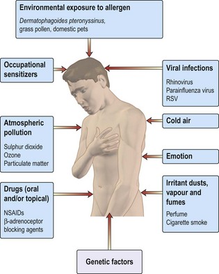
Figure 11.1 shows causes and triggers of asthma.

Figure 11.1 Causes and triggers of asthma. RSV, respiratory syncytial virus; NSAIDs, non-steroidal anti-inflammatory drugs.
Case history
A 24-year-old female was admitted to MAU with breathlessness for 6 h. She has had a ‘cold’ for 2 days. Past history revealed her being ‘chesty’ as a child. It was noticed that she could not complete sentences easily. Heart rate was 126/min and respiratory rate was 30/min. There was expiratory wheeze over the lung fields. This patient has severe acute asthma.
How would you manage this patient in A&E?
Immediate management should be:
• Reassurance that treatment will be effective
• Oxygen 40–60% (Keep SaO2 > 90%)
• Nebulised short acting β2 agonist (SAB), e.g. salbutamol 5 mg or terbutaline 10 mg) with oxygen. (NB only 10% of drug reaches the lungs)
• Arterial blood gases (ABGs):
• PEFR 4-hourly. Note: performing PEFR might be distressing for patient: do not demand unnecessary repeat testing
30 min later there has been no significant improvement in her breathlessness and PEFR. What would you do next?
• Continue with nebulised β2 agonist, now with added ipratropium (500 µg). Repeat every 30 min if necessary.
• Intravenous aminophylline infusion can be used if patient does not respond to repeated nebulisation with β2 agonist and ipratropium. However, do not use this if oral aminophylline has been taken.
There is now improvement in PEFR and breathlessness with nebulised β2 agonist and ipratropium. What treatment should be offered to her now?
Continuing management should be:
24–48 h before discharge
• Add inhaled corticosteroids, e.g. beclometasone, to oral steroids.
• Replace nebulised bronchodilators with inhalers.
• Introduce long-acting inhaled β2 agonists, e.g. salmeterol.
• Check inhaler technique (? might need spacer).
• Determine the cause of this attack (non-compliance, infection, allergen exposure).
Talk to asthma nurse
At discharge, patient should have:
• Oral and inhaled corticosteroids and long acting inhaled β2 agonist (LABA). SABAs can be used on an ‘as required’ basis.
Hyperventilation
Case history
A 15-year-old schoolgirl is brought by her teacher to the A&E department with dizziness and feeling faint. You notice that she is anxious, sighing and has erratic ventilation. There are no other physical signs on examination. Her teacher volunteered that the girl was anxious about impending examinations.
What should you do?
Full examination, CXR, PEFR or spirometry, and arterial blood gases (even if oximetry is normal).
You have decided that her symptoms are due to hyperventilation. This should be confirmed by demonstration of a respiratory alkalosis with a low PaCO2 and [H+] in the arterial blood. Reassure the patient and ask her to breathe into a closed paper bag: when settled she can be discharged with further reassurance. Note: mild asthma is a common provocative cause and might require further investigation.
Hyperventilation syndrome refers to a condition of recurrent attacks of anxiety, sometimes phobic in nature, and provoking such profound hyperventilation to cause a reduction in arterial pCO2 that tetany occurs. Other features include perioral and digital paraesthesia, carpopedal spasm, muscle weakness, dizziness and a sense of impending loss of consciousness or fear.
An attack of hyperventilation can be induced by a strong emotional experience in otherwise normal individuals, e.g. witnessing an accident.
In many patients, the label of hyperventilation syndrome is inappropriately given when mild asthma or other conditions, such as heart failure, lie behind the respiratory sensation. Indeed, hypocapnia resulting from hyperventilation might further provoke bronchoconstriction.
Clinically obvious hyperventilation can also result from a metabolic acidosis and will be recognised by arterial blood gas analysis: a reduced pH and bicarbonate contrary to the alkalosis of respiratory hyperventilation.
Tetany: see p. 451; overbreathing can cause tetany (p. 451).
Haemoptysis
Case history
A 63-year-old male smoker presents with a 4-month history of cough. Recently he has been coughing up blood in his sputum. He has also been breathless on exertion.
Coughing up blood is a dramatic symptom and can be frightening to patients and their families.
Colour of blood – this can differ with different causes
• Pink frothy sputum: pulmonary oedema.
• Make sure it is not haematemesis, which would be suggested by:
Enquire about epistaxis, which may cause confusion. Blood-stained saliva suggests bleeding from gums.
Common conditions presenting with haemoptysis
Less common causes of haemoptysis are:
Management of this case
A chest X-ray was taken (Fig. 11.2 and Information box) which showed a pleural effusion and a hilar mass. The pleural effusion was aspirated and sent for cytology. A pleural biopsy was taken under ultrasound control and showed no evidence of malignancy on histology.
Video-assisted thoracoscopy was then performed and this allowed visualisation of the pleura. A biopsy taken showed a squamous cell carcinoma.
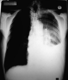
The patient was referred to the multidisciplinary team for discussion of treatment options.
Information
Chest X-rays
A standard chest X-ray (CXR) is taken posteroanteriorly (PA), with the patient facing the X-ray plate; the beam is directed at the patient’s back at a standard distance.
An emergency department film is often taken anteroposteriorly (AP), with the patient lying down (supine) on the X-ray plate; the beam is directed at the patient’s front – the distance from X-ray source can vary.
• Heart size and mediastinum are magnified.
• Pleural effusion which lies along back of chest cavity when patient supine might be missed.
Don’t request an AP ‘portable’ film unless it would be unsafe for the patient to have a PA film.
Management of massive haemoptysis
As little as 250 mL can fill the bronchial tree and be life threatening. Happily, this is uncommon but nevertheless frightening for everyone involved:
• Monitor: oxygen saturation with oximetry, blood pressure and pulse rate.
• Perform CXR. Exclude coagulation defects.
• Endotracheal intubation and suction might be required.
• Urgent bronchoscopy by an experienced doctor is sometimes required.
• A cuffed tube can be employed to protect the unaffected lung. It is inserted into the bronchus via a bronchoscope.
• Bronchial artery embolisation is highly effective if the bleeding vessel can be identified.
Chest pain
Diagnosing the cause of a chest pain is often difficult; it can be straightforward or take days to diagnose correctly. A careful history (eliciting site, character and radiation of the pain) is often more useful than tests:
• Exercise-induced central chest pain is usually cardiac in origin.
• Rest pain might be: cardiac, pleuritic, musculoskeletal, nerve root irritation, oesophageal, mediastinal or referred pain from abdomen.
• Lung diseases only cause pain if the pleura, mediastinum, intercostal nerves or bones are involved.
Is it cardiac pain?
Acute coronary syndrome
Pain at rest or on minimal exertion, sometimes very severe with sweating; pain persists.
Is it pleurisy?
Sharp pain in the sides of the chest, which ‘catches’ with breathing. This is often accompanied by fever, cough ± sputum or haemoptysis, indicating underlying lung disease.
Is it musculoskeletal?
• Muscles: Bornholm’s disease. Follows an upper respiratory tract infection; a low-grade fever can occur. Ache in muscles. Might be tender. Definite cases rare.
• Costochondral junction: Tietze’s disease; local pain on pressure over junctions. Responds to NSAIDs.
Is it oesophageal?
Reflux causes retrosternal burning pain, usually after food. Worse lying flat and eased by antacid. Can be severe and mimic myocardial infarction.
Is it referred pain from outside the chest?
Diaphragm irritation may cause shoulder tip pain. Localisation might be difficult for the patient. Several abdominal emergencies might have chest pain with or without abdominal pain.
Is it genuine?
Yes – nearly always. Exclude organic disease in all cases.
A tiny minority might be attention seeking or have psychiatric disease. Even patients with Munchausen’s syndrome might have genuine disease.
Respiratory failure
Respiratory failure (PaO2 < 8 kPa) is a common medical emergency often presenting with non-specific symptoms such as mild confusion or agitation. Recognition requires arterial blood gas (ABG) analysis (see below). Oximeters that estimate arterial oxygen saturation from the finger or ear lobe are useful in assessment or monitoring.
Oximeters might be falsely reassuring in the patient breathing oxygen. Importantly, they will not detect alveolar hypoventilation, producing high pCO2.
Respiratory failure commonly results from either a problem with the respiratory pump or because of intrinsic lung disease.
In intrinsic lung disease (apart from COPD) hypoxaemia is often combined with a reduced PaCO2
The hypoxaemia arises primarily from a mismatch of ventilation and perfusion in the pulmonary alveolar bed. Hypoxic stimulation of ventilation, coupled with abnormal respiratory sensation, then leads to a reduced arterial pCO2 (alveolar hyperventilation). A raised PaCO2 indicates impending respiratory arrest as it suggests either a reduction in ventilatory effort or failure of the respiratory pump.
In respiratory failure arterial blood gas sampling is necessary to
• Identify type, i.e. alveolar hypo- or hyperventilation
• Appreciate the degree of compensation (i.e. the chronicity of the condition)
• A coexisting metabolic acidosis commonly causes confusion and can be recognised by the base excess value (see below).
Information
ABG sampling is painful. Contrary to common belief, the use of local anaesthetic does not make the procedure more difficult. Heparin has a low pH and should be expelled from the syringe. Heparin-bonded microsamplers are available and small-diameter needles make local anaesthetic unnecessary. When taking ABG samples it is essential to note the inspiratory O2 concentration (FiO2).
Summary of acid–base changes (Fig. 11.3)
In a respiratory acidosis
Carbon dioxide clearance is reduced – there is alveolar hypoventilation. The PaCO2 and [H+] rise. The HCO3 is also increased due to renal compensation.

Figure 11.3 The Flenley acid–base nomogram. The bands show the 95% confidence limits representing the individual varieties of acid–base disturbance. The central white box shows the approximate limits of arterial pH and pCO2 in normal individuals.
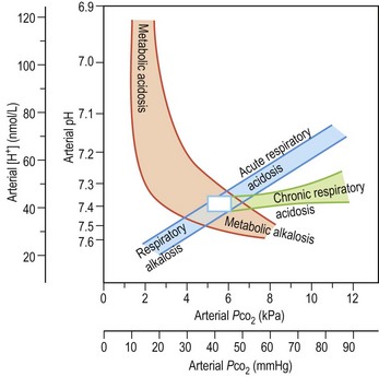
Examples: exacerbation of COPD, flail chest injury, Guillain–Barré syndrome.
In a respiratory alkalosis
There is alveolar hyperventilation and both the PaCO2 and [H+] are decreased. The HCO3 is slightly decreased. Examples: acute asthma, anxiety attack.
In a metabolic acidosis
There is disturbance of bicarbonate regulation or excessive H+ production. The HCO3 is reduced and the PaCO2 falls because of respiratory compensation.
Common ABG abnormalities
Life-threatening asthma
Supplemental O2 is being provided (note the high PaO2). There is a metabolic acidosis (note the pH and BE) as a result of metabolic demands exceeding O2 delivery and producing a lactic acidosis. Airflow limitation limits the normal respiratory compensation to this profound acidosis.
Acute or chronic respiratory failure in a patient with COPD
Acute or chronic respiratory acidosis exacerbated by a high FiO2 using variable performance mask (40–60% O2) (note high PaO2 and PaCO2). The high HCO3 results from renal compensation. The patient was changed to 28% oxygen.
Severe pneumonia (FiO2 60%)
Despite high FiO2 this patient is hypoxaemic because of ventilation: perfusion mismatch. The profound hypoxia despite oxygen and the associated metabolic acidosis indicate the need for urgent intubation and IPPV and are a reflection of circulatory failure resulting from septic shock.
Management
Respiratory failure can be difficult to assess or manage. Always review the CXR. Discuss with your consultant or other more senior staff. If you feel that the situation is unstable, do not hesitate to call an anaesthetist. Semi-elective intubation is much preferred to a respiratory arrest. It should be performed in the ward before transfer of the patient to the ICU.
How do I recognise impending respiratory arrest?
• Tachypnoea, respiratory rate > 30
• Sympathetic activation: pale and sweaty, agitation, confusion
• Progressive increase in PaCO2 or fall in PaO2
• Rapid desaturation on disconnection from O2, e.g. when drinking or coughing.
Evaluation is often at the ‘end of the bed’: is the patient getting tired? Be sensitive to subtle changes or a failure to improve.
Treating the cause
• Individual causes will require different actions: for instance, an intercostal drain for a tension pneumothorax or large pleural effusion (see p. 364).
• In neurological coma, intubation might be necessary for airway protection or to manage raised intracranial pressure by hyperventilation.
• Drug-induced respiratory failure can be confirmed by a therapeutic trial with a specific antidote, i.e. a bolus injection of naloxone for opiates and flumazanil for benzodiazepines. Infusions will be required if a positive response is obtained as antidotes have short half life.
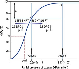
• Oxygen supplementation should be aimed at raising the PaO2 to beyond the steep part of the oxygen dissociation curve (Fig. 11.4). Very high PaO2 values are unnecessary but controlled O2 therapy via a fixed performance mask is only necessary in chronic respiratory failure resulting from COPD.

Figure 11.4 Oxygen dissociation curve. BPG, bisphosphoglycerate; a, arterial point; v, venous point; x, arterial venous difference; HbO2, oxygen saturation of haemoglobin.
Case history
A 65-year-old man with advanced COPD was admitted with a 1-week history of cough, breathlessness and purulent sputum. In the previous 24 h he had become mildly confused. He was agitated with a respiratory rate of 35, BP 170/90, sweaty with an oximeter reading on air of 72%. There was widespread wheeze and coarse crackles suggestive of retained secretions. ABG analysis revealed pH 7.32, PaO2 5.8, PaCO2 8.1, and HCO3 28.
What do these blood gases mean?
Hypoxaemia with mild acute respiratory acidosis. No evidence of chronic respiratory failure with chronically elevated pCO2 as HCO3 is normal, i.e. no compensation.
The initial treatment in A&E was:
• Nebulised bronchodilators (salbutamol 5 mg nebulised + ipratropium bromide 500 µg).
• IV amoxicillin (erythromycin if patient penicillin allergic).
• IV steroids (this is conventional treatment but there is limited evidence for effectiveness). Hydrocortisone 100 mg × 3 daily.
• Encouragement to clear secretions including sitting patient up and the attention of the physiotherapist.
• CXR to exclude pneumothorax or demonstrate associated pneumonia.
Information
Controlled oxygen via Venturi mask commonly leads to intermittent therapy as the mask is poorly tolerated by agitated, breathless patients. Oxygen via nasal prongs at 1 or 2 L/min is more effective and continuous but is not ‘controlled’.
Repeat ABGs were requested: pH 7.20, PaO2 6.5, PaCO2 12.5, HCO3 30.
These results show further CO2 retention and a deteriorating situation. Oxygen should not be removed – its removal will precipitate severe hypoxaemia. Alveolar ventilation must be increased. Despite using a nasal airway to stimulate cough and aid suction of respiratory secretions there was no improvement. Furosemide was given because it was difficult to exclude coexistent LVF. Intubation was considered appropriate but a trial of non-invasive ventilation (NIV), BiPAP with tight-fitting facial mask was first tried with repeat gases at 1 h. On NIV the respiratory rate slowed and the acute respiratory acidosis resolved.
Non-invasive ventilatory support employs a nose- or face-mask to provide ventilatory assistance to breathing (this is termed ‘spontaneous pressure support’) or timed breaths (‘pressure controlled ventilation’). An exhalation valve reduces re-breathing. NIV is successful in approximately 70% of patients with respiratory failure resulting from COPD. It should not be employed if intubation would be more appropriate.
Mechanical ventilation
In an unstable situation it is essential to maintain oxygenation. As intubation of an acutely unwell patient requires experience, refer to ITU outreach team.

Full access? Get Clinical Tree

