9 Respiratory disease
Respiratory symptoms are the most common cause of presentation to the family practitioner. Asthma occurs in more than 10% of British adults, and bronchial carcinoma is the most common fatal malignancy in the developed world. The lung is the major site of opportunistic infection in immunocompromised patients, and TB (including multiple drug-resistant strains) continues to increase, infecting one-third of the world’s population.
PRESENTING PROBLEMS
COUGH
Cough is the most common respiratory symptom and the underlying cause is often clear from other clinical features, particularly in more serious disease. Common causes of acute or transient cough are:
Chronic cough presents more of a diagnostic challenge, especially if physical examination, CXR and lung function are normal. In this context, consider:
While most patients with bronchogenic carcinoma have an abnormal CXR on presentation, fibreoptic bronchoscopy or spiral CT of the airways is advisable for an unexplained cough in adults (particularly smokers), as this may reveal a small endobronchial tumour or unexpected foreign body.
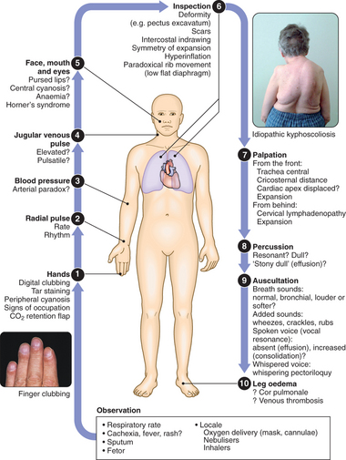
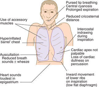
CLINICAL EXAMINATION OF THE RESPIRATORY SYSTEM
BREATHLESSNESS (DYSPNOEA)
Breathlessness or dyspnoea can be defined as the feeling of an uncomfortable need to breathe. It is unusual among sensations in having no defined receptors, no localised representation in the brain, and multiple causes both in health (e.g. exercise) and in diseases of the lungs, heart or muscles.
CHRONIC EXERTIONAL DYSPNOEA
Chronic obstructive pulmonary disease (COPD): Dyspnoea typically varies little day to day, but exercise capacity falls steadily over months or years. Patients usually report relief of dyspnoea at rest and overnight, a useful distinction from asthma. Orthopnoea may be present due to cranial displacement of the diaphragm on lying flat. Chronic cough and sputum (particularly in the morning) indicate bronchitis, but sputum may be absent when emphysema predominates. Recurrent acute exacerbations of breathlessness occur, usually in winter. Most but not all patients have a significant smoking history. In advanced disease, ankle swelling may develop as a result of cor pulmonale.
Asthma: Dyspnoea in asthma is associated with episodes of wheeze or chest tightness, varying in severity over time, but usually worse in the morning and often waking the patient overnight. There may be a history of childhood wheeze, or of wheeze or rhinitis provoked by pollens, dusts, household pets or occupational allergens. In exercise-induced asthma, wheeze and chest tightness typically come on immediately after exercise.
Heart disease: Impaired left ventricular function can cause exertional dyspnoea. Orthopnoea, cough and wheeze may also be present, as in lung disease. A history of angina or hypertension, or examination findings of displaced apex beat, raised JVP or cardiac murmurs may be useful in implicating a cardiac cause.
Interstitial or alveolar disease of the lung: Dyspnoea in these conditions is usually relentless and progressive. A detailed history should be obtained, including lifetime occupation and exposure to birds, dusts and other sources of organic agents which may provoke lung disease.
Diseases of the chest wall or respiratory muscles: May be obvious on history, examination and chest radiography. Other rarer causes of alveolar hypoventilation, e.g. brain-stem defects, and alveolar hypoventilation in gross obesity, may cause disordered breathing and cyanosis, but are not usually associated with breathlessness. Diaphragmatic weakness or palsy characteristically causes orthopnoea, with an abnormally large drop in the vital capacity on lying down. Respiratory failure tends to occur initially during sleep, with nocturnal hypoxaemia and hypercapnia, which resolve during the day.
Pulmonary thromboembolism: Often presents with acute breathlessness with or without chest pain. However, chronic thromboembolic disease should be suspected in patients who present with more gradual onset of breathlessness, particularly if there is a previous history of thromboembolic events or marked exertional breathlessness but a relatively normal examination and CXR. Leg swelling and an elevated JVP may be present, but are non-specific.
Psychogenic breathlessness and hyperventilation syndrome: Breathlessness not caused by organic disease of the heart or lungs is relatively common. It is particularly difficult to diagnose in patients with coexisting disease, such as asthma or heart disease. Suggestive features include an ‘inability to take a deep enough breath’, leading to extra deep sighs being taken, digital and perioral paraesthesiae, light-headedness, central chest discomfort or even carpopedal spasm due to acute respiratory alkalosis. Psychogenic breathlessness rarely disturbs sleep, frequently occurs at rest, may be provoked by stressful situations and may even be relieved by exercise. Arterial blood gases show normal PO2, low PCO2 and an alkalosis.
ACUTE SEVERE DYSPNOEA
This is one of the most common and dramatic medical emergencies.
It is important to ascertain the rate of onset and severity of the breathlessness, and whether associated cardiovascular symptoms (chest pain, palpitations, sweating and nausea) or respiratory symptoms (cough, wheeze, haemoptysis and stridor) are present. A previous history of repeated episodes of left ventricular failure, asthma or exacerbations of COPD is valuable. In children, the possibility of inhalation of a foreign body or acute epiglottitis should always be considered.
Common causes of acute breathlessness and their clinical features are in listed in Box 9.1.
CHEST PAIN
HAEMOPTYSIS
Coughing up blood, irrespective of the amount, is an alarming symptom and nearly always brings the patient to the doctor.
A clear history should be taken to establish that it is true haemoptysis and not haematemesis, gum bleeding or nosebleed. Haemoptysis must always be assumed to have a serious cause until proven otherwise. A history of repeated small haemoptyses, or blood-streaking of sputum, is highly suggestive of bronchial carcinoma. Fever, night sweats and weight loss suggest TB. Pneumococcal pneumonia is often the cause of ‘rusty’-coloured sputum but can cause frank haemoptysis, as can all the pneumonic infections which lead to suppuration or abscess formation. Bronchiectasis and intracavitary mycetoma can cause catastrophic bronchial haemorrhage and in these patients there may be a history of previous TB or pneumonia in early life. Pulmonary thromboembolism is a common cause of haemoptysis and should always be considered. Many episodes of haemoptysis are unexplained, even after full investigation, and are likely to be caused by simple bronchial infection.
Investigations and management
In severe acute haemoptysis, the patient should be nursed upright (or on the side of the bleeding if this is known), given oxygen and haemodynamically resuscitated. In the vast majority of cases, however, haemoptysis itself is not life-threatening and it is possible to follow a logical sequence of investigations which include: CXR, FBC, clotting screen, bronchoscopy and CT pulmonary angiography (CTPA).
THE SOLITARY RADIOGRAPHIC PULMONARY LESION
The incidental finding of a solitary pulmonary nodule (SPN) on a plain CXR in an adult patient is a common dilemma and the differential diagnosis is broad (Box 9.2). Between 20 and 30% of all cancers present in this way; the incidence increases with age and accounts for >50% of nodules in patients aged >50 yrs.
Radiology
Management
Percutaneous needle biopsy under CT guidance has proved to be the most effective procedure for the diagnosis of SPNs, which are usually beyond the reach of the bronchoscope. However, if the lesion has a high probability of malignancy and the patient is fit for surgery, the best option may be to proceed to resection. If bacterial infection is included in the differential diagnosis, an antibiotic should be given during the period in which the investigations are being performed. In elderly patients in whom a primary malignant lesion is suspected but who are considered unfit for any form of curative treatment, an interval CXR may be the most appropriate management.
PLEURAL EFFUSION
The accumulation of fluid within the pleural space is termed pleural effusion. Accumulations of frank pus (empyema) or blood (haemothorax) represent separate conditions. Pleural fluid accumulates due either to increased hydrostatic pressure or decreased osmotic pressure (‘transudative effusion’ as seen in cardiac, liver or renal failure), or to increased microvascular permeability caused by disease of the pleural surface itself, or injury in the adjacent lung (‘exudative effusion’). Some causes of pleural effusion are shown in Box 9.3. Particular attention should be paid to a recent history of respiratory infection, the presence of heart, liver or renal disease, occupation (e.g. exposure to asbestos), contact with TB, and risk factors for thromboembolism.
Symptoms and signs of pleurisy often precede the development of an effusion, especially in patients with underlying pneumonia, pulmonary infarction or connective tissue disease. However, the onset may be insidious. Breathlessness is often the only symptom related to the effusion and its severity depends on the size and rate of accumulation.
Radiology
The classical appearance of pleural fluid on CXR is of a curved shadow at the lung base, blunting the costophrenic angle and ascending towards the axilla. Fluid appears to track up the lateral chest wall. In fact, fluid surrounds the whole lung at this level, but casts a radiological shadow only where the X-ray beam passes tangentially through the fluid against the lateral chest wall. Around 200 ml of fluid is required to be detectable on a PA CXR, but smaller effusions can be identified by USS or CT. Previous scarring or adhesions in the pleural space can cause localised effusions. USS is more accurate than plain CXR for determining the volume of pleural fluid and frequently provides additional helpful information. The presence of loculation suggests an evolving empyema or resolving haemothorax. CT displays pleural abnormalities more readily than either plain radiography or USS, and may distinguish benign from malignant pleural disease.
Pleural aspiration and biopsy
Pleural aspiration reveals the colour and texture of fluid and on appearance alone may immediately suggest an empyema or chylothorax. The presence of blood is consistent with pulmonary infarction or malignancy, but may represent a traumatic tap. Biochemical analysis allows classification into transudate and exudate. An exudate usually has a protein concentration of >30 g/l or can be distinguished using Light’s criteria (Box 9.4). The predominant cell type provides useful information and cytological examination is essential. A low pH suggests infection but may also be seen in rheumatoid arthritis, ruptured oesophagus or advanced malignancy. In some clinical settings (e.g. left ventricular failure) it should not be necessary to sample fluid unless atypical features are present. Combining pleural aspiration with biopsy (using an Abrams needle) increases the diagnostic yield, but the best results are obtained from CT-guided biopsy or video-assisted thoracoscopy, allowing the operator to visualise the pleura and guide the biopsy directly.
Management
Therapeutic aspiration may be required to palliate breathlessness, but removing >1.5 litres in one episode is inadvisable as this can cause re-expansion pulmonary oedema. An effusion should never be drained to dryness before establishing a diagnosis as further biopsy may be precluded until further fluid accumulates. Treatment of the underlying cause—e.g. heart failure, pneumonia, pulmonary embolism or subphrenic abscess—will often be followed by resolution of the effusion. The management of pleural effusion in association with pneumonia, TB and malignancy is dealt with later in this chapter.
SLEEP-DISORDERED BREATHING
A variety of respiratory disorders manifest themselves during sleep, e.g. nocturnal cough and wheeze in asthma. Nocturnal hypoventilation may exacerbate respiratory failure in patients with restrictive lung disease such as kyphoscoliosis, diaphragmatic palsy, muscle weakness (e.g. muscular dystrophy) or intrinsic lung disease (e.g. COPD, pulmonary fibrosis). In contrast, a small but important group of disorders cause problems only during sleep due to upper airway obstruction (obstructive sleep apnoea) or abnormalities of ventilatory drive (central sleep apnoea).
THE SLEEP APNOEA/HYPOPNOEA SYNDROME (SAHS)
It is now recognised that 2–4% of the middle-aged population suffers from recurrent upper airway obstruction during sleep, which causes sleep fragmentation leading to daytime sleepiness. This results in a threefold increased risk of road traffic accidents.
A reduction in upper airway muscle tone during sleep results in pharyngeal narrowing, which often manifests as snoring. Negative pharyngeal pressure during inspiration can then cause complete upper airway occlusion, usually at the level of the soft palate. This leads to transient wakefulness and recovery of upper airway muscle tone. The subject rapidly returns to sleep, snores and becomes apnoeic once more. This cycle repeats itself many times, causing severe sleep fragmentation. Predisposing factors include:
Clinical assessment
Investigations
Differential diagnosis
Narcolepsy is a rare cause of sleepiness, occurring in 0.05% of the population and is associated with cataplexy (when muscle tone is lost in fully conscious people in response to emotional triggers), hypnagogic hallucinations (hallucinations at sleep onset) and sleep paralysis.
Idiopathic hypersomnolence occurs in younger individuals and is characterised by long nocturnal sleeps.
Management
RESPIRATORY FAILURE
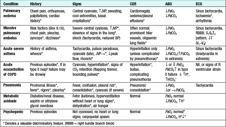
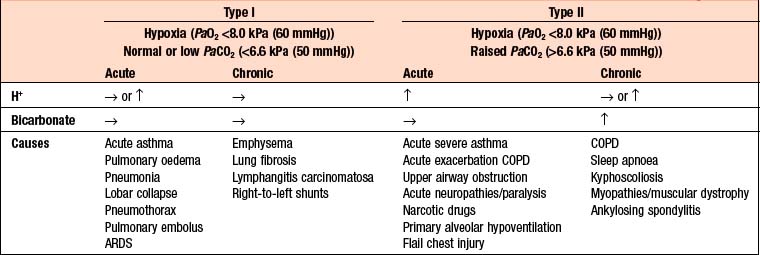
The term respiratory failure is used when pulmonary gas exchange fails to maintain normal arterial oxygen and carbon dioxide levels. Its classification into type I and type II relates to the absence or presence of hypercapnia (raised PaCO2). The main causes are shown in Box 9.5.
Management
The consequences of untreated severe hypoxaemia include:
Oxygen therapy
Oxygen delivery depends on many factors:
Normally, high-flow oxygen (35–60%) is appropriate treatment in respiratory failure (e.g. severe asthma, pulmonary oedema or pneumonia) because respiratory drive is high. A small percentage of patients with severe chronic COPD and type II respiratory failure develop abnormal tolerance of raised CO2 and may become dependent on hypoxic drive to breathe. In these patients only, lower concentrations of oxygen (24–28% via a Venturi mask) may be needed to avoid precipitating worsening respiratory depression.
Close monitoring of ABGs is essential to ensure acceptable PaO2 levels. If there is persistent hypoxia, progressive respiratory acidosis, or the patient becomes exhausted, an early decision should be made about whether it is appropriate to commence non-invasive ventilation or formal intubation and mechanical ventilation. Very ill patients may require immediate ventilatory support on presentation.
CHRONIC AND ‘ACUTE ON CHRONIC’ TYPE II RESPIRATORY FAILURE
The most common cause of chronic type II respiratory failure is COPD. When CO2 retention is chronic, the acidaemia is corrected by renal retention of bicarbonate, which results in a plasma pH within the normal range. This ‘compensated’ pattern, which is also seen in some patients with chronic neuromuscular disease or kyphoscoliosis, is maintained until there is a further pulmonary insult, such as an exacerbation of COPD which precipitates an episode of ‘acute on chronic’ respiratory failure. The further acute increase in PaCO2 results in acidaemia and worsening hypercapnia, and may lead to drowsiness and eventually to coma.
The principal aim of treatment in acute on chronic type II respiratory failure is to achieve a safe PaO2 (>7.0 kPa (52 mmHg)) without increasing PaCO2 and acidosis, while identifying and treating the precipitating condition. These patients usually have severe pre-existing lung disease, and only a small insult may be required to tip the balance towards severe respiratory failure. Moreover, in contrast to acute severe asthma, a patient with ‘acute on chronic’ type II respiratory failure due to COPD may not feel overtly distressed despite being critically ill with severe hypoxaemia, hypercapnia and acidaemia.
If controlled oxygen treatment causes a further increase in the PaCO2 associated with a reduction in pH, non-invasive or invasive ventilatory support is usually indicated. In these patients the decision regarding invasive ventilation can be particularly complex and difficult. Ideally, an early decision should be made, based on whether there is a potentially remediable precipitating condition and whether the patient is likely to regain an acceptable quality of life.
LUNG TRANSPLANTATION
Lung transplantation is now an established treatment for carefully selected patients with advanced lung disease unresponsive to medical treatment. Single-lung transplantation may be used for older patients with emphysema and patients with intrapulmonary restrictive disorders such as lung fibrosis. It is contraindicated in patients with chronic bilateral pulmonary infection, such as cystic fibrosis and bronchiectasis, where bilateral lung transplantation is the favoured option. Heart–lung transplantation remains necessary for the treatment of patients with advanced congenital heart disease such as Eisenmenger’s syndrome and is preferred by some surgeons for the treatment of primary pulmonary hypertension unresponsive to medical therapy. Although prognosis is good with modern immunosuppressive drugs, the availability of lung transplants remains limited due to shortage of donor lungs.
OBSTRUCTIVE PULMONARY DISEASES
ASTHMA
Asthma is characterised by chronic airway inflammation and increased airway hyper-responsiveness leading to wheeze, cough, chest tightness and dyspnoea. Airflow obstruction in asthma is variable over time and reversible with treatment. Around 300 million people world-wide suffer from asthma and the prevalence is increasing, particularly in countries with a Western lifestyle. In childhood, asthma is more common in boys, but following puberty females are more frequently affected. Multiple environmental and genetic determinants are implicated in the aetiology of asthma. The ‘hygiene hypothesis’ proposes that decreased infections in early life bias the immune system towards an allergic phenotype. There is a strong association between atopy—a propensity to produce IgE—and asthma.
Clinical features
Asthma is not a uniform disease but a dynamic clinical syndrome characterised by episodes of wheezing, chest tightness, breathlessness and cough. There is diurnal variation in symptoms (worse in early morning); sleep is often disturbed by cough and wheeze. In mild intermittent asthma, patients may be asymptomatic between exacerbations. In persistent asthma the pattern is one of chronic wheeze and breathlessness.
Symptoms may be precipitated by:
Investigations
Pulmonary function tests: Diagnosis is based on a compatible history and an increase in FEV1 of ≥15% and at least 200 ml following bronchodilator/trial of corticosteroids; or >20% diurnal variation on ≥3 days in a week for 2 wks on PEF (peak expiratory flow) diary; or a decrease in FEV1 of ≥15% after 6 mins of exercise.
Radiological examination: Often normal. There is lobar collapse if mucus has occluded a large bronchus. Hyperinflation is present in acute asthma. Flitting infiltrates may be seen in allergic bronchopulmonary aspergillosis (ABPA).
Measurement of allergic status: Total IgE ± allergen-specific IgE and/or skin prick tests are carried out. There may be a peripheral blood eosinophilia or sputum eosinophilia (the latter also used to measure airway inflammation).
Management
The goals of asthma therapy are to:
Patient education: Patients should be taught the relationship between symptoms and inflammation, the importance of key symptoms (e.g. nocturnal waking), about different types of medication and the use of PEF to guide management decisions. Written action plans may be helpful.
Avoidance of aggravating factors: Asthma control may be improved by reducing exposure to trigger antigens, e.g. household pets. In occupational asthma, removal from the offending agent may lead to cure. Many patients are sensitised to several antigens, making avoidance almost impossible. Patients should be advised not to smoke.
Pharmacological treatment
Step 1—Occasional use of inhaled short-acting β2-adrenoreceptor agonist bronchodilators: This is used for patients with mild intermittent asthma (symptoms < once/wk for 3 mths and <2 nocturnal episodes/mth). Patients often underestimate the severity of asthma.
Step 2—Regular anti-inflammatory therapy (preferably inhaled corticosteroids (ICS)): This is used for any patient who has experienced an exacerbation in the last 2 yrs, uses inhaled β2-agonists >3 times/wk, reports symptoms >3 times/wk, or is awakened by asthma 1 night/wk.
Step 3—Add-on therapy: If poor control remains, despite regular ICS up to a dose of 800 μg/day BDP or equivalent, add-on therapy should be considered with either long-acting β2-agonists (LABAs), such as salmeterol and formeterol, or leukotriene receptor antagonists (e.g. montelukast 10 mg daily). LABAs have consistently been demonstrated to improve asthma control and reduce exacerbations compared to increasing the dose of ICS alone. Fixed-combination inhalers of ICS and LABAs are convenient and help compliance.
Step 4—Poor control on a moderate dose of inhaled steroid and add-on therapy: The ICS dose may be increased to 2000 μg BDP or equivalent daily. A nasal corticosteroid should be used if upper airway symptoms are prominent. Consider trials of leukotriene receptor antagonists or theophyllines. Further studies on new therapies, such as monoclonal antibodies directed against IgE, are awaited.
Step 5—Continuous or frequent use of oral steroids: Prednisolone therapy should be prescribed in the lowest amount necessary to control symptoms. Long-term corticosteroid tablets (>3 mths) or receiving more than three or four courses per year put patients at risk of systemic side-effects. Osteoporosis may be prevented using bisphosphonates.
Step-down therapy: Once asthma control is established, inhaled (or oral) corticosteroid dose should be titrated to the lowest dose at which effective control of asthma is maintained.
Exacerbations of asthma
Asthma exacerbations are characterised by increased symptoms, deterioration in PEF and an increase in airway inflammation. They may be precipitated by infections (most commonly viral), moulds (Alternaria and Cladosporium) and, on occasion, pollen (particularly following thunderstorms). Most attacks are characterised by a gradual deterioration over several hours to days, but some appear to occur with little or no warning: so-called brittle asthma.
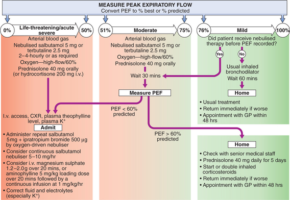
Management of mild–moderate exacerbations
CHRONIC OBSTRUCTIVE PULMONARY DISEASE (COPD)
COPD is a heterogeneous condition embracing several overlapping pathological processes including chronic bronchitis, chronic bronchiolitis (small airway disease) and emphysema. Many patients also exhibit a systemic component characterised by impaired nutrition, weight loss and skeletal muscle dysfunction. COPD is defined by the presence of airways obstruction, which does not change markedly over several months and, unlike asthma, is not fully reversible. The prevalence of COPD in the UK is estimated at 1–2%, but is probably greater. Exacerbations of COPD account for 10% of hospital admissions in the UK, and with ∼30 000 deaths/yr it represents the sixth most common cause of death in the UK. Following the marked increase in tobacco consumption in developing countries, COPD is gaining in global importance. Cigarette smoking is the most important risk factor. Susceptibility to cigarette smoke varies but both the dose and duration of smoking appear to be important. Other risk factors are solid fuel fires, air pollution, occupation (coal miners) and low socio-economic class. Rarely, an inherited deficiency of α1 antitrypsin predisposes to premature emphysema.
Clinical features
COPD should be suspected in any patient over the age of 40 yrs who has persistent cough and sputum production and/or breathlessness.
9.7 MODIFIED MRC DYSPNOEA SCALE
Grade | Degree of breathlessness related to activities |
---|---|
0 | No breathlessness except with strenuous exercise |
1 | Breathlessness when hurrying on the level or walking up a slight hill |
2 | Walks slower than contemporaries on level ground because of breathlessness or has to stop for breath when walking at own pace |
3 | Stops for breath after walking about 100 m or after a few mins on level ground |
4 | Too breathless to leave the house, or breathless when dressing or undressing |
Two classical phenotypes have been described:
Investigations
Management and prognosis
Pessimism is unjustified, as it is usually possible to improve breathlessness, reduce the frequency and severity of exacerbations, and improve health status and prognosis.
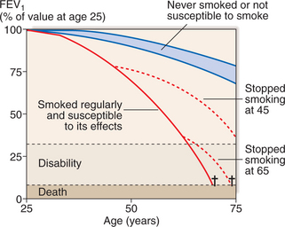
Smoking cessation: Offer help to stop smoking at every opportunity. Combine pharmacotherapy with appropriate support as part of a programme. It is the only intervention proven to decelerate the decline in FEV1 (Fig. 9.3).

Fig. 9.3 Model of annual decline in FEV1 with accelerated decline in susceptible smokers. When smoking is stopped, subsequent loss is similar to that in healthy non-smokers.
Bronchodilators: Short-acting bronchodilators for mild disease (β2-agonist or anticholinergic). Longer-acting bronchodilators are more appropriate for patients with moderate to severe disease. From the wide range available, select a device which the patient can use effectively. Significant improvements in breathlessness may be reported despite minimal changes in FEV1, probably reflecting reduced hyperinflation. Theophylline preparations improve breathlessness, but use is limited by side-effects. Oral bronchodilators may be contemplated in patients who cannot use inhaled devices efficiently.
Inhaled corticosteroids: Reduce the frequency and severity of exacerbations; they are recommended in patients with moderate to severe disease (FEV1 <50%) who report >2 exacerbations requiring antibiotics or oral steroids/yr. Regular use leads to a small improvement in FEV1 but no change in the rate of decline of lung function. ICS/LABA combinations produce further improvements in breathlessness and exacerbation rate. Oral corticosteroids are useful during exacerbations, but maintenance therapy contributes to osteoporosis and impaired muscle function and should be avoided.
Pulmonary rehabilitation: Encourage exercise. Multidisciplinary programmes (usually 6–12 wks’ duration) incorporating physical training, education and nutritional counselling reduce symptoms, improve health status and enhance confidence.
Oxygen therapy: Long-term domiciliary oxygen therapy (LTOT) improves survival, prevents progression of pulmonary hypertension, decreases the incidence of secondary polycythaemia and improves neuropsychological health. A minimum of 15 hrs/day is recommended, to keep PaO2 >8 kPa (60 mmHg) or SaO2 >90%. Ambulatory oxygen therapy should be considered in patients who desaturate on exercise and show objective improvement in exercise capacity and/or dyspnoea with oxygen. Short-burst oxygen therapy is widely prescribed but is expensive and of unproven benefit.
Surgical intervention: In highly selected patients, lung volume reduction surgery (removing non-functioning emphysematous lung tissue) reduces hyperinflation and decreases work of breathing. Bullectomy is occasionally performed to remove large bullae that compress surrounding lung tissue.
Other measures: Influenza vaccination; pneumococcal vaccination; trial of mucolytic therapy.
COPD has a variable natural history. Prognosis is inversely related to age and directly related to FEV1. Poor prognostic indicators include weight loss and pulmonary hypertension.
Acute exacerbations of COPD
These are characterised by an increase in symptoms and deterioration in lung function. They are more common in severe disease and may be caused by bacteria, viruses or a change in air quality. Respiratory failure and/or fluid retention may be present. Many patients can be managed at home with the use of increased bronchodilator therapy, a short course of oral corticosteroids and, if appropriate, antibiotics. Cyanosis, peripheral oedema or altered conscious level should prompt hospital referral.
Oxygen therapy: High concentrations of oxygen may cause respiratory depression and worsening acidosis (p. 280). Controlled oxygen at 24% or 28% should be used, aiming for PaO2 >8 kPa (60 mmHg) (or an SaO2 >90%) without worsening acidosis.
Corticosteroids: Oral prednisolone (usually 30 mg for 5–10 days) reduces symptoms and improves lung function. Prophylaxis against osteoporosis should be considered if frequent courses are needed.
Antibiotic therapy: Recommended for an increase in sputum purulence, sputum volume or breathlessness. An aminopenicillin or a macrolide should be used. Co-amoxiclav is only required in regions where β-lactamase-producing organisms are known to be common.
Non-invasive ventilation (NIV): If patients have persistent tachypnoea and a respiratory acidosis (H+ ≥ 45/pH < 7.35), NIV is associated with reduced requirements for mechanical ventilation and reductions in mortality. Consider mechanical ventilation where there is a reversible cause for deterioration (e.g. pneumonia), or if there is no prior history of respiratory failure.
Additional therapy: Evidence for i.v. aminophylline is limited and there is a risk of arrhythmias and drug interactions. The respiratory stimulant doxapram has been largely superseded by NIV. Diuretics should be administered if peripheral oedema has developed.
BRONCHIECTASIS
Bronchiectasis is defined as abnormal dilatation of the bronchi due to chronic airway inflammation and infection. It is usually acquired, but may result from an underlying genetic or congenital defect of airway defences (Box 9.8).
Investigations
Sputum: May reveal common respiratory pathogens. As disease progresses, Pseudomonas aeruginosa or fungi such as Aspergillus and various mycobacteria may be seen. Frequent cultures assist appropriate antibiotic selection.
Radiology: CXR may be normal in mild disease. In advanced disease, thickened airway walls, cystic bronchiectatic spaces, and associated areas of pneumonic consolidation or collapse may be seen. CT is much more sensitive, and shows thickened dilated airways.
Assessment of ciliary function: Saccharin test or nasal biopsy may be used.

Stay updated, free articles. Join our Telegram channel

Full access? Get Clinical Tree

