Reactive Mesothelial Hyperplasia
Alvaro C. Laga
Timothy C. Allen
Carlos Bedrossian
Rodolfo Laucirica
Philip T. Cagle
Reactive mesothelial cell hyperplasia is often seen in association with pleural effusion, including infections, collagen-vascular diseases, drug reactions, pneumothorax, chest surgery, and trauma. Reactive mesothelial cells proliferate along the pleural surface and may shed into pleural fluid. Benign reactive mesothelial cells may have cytologic atypia and mitoses, may proliferate in papillary tufts or other structures, and may be entrapped by overlying organizing pleuritis or pleural fibrosis mimicking invasion. Distinguishing reactive mesothelial cell hyperplasia from diffuse malignant mesothelioma may be a diagnostic dilemma, especially with small biopsies.
Histologic Features
The mesothelial cells are generally cuboidal, with round nuclei, prominent nucleoli, and abundant eosinophilic cytoplasm.
Depending on the severity of the reaction, the mesothelial cells may exhibit cytologic atypia and mitoses.
Entrapment of proliferating reactive mesothelial cells by fibrin, granulation tissue, and/or fibrous tissue overlying the pleural surface in pleuritis may mimic invasion; the entrapped mesothelial cells are typically arranged linearly parallel to the pleural surface and lack invasion into the deeper pleural and underlying tissues.
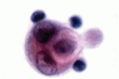
 Figure 130.1 Cluster of enlarged, cytologically atypical reactive mesothelial cells in a pleural fluid may mimic neoplastic mesothelial cells.
Stay updated, free articles. Join our Telegram channel
Full access? Get Clinical Tree
 Get Clinical Tree app for offline access

|