FIGURE 22-1
(A) Blastomyces
(B) Corpora amylacea
(C) Hamazaki-Wesenberg bodies
(D) Hemosiderin granules
(E) Histoplasma organisms
3. A specimen from a patient who died of septicemia is shown here. The approximate age of the form of lung injury depicted in Figure 22-2 is

(A) 12 hours
(B) 2 days
(C) 3–5 days
(D) 1 month
(E) 3 months
4. Which form of lung injury is similar to diffuse alveolar damage, is localized to the peribronchiolar parenchyma, and is characterized by fibroblastic plugs?
(A) Acute interstitial pneumonia
(B) Bronchiolitis obliterans-organizing pneumonia
(C) Lymphocytic interstitial pneumonia
(D) Non-specific interstitial pneumonia
(E) Pulmonary alveolar proteinosis
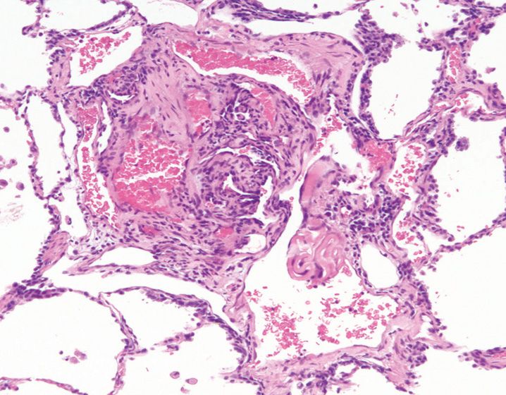
5. A 70-year-old man presents with dyspnea and progressive worsening of symptoms over the past few months. High-resolution CT scan shows bilateral, interstitial, reticular markings prominent at the bases and periphery of the lungs. Based on Figure 22-3, what is the most likely diagnosis?
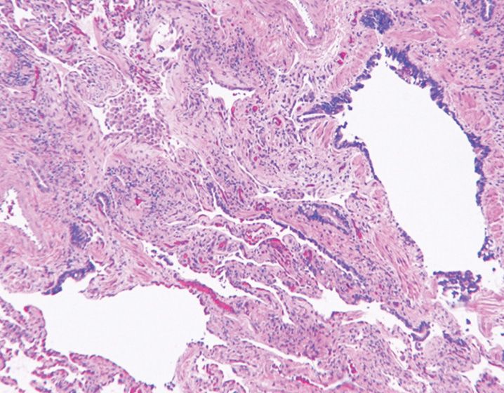
(A) Acute interstitial pneumonia
(B) Bronchiolitis obliterans-organizing pneumonia
(C) Desquamative interstitial pneumonia
(D) Non-specific interstitial pneumonia
(E) Usual interstitial pneumonia
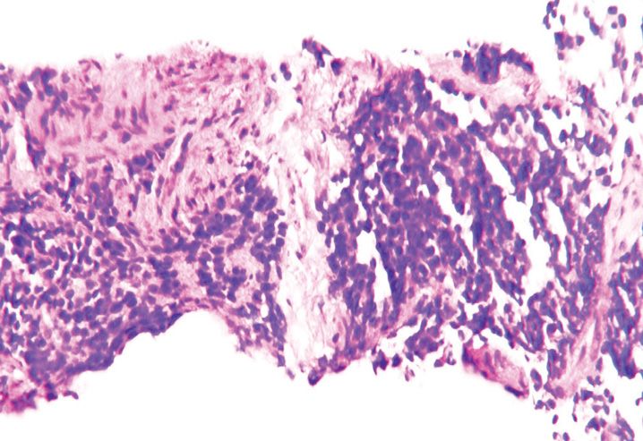
6. A wedge biopsy from a patient with interstitial lung disease is shown here (Figure 22-4). Based on the histologic features, what is the best diagnosis?
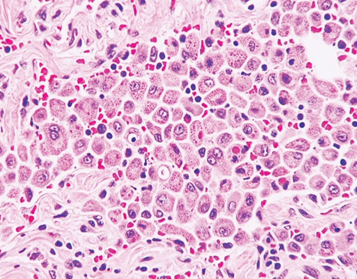
(A) Acute interstitial pneumonia
(B) Bronchiolitis obliterans-organizing pneumonia
(C) Desquamative interstitial pneumonia/Respiratory bronchiolitis interstitial lung disease
(D) Non-specific interstitial pneumonia
(E) Usual interstitial pneumonia
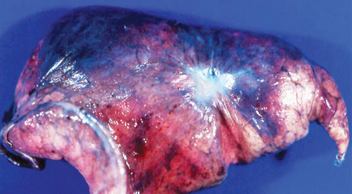
7. The gross appearance of lung shown in Figure 22-5 may be associated with all the following conditions, except
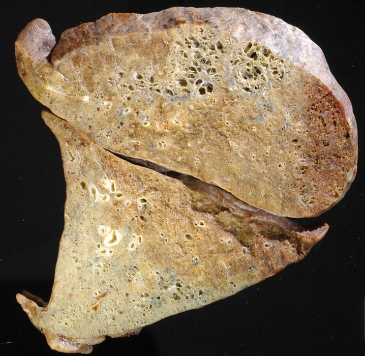
(A) Diffuse alveolar damage
(B) Eosinophilic pneumonia
(C) Idiopathic interstitial pneumonia
(D) Inorganic dust exposure
(E) Lymphoma
8. Which of the following histologic reactions/findings is least likely to be encountered in a quarry worker?
(A) Concentric hyaline nodules
(B) Dust macule
(C) Diffuse interstitial fibrosis
(D) Pulmonary alveolar proteinosis
(E) Stellate interstitial fibrosis
9. Which of the following statements about asbestos fibers is incorrect?
(A) By definition, their length is three times their diameter
(B) Heavy exposure may lead to development of carcinoma and mesothelioma
(C) Serpentine and amphibole groups are the commonly encountered classes
(D) They are composed of silica plus iron, magnesium, sodium, and other metals
(E) They cause a more localized reaction compared to silicosis
10. Coating with which of the following particles leads to formation of an asbestos body?
(A) Cadmium
(B) Calcium
(C) Copper
(D) Iron
(E) Lead
11. In contrast to simple coal worker’s pneumoconiosis, complicated coal worker’s pneumoconiosis is usually associated with which of the following features?
(A) Dust-filled macrophages in the interstitium surrounding respiratory bronchioles
(B) Hyalinized and collagenous stroma
(C) Minimal radiographic changes
(D) Progressive massive fibrosis
(E) Reaction to inhaled coal dust particles
12. The finding of giant cell interstitial pneumonia is most likely associated with which of the following conditions?
(A) Berylliosis
(B) Coal workers’ pneumoconiosis
(C) Hard metal pneumoconiosis
(D) Siderosis
(E) Silicosis
13. The pathologic triad of temporally uniform chronic interstitial pneumonia with peribronchiolar accentuation, non-necrotizing granulomas in peribronchiolar interstitium, and foci of bronchiolitis obliterans is diagnostic of which of the following pathologic entities?
(A) Diffuse alveolar damage
(B) Hypersensitivity pneumonia
(C) Lymphoid interstitial pneumonia
(D) Sarcoidosis
(E) Usual interstitial pneumonia
14. A 25-year-old man presents with acute onset hemoptysis, anemia, azotemia, and diffuse pulmonary infiltrates. A kidney biopsy shows membranoproliferative glomerulonephritis with linear IgG deposits. What is the most likely cause of this patient’s symptoms?
(A) Cocaine intoxication
(B) Drug reaction
(C) Goodpasture’s syndrome
(D) Heart failure
(E) Idiopathic pulmonary hypertension
15. A 35-year-old woman of African descent presents with dyspnea and cough. She is found to have extensive hilar adenopathy. Infectious processes and neoplasms have been clinically excluded. All of the following are associated with this condition, except
(A) Asteroid bodies
(B) Granulomatous vasculitis
(C) Granulomas within airspaces
(D) Hyalinized fibrosis
(E) Well-formed epithelioid granulomas
16. A 45-year-old man presents with history of fever, weight loss, hemoptysis, and sinusitis. Preliminary investigation reveals elevated serum antineutrophil cytoplasmic antibodies (ANCA) targeted against proteinase-3 and cavitary pulmonary nodules. Which of the following clinicopathologic features is not associated with this condition?
(A) Can be fatal if not treated promptly
(B) Geographic areas of parenchymal necrosis bound by epithelioid histiocytes
(C) Glomerulonephritis
(D) Necrotizing vasculitis
(E) Blood eosinophilia
17. Churg-Strauss syndrome is characterized by all of the following clinicopathologic features, except
(A) Asthma
(B) Blood eosinophil count of <500/μL
(C) Elevated p-ANCA
(D) Mild segmental glomerulonephritis
(E) Necrotizing vasculitis
18. A 75-year-old man presents with hemoptysis and is found to have a 4.5 cm nodular lesion in the left lobe of the lung (see Figure 22-6). The cells of interest in the lesion are diffusely positive for CD20 and coexpress CD43. They are negative for CD5 and CD10. These findings are diagnostic of
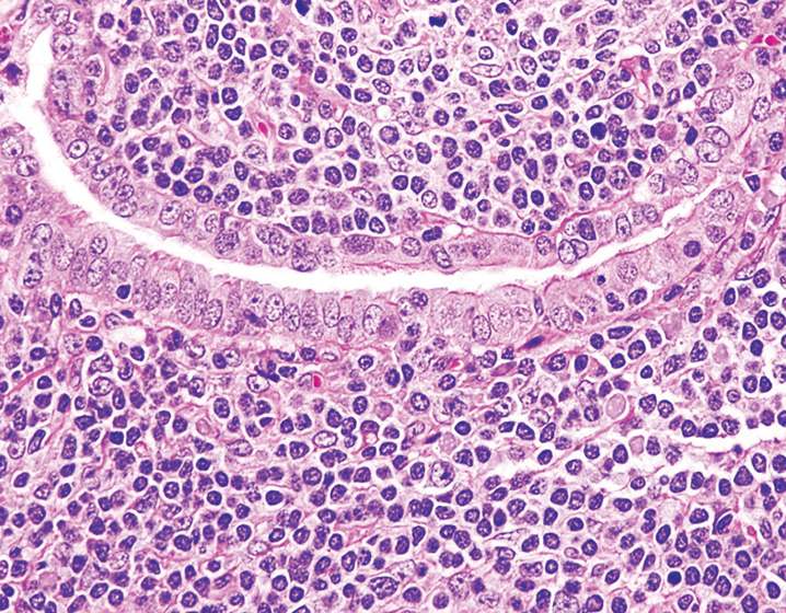
(A) Follicular bronchiolitis
(B) Low-grade B-cell lymphoma
(C) Lymphoid interstitial pneumonia
(D) Lymphomatoid granulomatosis
(E) Nodular lymphoid hyperplasia
19. A 57-year-old man presents with hemoptysis and is found to have bilateral necrotizing nodules in the lung. Histologically, they are composed of small lymphocytes, histiocytes, plasma cells, and large atypical lymphocytes with prominent vascular involvement. The large lymphoid cells are CD20-positive and the background cells are CD3-positive. What is the most likely diagnosis?
(A) Hodgkin disease
(B) Lymphomatoid granulomatosis
(C) Lymphoid interstitial pneumonia
(D) Tuberculosis
(E) Wegener’s granulomatosis
20. All the following viruses cause organizing pneumonia with multinucleated giant cells, except
(A) Adenovirus
(B) Measles virus
(C) Parainfluenza virus
(D) Respiratory-syncytial virus
(E) Varicella-zoster virus
21. Which of the following fungal organisms causes a “target-like lesion” characterized by hemorrhagic infarction with a dark rim surrounding a central yellow-gray area?
(A) Aspergillus
(B) Candida
(C) Fusarium
(D) Pseudoallescheria boydii
(E) Mucor
22. All the following morphologic features favor the presence of mucormycosis over Aspergillosis, except
(A) Haphazard tissue distribution
(B) 90° branching
(C) Non-septate hyphae
(D) Regular branching points
(E) Wide hyphae
23. Which of the following types of crystals is Aspergillus niger mycetoma associated with?
(A) Calcium oxalate
(B) Calcium phosphate
(C) Calcium silicate
(D) Triple phosphate
(E) Tyrosine
24. A 25-year-old young man with a history of renal transplantation develops a pulmonary abscess. Histologic evaluation shows poorly formed granulomas surrounding necrotic debris. A modified Ziehl-Neelsen preparation is shown here (Figure 22-7). What is the etiology of his pulmonary abscess?
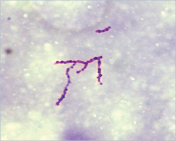
(A) Actinomycosis
(B) Botryomycosis
(C) Sporotrichosis
(E) Tuberculosis
25. A 45-year-old woman with a history of systemic lupus erythematosus presents with fever and dyspnea. The histologic changes in her lung biopsy (Figure 22-8) are pathognomonic of which of the following entities?
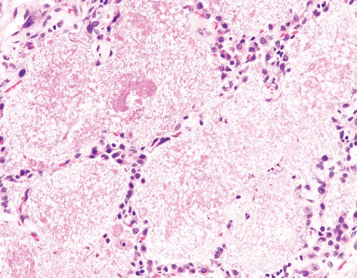
(A) Bronchopneumonia
(B) Diffuse alveolar damage
(C) Pulmonary alveolar proteinosis
(D) Pulmonary edema
(E) Pneumocystis jiroveci infection
26. A farmer from Southwest region of United States is found to have a 2.5 cm lung nodule. The wedge biopsy findings shown in Figure 22-9 are diagnostic of
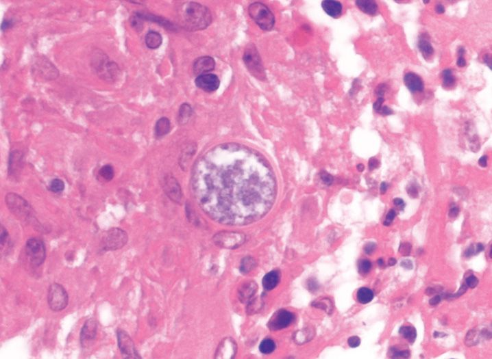
(A) Blastomycosis
(B) Coccidioidomycosis
(C) Cryptococcosis
(D) Histoplasmosis
(E) Paracoccidioidomycosis
27. The pulmonary infection depicted in Figure 22-10 is associated with all of the following features, except
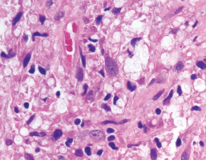
(A) Approximately a third of patients are asymptomatic
(B) Hilar lymph node enlargement is common
(C) Non-necrotizing granulomas
(D) Oval yeasts without a capsule
(E) Pigeon breeders
28. A biopsy from a patient exposed to hot tub water shows a combination of well-formed non-necrotizing granulomas and organizing pneumonia. What is the most likely cause of patient’s lung injury?
(A) Mycobacterium avium-intracellulare complex
(B) Mycobacterium chelonei
(C) Mycobacterium fortuitum
(D) Mycobacterium kansasii
(E) Mycobacterium tuberculosis
29. The filariform larval form of which of the following intestinal nematodes is associated with hyperinfection syndrome and bronchopneumonia?
(A) Ascaris lumbricoides
(B) Ankylostoma duodenale
(C) Enterobius vermicularis
(D) Trichuris trichiura
(E) Strongyloides stercoralis
30. A pneumonectomy specimen from a 36-year-old woman with long-standing history of exertional dyspnea and pulmonary hypertension shows this lesion (Figure 22-11). What is the best diagnosis?
(A) Concentric laminar fibrosis
(B) Medial hypertrophy of arteries
(C) Muscularization of arteries
(D) Necrotizing arteritis
(E) Plexiform lesion
31. Pulmonary sequestration is characterized by all the following pathologic findings, except
(A) Lacks communication with the tracheobronchial tree
(B) May rarely have a connection to the esophagus or stomach
(C) May occur within or outside of the visceral pleural lining
(D) Most commonly occurs in the right lobe
(E) Receives blood supply from one or more anomalous systemic arteries
32. Which of the following statements does not apply to congenital cystic adenomatoid malformation (CCAM)?
(A) It has features of immaturity and malformation of small airways and distal lung parenchyma
(B) It is most commonly seen in stillborn or premature infants with anasarca
(C) It is often associated with spontaneous pneumothorax
(D) It is sometimes associated with elevated maternal serum alpha-fetoprotein
(E) Most CCAMs communicate with the normal tracheobronchial tree
33. A wedge biopsy from a nodular lesion in a 50-year-old man with history of smoking is shown in Figure 22-12. Which immunohistochemical stain is most useful in highlighting the cells of interest?
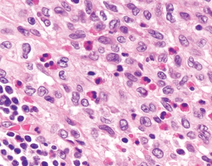
(A) CD1a
(B) CD4
(C) CD10
(D) Smooth muscle actin
(E) Chromogranin
34. The lesion depicted in Figure 22-13 is associated with which of the following gene mutations?
(B) β-catenin
(C) KIT
(D) NF1
(E) TSC1
35. A 50-year-old woman has a history of autoimmune pancreatitis and is found to have a nodular lesion in the lung. All the following features would confirm a diagnosis of IgG4-related sclerosing lesion, except
(A) Fibrosis
(B) Increased numbers of IgG4-positive plasma cells
(C) Intimal and mural inflammation involving the pulmonary arteries
(D) Lymphoplasmacytic infiltrate
(E) Serum IgG4 <10 mg/dL
36. Which is the most common organism isolated from cystic fibrosis patients presenting with recurrent pulmonary infections?
(A) Chlamydia pneumoniae
(B) Haemophilus influenzae
(C) Mycoplasma pneumoniae
(D) Pseudomonas aeruginosa
(E) Streptococcus pneumoniae
37. A wedge biopsy of lung shows a small lesion composed of ovoid cells with stippled chromatin pattern, arranged as nests within the alveolar septa and around pulmonary veins. Additional work-up shows that the cells are strongly positive for EMA and vimentin, and do not express cytokeratin, chromogranin, S-100, and actin. What is the best diagnosis for this lesion?
(A) Carcinoid tumorlet
(B) Granuloma
(C) Minute meningothelial-like lesion
(D) Paraganglioma
(E) Sclerosing hemangioma
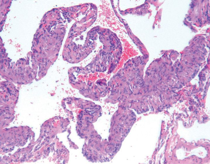
38. A lobectomy specimen from a 45-year-old woman with a lung mass is shown in Figure 22-14. What is the most likely diagnosis?
(A) Adenocarcinoma
(B) Bronchioloalveolar carcinoma
(C) Carcinoid tumor
(D) Large cell carcinoma
(E) Squamous cell carcinoma
39. Per the 2011 International Association for the Study of Lung Cancer (IASLC)/American Thoracic Society (ATS)/European Respiratory Society (ERS) international multidisciplinary classification of lung adenocarcinoma, which of these patterns is not categorized as a distinct histologic pattern of invasive adenocarcinoma?
(A) Acinar
(B) Micropapillary
(C) Mucinous
(D) Papillary
(E) Solid
40. A 65-year-old man with a significant smoking history is found to have an exophytic, cavitary mass obstructing the main bronchus. Based on this information, what is the most likely diagnosis?
(A) Adenocarcinoma
(B) Carcinoid tumor
(C) Large cell carcinoma
(D) Small cell carcinoma
(E) Squamous cell carcinoma
41. A transbronchial lung biopsy of a perihilar mass in a 55-year-old man with significant smoking history is depicted in Figure 22-15. Which of these morphologic findings is not typical of this entity?
Stay updated, free articles. Join our Telegram channel

Full access? Get Clinical Tree


