176 Pseudoxanthoma elasticum
Patient 1
Salient features
History
• Family history (either autosomal recessive, which is most common, or autosomal dominant; the gene for both forms has been mapped to chromosome 16, Human Mol Genet 1997;6:1823)
• Upper GI haemorrhage, myocardial infarction, stroke and intermittent claudication, visual loss
• Hypertension (from involvement of renal vasculature)
Examination
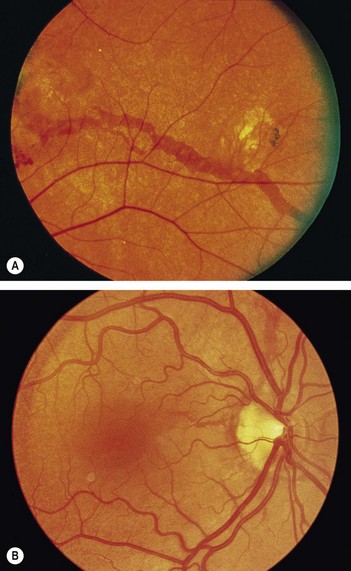
• Fundus shows angioid streaks: linear grey or dark red streaks with irregular edges lying beneath the retinal vessels (Fig. 176.1) (roughly 50% of patients with angioid streaks have pseudoxanthoma elasticum, whereas 85% of those with pseudoxanthoma have angioid streaks).
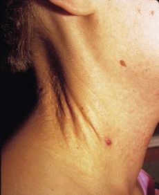
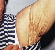
• Look at the neck (Fig. 176.2), antecubital fossae, axillae (Fig. 176.3), groin and periumbilical region for loose ‘chicken skin’ appearance of skin
• Examine peripheral pulses: absent pulses from peripheral arterial involvement but acral ischaemia is uncommon because of development of collaterals.
Stay updated, free articles. Join our Telegram channel

Full access? Get Clinical Tree


