Protocol for the Examination of Specimens From Patients With Primary Pituitary Tumors

Image Gallery
Pituitary Gland, Sella, and Adjacent Structures
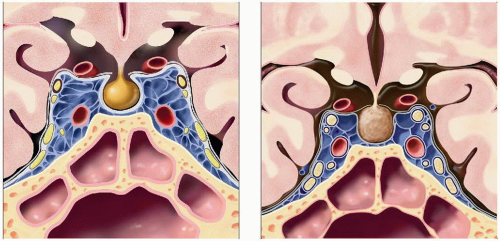
 (Left) The adenohypophysis is comprised of the pars tuberalis, pars intermedia, and pars distalis. The neurohypophysis is comprised of the median eminence of hypothalamus, infundibulum, and pars nervosa. (Right) The pituitary is composed of neural tissue (the pituitary stalk and posterior lobe [PL]), epithelial tissue (the anterior lobe [AL]), and the cystic remnants of the intermediate lobe (IL). Normal distribution of ACTH, GH, and PRL cells is shown (right figure). |
 (Left) The cranial nerves traverse the cavernous sinus within the lateral wall of the cavernous sinus: Oculomotor, trochlear, and first and second divisions of trigeminal nerves. The only cranial nerve within the sinusoids of the cavernous sinus is the abducens nerve. (Right) Coronal graphic shows physiologic pituitary hyperplasia. The gland is uniformly enlarged and has a mildly convex superior margin. |
 (Left) Submentovertex graphic shows the typical sites for miscellaneous nonparenchymal CNS metastases. These include the choroid plexus and ventricles
 , pituitary gland, infundibular stalk , pituitary gland, infundibular stalk  , & eye , & eye  . (Right) The COW is located in the suprasellar cistern just below the diencephalon. The hypothalamus, infundibular stalk, and optic chiasm lie in the middle of the COW. The horizontal ACA segment passes above the optic nerves. The PCoA passes above the oculomotor nerves. . (Right) The COW is located in the suprasellar cistern just below the diencephalon. The hypothalamus, infundibular stalk, and optic chiasm lie in the middle of the COW. The horizontal ACA segment passes above the optic nerves. The PCoA passes above the oculomotor nerves.Stay updated, free articles. Join our Telegram channel
Full access? Get Clinical Tree
 Get Clinical Tree app for offline access

|