Protocol for the Examination of Specimens From Patients With Carcinoma of the Thyroid Gland
Image Gallery
Anatomic and Tumor Staging Graphics
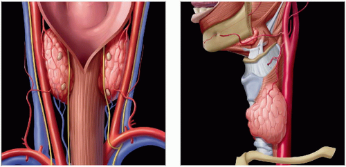 (Left) Coronal posterior graphic demonstrates the anatomic position of the thyroid gland lobes along with the embedded parathyroid glands. The relationship to the larynx and vessels, along with the nerves, is illustrated. (Right) This sagittal view highlights the left lobe of the thyroid gland and its relationship to the tracheal cartilages, membranes, and vessels of the neck. These anatomic landmarks are helpful in the staging of tumors. |
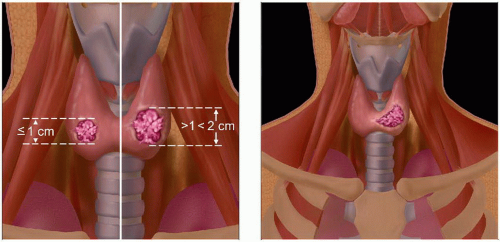 (Left) Coronal graphic shows T1 bilateral thyroid carcinomas, which are confined to the thyroid gland. One tumor is called “microscopic” since it is < 1 cm (left). The presence of bilateral disease is given an (m) designation in the staging system to represent bilateral tumors. (Right) Coronal graphic shows a T2 thyroid carcinoma defined as > 2 cm but ≤ 4 cm and confined to the thyroid gland.
Stay updated, free articles. Join our Telegram channel
Full access? Get Clinical Tree
 Get Clinical Tree app for offline access

|