Protocol for the Examination of Specimens From Patients With Carcinoma of the Adrenal Gland
Image Gallery
Tumor Staging Graphics
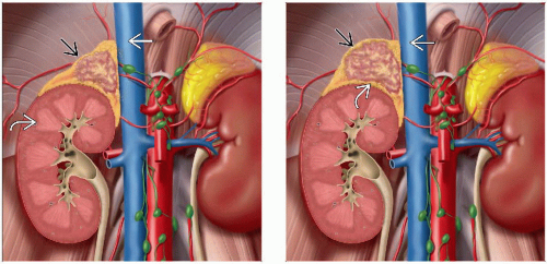 (Left) Coronal graphic demonstrates T1 disease. The primary tumor
 is ≤ 5 cm in greatest dimension, without invasion of adjacent organs, including kidney is ≤ 5 cm in greatest dimension, without invasion of adjacent organs, including kidney  or inferior vena cava or inferior vena cava  (Right) Coronal graphic demonstrates T2 disease. The primary tumor (Right) Coronal graphic demonstrates T2 disease. The primary tumor  is > 5 cm in greatest dimension, without invasion of adjacent organs, including kidney is > 5 cm in greatest dimension, without invasion of adjacent organs, including kidney  or inferior vena cava or inferior vena cava  . .Stay updated, free articles. Join our Telegram channel
Full access? Get Clinical Tree
 Get Clinical Tree app for offline access

|