Proliferative Fasciitis/Myositis
Elizabeth A. Montgomery, MD
Key Facts
Terminology
Tumefactive subcutaneous (fasciitis) or intramuscular (myositis) proliferation featuring ganglion-like fibroblasts
Macroscopic Features
Usually 2-3 cm
Microscopic Pathology
Mostly plump stellate to spindled fibroblasts and myofibroblasts
Large ganglion-like fibroblasts
Macronucleoli, abundant amphophilic cytoplasm
Mitotic activity common
Pediatric examples can display exuberant mitotic activity, which can lead to misinterpretation as sarcomas
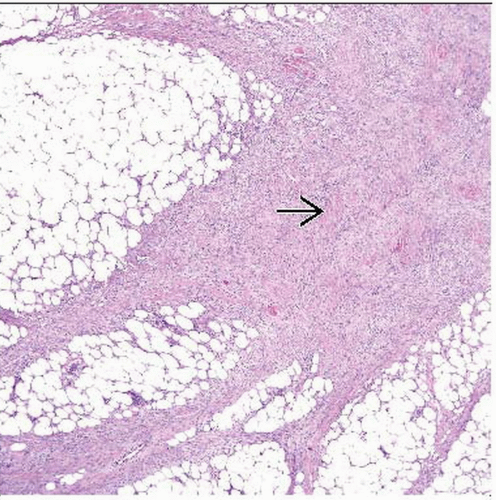 Hematoxylin & eosin shows proliferative fasciitis at low magnification. It tracks along fibrous septa and is somewhat less cellular in the center  , where there is keloid-like collagen. , where there is keloid-like collagen. |
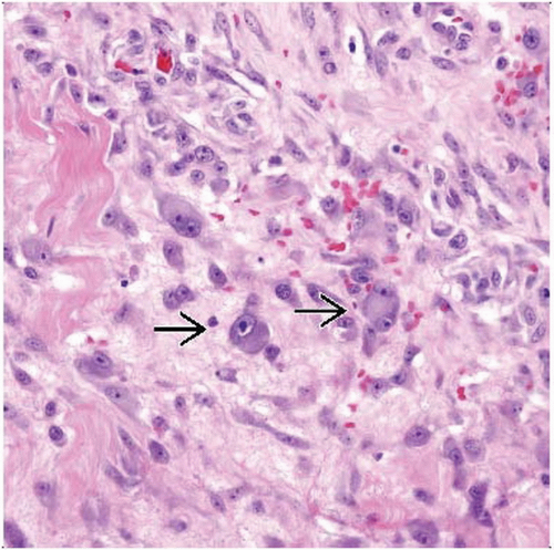 Hematoxylin & eosin shows the so-called ganglion-like cells  of proliferative fasciitis. These are fibroblasts, but their macronucleoli are reminiscent of those found in ganglion cells. of proliferative fasciitis. These are fibroblasts, but their macronucleoli are reminiscent of those found in ganglion cells. |
TERMINOLOGY
Definitions
Tumefactive subcutaneous (fasciitis) or intramuscular (myositis) proliferation featuring ganglion-like fibroblasts
Background of myofibroblasts and fibroblasts similar to those in nodular fasciitis
CLINICAL ISSUES
Epidemiology
Incidence
Rare; less common than nodular fasciitis
Age
Middle-aged and older adults; rare in children
Gender
No predominance
Site
Proliferative fasciitis: Upper extremity (forearm) > lower extremity > trunk
Proliferative myositis: Trunk > shoulder girdle > upper arm > thigh
Presentation
Rapidly growing painless mass; more likely to be painful than nodular fasciitis
Usually no history of trauma


