Principles of diagnosis
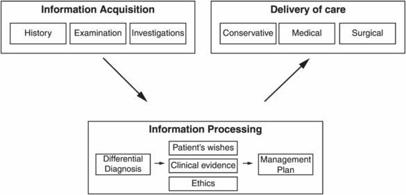
How is a diagnosis reached?
Three steps:
1. History taking
2. Physical examination
3. Investigations

Differential diagnoses
How can a differential diagnosis be made?
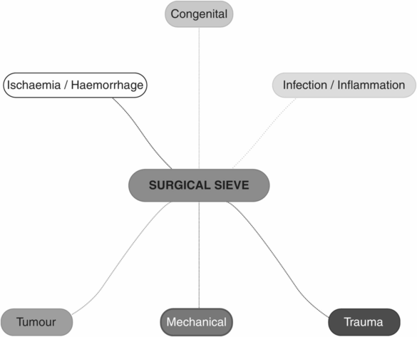
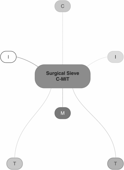
A logical differential diagnosis can be formulated by applying a list of pathological processes to any organ in an anatomical area. The surgical sieve used in this book is C-MIT:
Congenital
Mechanical: extrinsic, mural, intraluminal
Infective or Inflammatory: autoimmune, bacterial, viral, fungal, protozoal
Ischaemic or haemorrhagic (vascular): stenosis, embolism, thrombosis, haemorrhage, dissection, aneurysmal change, vasospasm
Tumour: primary, secondary (metastases), lymphoproliferative
Trauma: penetrating, blunt, chemical, electrical, thermal
All the differential diagnoses in the book are conveyed in the same diagrammatic manner, based on the pathological processes mentioned above.


Principles of examination
What checks must be made prior to beginning the physical examination?
The surgeon must prepare for the examination.
Check physiological parameters (vital signs) and fluid balance from the observations chart.
Examine patient’s bedside or environment.
General examination of the patient from the end of the bed.
How should the examining surgeon prepare before commencing an examination?
WIPER
Wash hands, remove all jewellery and watches and expose arms above elbows.
Introduce yourself.
Purpose and Permission: explain purpose and gain verbal consent (permission) to proceed with the examination.
Expose the patient appropriately for the examination you are about to perform.
Recline: position the patient appropriately.
‘Good morning, Mrs Taylor. My name is Mr Smith. I am a surgical registrar working for Mr Johnson. I understand you have pain in your calf. I would like to examine you to find out the cause. Would that be OK?’
Find a chaperone.
Expose the patient’s legs, removing shoes and socks. Cover the inguinal area and perineum with a sheet.
Place the patient flat with one pillow on an examination couch.
What physiological parameters should be checked in every examination?
The first part of the physical examination is to inspect the patient’s observation chart for physiological parameters (vital signs). If the observation chart contains inaccurate, incomplete or old vital signs it is the examiner’s responsibility to assess and measure these at the time of examination.
The vital signs or physiological parameters are grouped in the following manner and must always be documented:
Cardiac: heart rate and rhythm (HR) and blood pressure (BP)
Respiratory: respiratory rate (RR) and oxygen saturation (Sats)
Temperature (degrees Celsius)
Glasgow Coma Scale (GCS) score
Blood sugar levels (if available)
Fluid balance (positive or negative), including urine output (ml/h in the last 3 hours), drain, NG and stoma output, as well as oral intake
What ‘bedside’ observations should be made?
Look for any signs in the patient’s immediate environment that would hint at the patient’s health:
Evidence of acute illness: attachment to cardiac monitors, defibrillators or infusion pumps
Floor: purulent discharge, bleeding, incontinence, vomitus
Surgical appliances: drains, catheters, vacuum suction devices, blood transfusions, parenteral feeding bags
Orthopaedic appliances: prostheses, walking aids, external fixation devices
Inspect the bedside for clues, ‘MD’:
3 Ms: monitors, mobility aids, medical equipment (e.g. ventilators, dialysis machines)
3 Ds: drips, drains, drug infusions
How is the general examination performed from the end of the bed?
The surgeon stands at the end of the bed keeping her/his hands in a neutral position (e.g. behind the back) so as to emphasise the lack of patient contact and focus on the global observation of the patient. Only once this is completed can the examiner move to the right-hand side of the patient’s bed (for right-handed examiners) and proceed with the organ-system-based assessment. Also assess the patient’s general appearance and behaviour:
Pain
Lethargy, confusion or agitation
Malnutrition, obesity
How can the peripheral stigmata of disease be remembered?
Airway – stridor, anaphylaxis
Breathing – dyspnoea, cyanosis, use of accessory muscles, patient position to facilitate breathing
Circulation – external bleeding
Deficit of neurology – consciousness, comfort (pain), cognition (3Cs)
Dermatology – jaundice, rashes, pallor
Emaciation – cachexia, malnourishment, evidence of catabolism
Fluid status – dehydrated, overloaded/oedematous
Facies – syndromes
Foul smell – foetor, melaena, urine/faecal incontinence, infected tissue/pus, alcohol
JACCOL is a quick method of remembering the peripheral stigmata of systemic disease from the end of the bed:
Jaundice, Anaemia, Clubbing, Cyanosis, Oedema, Lymphadenopathy
Stay updated, free articles. Join our Telegram channel

Full access? Get Clinical Tree


