nerve tissue
Nerve tissue and the nervous system
The nervous system has two structural components. Nerve tissue located in the brain and spinal cord is the major component of the central nervous system (CNS). Other primary tissues are present in the CNS though they are sparse, e.g. connective tissue provides some support. The peripheral nervous system (PNS) comprises nerve tissue which is not in the CNS, e.g. cranial and spinal nerves and their branches, and autonomic nerves (see below). Connective tissue also has a supporting role in the PNS.
The nervous system has two functional components, somatic and autonomic. The somatic nervous system involves the detection of sensations and the control of skeletal muscle contraction. Some sensations are not consciously perceived, such as a change in tension in a tendon, though the change detected can cause a response, such as contraction of skeletal muscle(s). The functions of the autonomic nervous system (ANS) involve the control of contraction of smooth and cardiac muscle cells and the secretory activity of some gland cells. The ANS, in general, is not under conscious control. However, an important exception is that the autonomic nerves involved in controlling the smooth muscle cells in the wall of the urinary bladder are normally under conscious control by the age of 2–3years.
Neurons
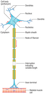
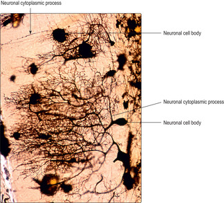
Neurons are derived from the ectoderm germ layer of the embryo (see Mitchell B, Sharma R. Embryology: An Illustrated Colour Text. Elsevier: 2004). Most neurons have a large mass of cytoplasm and large nucleus forming the body of the cell (the perikaryon) and a variable number of cytoplasmic processes (Fig. 6.1). Perikarya may measure up to 150μm in diameter, which is about 20 times larger than a red blood cell. The cytoplasmic processes are of two forms, dendrites and axons. Most neurons have several short dendrites that receive signals which they then pass to their own cell body. In general, each neuron has a long, single axon which takes signals away from the perikaryon either to other neurons or to other cell types, e.g. muscle cells. Axons may be up to about a metre in length in humans, i.e. the length from the lower regions of the spinal cord to the foot.
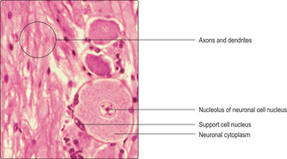
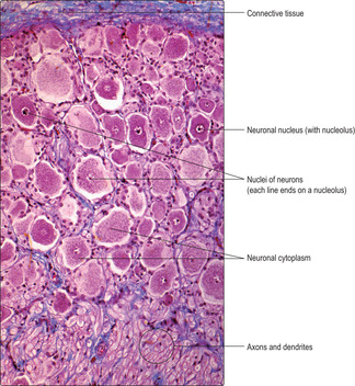
In routine H&E-stained sections, neuronal nuclei have large, palely stained areas (Fig. 6.2). Such areas represent uncoiled euchromatin, the hallmark of DNA transcription and a prerequisite for protein synthesis. The cytoplasm in neuronal cell bodies often appears granular, especially if special stains have been used (Fig. 6.3). The granules are referred to as Nissl granules and they are due to large accumulations of rough endoplasmic reticulum (RER), which are involved in the synthesis of various proteins. Some of these proteins provide the structural tubules and filaments in axons and dendrites. Others are enzymes involved in producing molecules which aid the transmission of signals.
Neurons vary in shape and size, and according to function and location. The complexity of their cytoplasmic processes is vast and revealed by special stains (Fig. 6.4).
■ Variation in shape. Neurons are classified as multipolar (Fig. 6.1), bipolar or pseudounipolar (Fig. 6.5). The polarity is related to the number of cytoplasmic processes projecting from the perikaryon. Many neurons are multipolar and, typically, one axon and many dendrites extend from the perikaryon (Fig. 6.1). Some neurons in the brain and ventral regions (horns) of the spinal cord are multipolar. Bipolar neurons are present in organs of the special senses (e.g. the eye) and they have two cytoplasmic processes, one an axon and one a dendrite. Pseudounipolar neurons possess one process, though it bifurcates and one branch receives signals and the other branch transmits them away from the cell body. Examples of pseudounipolar neurons are those that transmit sensory information from the skin.
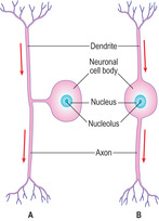
The rate at which nerve impulses are transmitted along axons varies. In general, the wider the diameter of an axon, the faster the nerve impulse travels. The speed of transmission of nerve impulses is also higher in axons that are sheathed in myelin (Fig. 6.1). Myelin is a lipid bilayer arranged as concentric, multiple layers around some axons (Figs 6.6A, 6.7 and 6.8). The layers are formed from the cell membranes of Schwann cells in the PNS and, in the CNS, from the cell membranes of certain glial cells (oligodendrocytes). In places, the myelin sheaths around axons are not layered and these regions are described as nodes of Ranvier (Figs 6.1 and 6.9). The nodes are the interface between two adjacent myelin-producing cells and they are important in ensuring a relatively rapid rate of transmission of the nerve impulse. Other axons are not ensheathed in myelin, but several axons are indented into the cytoplasm of one myelin-producing Schwann cell (Fig. 6.6B). Transmission of impulses along these non-myelinated axons is relatively slow.
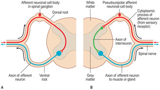
■ Variation in location. Afferent neurons carry sensory signals to the CNS and efferent neurons carry signals away from the CNS to various cells, e.g. muscle and gland cells. Afferent nerves enter the spinal cord by dorsal roots and efferent nerves leave by ventral roots (Fig. 6.10). Some afferent neurons communicate directly in the CNS with efferent neurons and form a two-neuron reflex arc (pathway) (Fig. 6.10A). Others transmit information to interneurons in the spinal cord which in turn communicate with efferent neurons in a three-neuron arc (Fig. 6.10B). In both types of reflex arc other neurons transmit impulses to the brain.
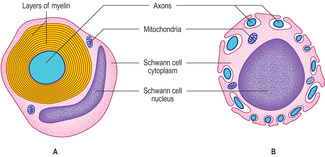 |
Fig. 6.6
Stay updated, free articles. Join our Telegram channel
Full access? Get Clinical Tree
 Get Clinical Tree app for offline access

|
