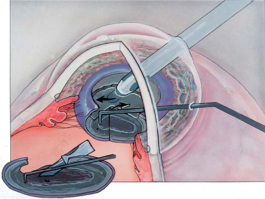
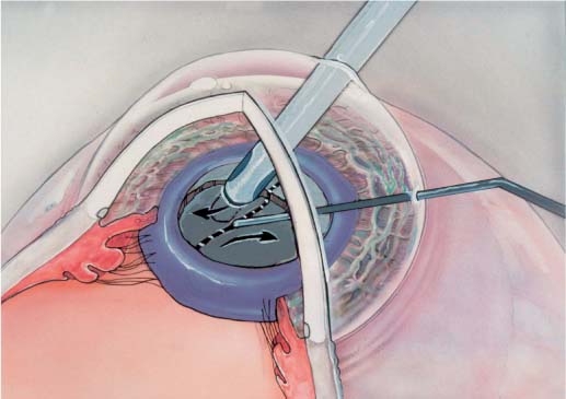
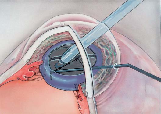
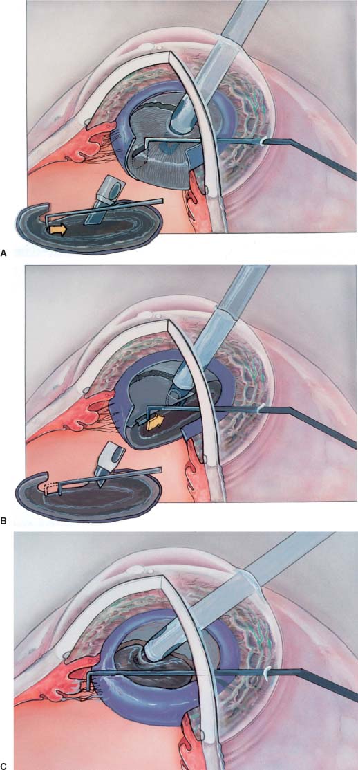
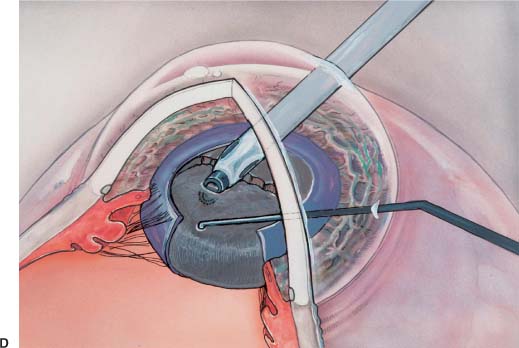
Chapter 31 Compared to other methods of nucleus disassembly, phaco chop reduces ultrasound power, total ultrasound time, and stress on the zonules. This chapter focuses on preventing complications in two areas—in high-risk cases or situations, and when learning or performing phaco chop. In 1993, Kunihiro Nagahara (Japan) introduced the concept of phaco chop at the Third American-International Congress on Cataract, IOL and Refractive Surgery, in Seattle, Washington. Nagahara observed that a chopping force oriented parallel to the lens lamellae (“with the grain”) splits the nucleus along a natural cleavage plane. Other variations such as “snap and split” and “phaco quick chop” utilize the same principle1 (see Chapter 13). Nagahara’s technique is “nonstop” phaco chop because it eliminates the initial groove and crack required by the four-quadrant divide and conquer2,3 and “stop and chop”4 techniques. Like sawing through a log lying on its side, multiple sculpting passes—against the grain—are needed to create an adequately deep groove for cracking. Phaco chop would be analogous to placing the log upright on one end and chopping it with an ax. One strike, parallel to the grain, splits the log in half. The initial chop completely bisects the nucleus without any sculpting. The chopping instrument passes peripherally beneath the anterior capsule, and hooks the equator of the nucleus (Fig. 31–1). I use a Lieberman microfinger for chopping, because its slender, curved tip is ideally shaped for this maneuver. The proximal nucleus is deeply impaled with the phaco tip. The chopping instrument is pulled toward the phaco tip causing an initial compression, followed by a fracture (Fig. 31–2). Upon contact, there is a slight lateral separation of the two tips to propagate the fracture through the proximal remainder of the nucleus (Fig. 31–2). This manual energy, generated by one instrument pushing against the other, replaces the need for ultrasound energy to subdivide the nucleus. After the nucleus is rotated, a pie-shaped piece is created by the second chop (Fig. 31–3). Although this first piece is the most difficult to remove, the strong holding force afforded by high vacuum will usually elevate it out of the bag. Alternatively, the curved tip of the microfinger can slip behind the equator of this nuclear piece to manually tumble it forward into the anterior chamber. Each subsequent piece is chopped and removed by repeating these steps. In addition to requiring less phaco power and time,5–7 chopping minimizes the stress placed on the zonules. Like a vise holding a piece of wood, it is the zonules and capsule that anchor and fixate the nucleus as the groove is sculpted by the phaco tip. With chopping, however, it is the phaco tip against which the chopping instrument pushes. These manual forces are directed centrally inward rather than outward toward the zonules and capsule. These differences in zonular stress can be clearly appreciated when sculpting and chopping are filmed in cadaver eyes from the Miyake-Apple viewpoint. FIGURE 31–1 Microfinger (or chopper) passes beneath anterior capsule edge to hook nucleus equator. Phaco tip impales nucleus proximally. The phaco tip will move rightward with a slight lifting motion. The chopper will move leftward with slight depression. The attributes of decreased zonular stress and decreased phaco power define the indications for phaco chop. Although advantageous for routine cases, phaco chop is most valuable for complicated cases or situations—those that entail greater risk of posterior capsule rupture or corneal decompensation. Small pupils increase the complication rate for two reasons.8 First, physical working space within the pupillary plane is limited, making it easier to aspirate or phaco the iris. Nonstop phaco chop eliminates the need to phaco near or behind the iris because the phaco tip stays within the central 3 mm of the pupil. Second, visualization of the lens is impaired.9 The peripheral lens is hidden by the iris, and the intensity of the red reflex is significantly reduced with each millimeter decrease in pupil diameter. FIGURE 31–2 Microfinger (or chopper) chops against the phaco tip. FIGURE 31–3 Chopping steps repeated after slight rotation. A poor red reflex makes it difficult to judge the depth at which the phaco tip is sculpting. This is a problem for the cracking technique, which requires an adequately deep central trough. In contrast to sculpting, phaco chop is a more kinesthetic technique in which visualization of the depth of the two instrument tips is less important. It is only necessary to visualize that the chopper is passing beneath the anterior capsule as it hooks the equator of the nucleus. Mature white cataracts present multiple problems.10 The absence of a red reflex makes it very difficult to visualize, and therefore perform a capsulorrhexis. The best solution is the use of indocyanine green11 or trypan blue12 dye to stain the anterior capsule. An absent red reflex also precludes visualization of the depth of the phaco tip during sculpting. One lacks the increasingly brighter red reflex that appears as the posterior capsule is neared. As stated earlier, chopping relies more on tactile than on visual clues. Finally, these nuclei are often very large and brunescent. Firm, solid nuclei more effectively transmit the forces of phacoemulsification and other manipulation directly to the zonules and posterior capsule. This includes the forces of nuclear sculpting, cracking, and rotation. There is correspondingly less epinucleus present to cushion the capsule from these stresses. The added phaco time and energy necessary to remove these brunescent nuclei also increase the potential for endothelial cell loss.13 The ability of phaco chop to reduce the amount of nuclear sculpting, the total phaco time and energy, and the stress on the zonules is a particular advantage in these high-risk eyes.14 If a can opener capsulotomy is necessary, outward cracking motions should be minimized as much as possible, in favor of chopping maneuvers. Cataracts with loose zonules are among the most challenging to emulsify.15–18 Predisposing factors include exfoliation, advanced age, trauma, retinopathy of prematurity, and prior intraocular surgery (e.g., prior vitrectomy or trabeculectomy). Loose zonules pose three sets of problems for the phaco surgeon. First, the nucleus, epinucleus, and cortex do not easily separate from a capsule that is not firmly anchored. This makes it difficult to rotate the nucleus, and subsequent aspiration of the epinucleus and cortex may pull the anterior or posterior capsule centrally together with the lens material. Second, the weakened zonules may break more easily.19,20 Aspirating the anterior capsule, or adherent lens material may dehisce the zonules in that region. Pushing the nucleus against the capsular bag (as with sculpting), or forceful nuclear rotation may shear zonules 180 degrees away. Finally, less centrifugal tension by the zonules allows the flaccid central posterior capsule to trampolene forward. Folds of redundant posterior capsule may be aspirated by the phaco or irrigation and aspiration (I&A) instruments. FIGURE 31–4 (A) Incorrect: The chopper is positioned too high. It will skim over the nucleus. An adequate chop is therefore impossible. (B) Incorrect: Both the chopper and phaco tip are too high. (C) Incorrect: The chopper is placed outside the capsular bag, which will create a zonular dialysis on attempted chopping. (D) Incorrect: The phaco tip is not deep enough into the nuclear material, and in addition is too central to adequately create a chop. Phaco chop greatly reduces the stress placed on the zonules and capsule by replacing sculpting and cracking forces with the manual forces of one instrument pushing against another.21 Unlike with cracking, these manual forces are directed centripetally inward, rather than outward toward the zonules. Failure to properly complete these preliminary maneuvers predisposes the eye to complications. Because the capsulorrhexis resists tearing,22
PREVENTION PEARLS AND
DAMAGE CONTROL: PART 4
“NONSTOP” PHACO CHOP—TECHNIQUE AND RATIONALE
PHACO CHOP FOR HIGH-RISK CASES AND SITUATIONS
SMALL PUPILS
MATURE WHITE OR BROWN CATARACTS
LOOSE ZONULES
PROBLEMS WITH CAPSULORRHEXIS, POSTERIOR CAPSULE INTEGRITY, OR HYDRODISSECTION
![]()
Stay updated, free articles. Join our Telegram channel

Full access? Get Clinical Tree


