Postoperative hypotension

Problem 3 Postoperative hypotension


With the various possibilities in mind, you head towards the ward.


On arrival at the ward you quickly read the patient’s notes to aid your assessment. The man had a 2 month history of dysphagia prior to his diagnosis of oesophageal adenocarcinoma. He was treated with a 6 week course of chemotherapy prior to his surgery. His past medical history included controlled hypertension and reflux, for which he was taking bendroflumethiazide and esomeprazole respectively. He is an ex-smoker with a 40 pack-year history.


He had been making an uneventful recovery from his surgery although his gut has not yet started to function. You note from his charts that a tachycardia has developed over the last 24 hours. Over the last 3 hours his urine output has dropped from over 30 mL/hour to 10 mL/hour. His temperature is 38.4°C. You read the operation record and note that the procedure was uncomplicated and a primary anastomosis was performed. A broad-spectrum antibiotic was given prophylactically and continued for 48 hours.


You attend the patient. He is overweight and you estimate his body mass index (BMI) to be 30. He is slouched in the bed with an oxygen mask on his forehead. He has a triple-lumen central line with a bag of saline attached. An epidural catheter is in situ. His chest drain contains a total of 200 mL of sero-sanguinous material, and there is 300 mL of bile-stained fluid in the nasogastric drainage bag and 400 mL in the urinary catheter bag. All the bags were emptied 12 hours earlier. A feeding jejunostomy is running at 40 mL/hour. Little history is available from the patient because of his confusion.


Before you examine the patient more closely you replace his oxygen mask over his mouth and nose and check that his intravenous fluid is running freely. He is mildly confused and is unable to answer your questions appropriately. His skin is pale and clammy and his pulse is faint but regular at 120/min. You confirm his blood pressure to be 85/65 mmHg. He has a tachypnoea of 28 breaths per minute and oxygen saturations are 87%. He already has peripheral venous access and a triple-lumen central line that was inserted during the operation. Both insertion sites look clean and dry. His JVP is not visible and his tongue is dry. His heart sounds are normal.


His lung examination reveals a dull percussion note and absent breath sounds in the lower third of both lung bases. There are associated crepitations at both mid and basal zones. The upper lung fields are clear. The thoracotomy wound looks clean. The chest drain sites are erythematous but dry (the apical drain was removed 2 days earlier). The drainage bottle contains turbid sero-sanguinous fluid and the fluid level swings with deep respiration.


His abdomen is mildly distended, but soft, with minimal wound tenderness to deep palpation. There are no localizing signs. The abdominal wound looks clean and dry, as are the drain and jejunostomy sites. Bowel sounds are absent.


His urine appears concentrated but clear. He has bilateral lower limb oedema, although his calves are not tender.


The patient is transferred to the high dependency unit for resuscitation and further management. A bolus of intravenous isotonic saline is given and blood samples collected for laboratory analysis.


The following results become available:


Investigation 3.2 Arterial blood gas analysis on room air


pO2 52 mmHg pCO2 34 mmHg
pH 7.29 Base excess −9.5


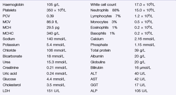
A portable chest X-ray is performed (Figure 3.1).


The patient responds well to your resuscitation measures and his hypotension corrects with intravenous fluid. He is started on broad-spectrum intravenous antibiotics, avoiding aminoglycosides due to concern about nephrotoxicity.


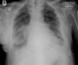
The chest effusions are further visualized with a contrast-enhanced CT scan of the chest and abdomen. One sequence is shown (Figure 3.2).


The right-sided pleural collection is drained percutaneously under radiological control. The material is sanguinous and a sample sent for culture grows enterococci, sensitive to the antibiotics you have prescribed. A chest drain is left in the cavity (and removed after a week). His condition rapidly improves over the next 48 hours. An oral contrast study is performed which does not show any evidence of an anastomotic leakage. The postoperative recovery progresses slowly, but without further mishap.


Answers


A.1 The confusion and hypotension suggest this patient is suffering from shock. Shock is defined as inadequate tissue perfusion. The common signs are related to a reduced oxygen delivery to the tissues. There is commonly a loss in effective circulating blood volume. As this occurs, physiological compensatory mechanisms are initiated, with the primary aim of maintaining oxygen delivery to the vital organs, namely the brain, heart and kidneys. As this compensation fails, so the signs of shock develop.


The causes of shock can be classified into those caused by a loss in total blood volume, or where an effective loss occurs due to volume redistribution. Loss of volume can occur with internal or external blood loss, loss in plasma volume secondary to burns, or fluid loss most commonly from the gastrointestinal tract. Volume redistribution can occur secondary to vasodilatation and increased capillary permeability, most frequently seen in sepsis, anaphylaxis or following acute spinal injury.


Cardiogenic shock occurs secondary to a primary pump failure.


There are several potential causes in this case:


Signs of shock differ according to the cause and its severity. A previously fit adult can lose 15% of their effective blood volume with minimum, transient effects such as mild tachypnoea. Physiological mechanisms will restore blood volume within 24 hours. This is equivalent to donating blood.


Loss of 30% of blood volume can result in tachycardia and narrowing of pulse pressure (the difference between systolic and diastolic blood pressure). This reduces due to compensatory vasoconstriction producing a rise in diastolic pressure. Urine output may reduce and become more concentrated, due to an increase in ADH (antidiuretic hormone).


Loss of more than 40% of effective blood volume (2000 mL) will normally result in a falling systolic blood pressure and tachycardia, as physiological mechanisms fail to compensate adequately. Patients often become confused owing to reduced cerebral perfusion. Urgent action is required as further loss in circulating volume may be fatal.


You have these thoughts in mind as you head towards the ward.


A.2 Additional information may be available from several sources:


Apr 2, 2017 | Posted by in GENERAL SURGERY | Comments Off on Postoperative hypotension

Full access? Get Clinical Tree

Get Clinical Tree app for offline access