9 Postoperative care and complications
Introduction
Following an operation, there are three phases of patient care. After a short period of immediate postoperative care in a recovery room to ensure the full return of consciousness, the patient is returned to surgical ward care, unless there are indications for transfer to a high-dependency unit or intensive therapy unit. On discharge from ward care, patients may still require rehabilitation and convalescence before they are ready to resume domestic or other activities. This chapter discusses the first two phases, during which attention focuses on the regulation of homeostasis and the prevention, detection and management of complications.
The major life-threatening complications that may arise in the recovery room are airway obstruction, myocardial infarction, cardiac arrest, haemorrhage and respiratory failure. These complications can also arise during ward care, but except for haemorrhage and cardiopulmonary catastrophe, many of the problems arising in this phase do not threaten life and are often specific to the operation performed.
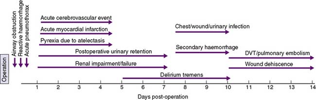
A timeline showing typical times for the development of postoperative complications is given in Figure 9.1.
Immediate postoperative care
Patients who have received a general anaesthetic should be observed in the recovery room until they are conscious and their vital signs are stable. Acute pulmonary, cardiovascular and fluid derangements are the major causes of life-threatening complications in the early postoperative period, and the recovery room provides specially trained personnel and equipment for the observation and treatment of these problems.
In general, the anaesthetist exercises primary responsibility for the patient’s cardiopulmonary function and the surgeon is responsible for the operative site, the wound and any surgically placed drains. Clinical notes should accompany the patient. These include an operation note describing the procedure performed, an anaesthetic record of the patient’s progress during surgery, a postoperative instruction sheet with regard to the administration of drugs and intravenous fluids, and a fluid balance sheet.
Monitoring of airway, breathing and circulation is the main priority in the immediate postoperative period (EBM 9.1). The nature of the surgery and the patient’s premorbid medical condition will determine the intensity of postoperative monitoring required; however, the patient’s colour, pulse, blood pressure, respiratory rate, oxygen saturation and level of consciousness will be routinely observed. The nature and volume of drainage into collecting bags or wound dressings, and urinary output are also monitored, if appropriate. Continuous electrocardiogram (ECG) monitoring is undertaken and oxygenation is assessed by the use of a pulse oximeter. Monitoring of central venous pressure (CVP) may be indicated if the patient is hypotensive, has borderline cardiac or respiratory function, or requires large amounts of intravenous fluids.
The patient may initially remain intubated, but following extubation should receive supplemental oxygen by face mask or nasal prongs and should be encouraged to take frequent deep breaths. The patient must breathe adequately and maintain a good colour. Shallow breathing may mean that the patient is still partially paralyzed. A dose of neostigmine can reverse the residual effects of curariform agents. Cyanosis is an ominous sign indicating hypoxaemia due to inadequate oxygenation, and may be due to airway obstruction or impaired ventilation. Respiratory depression later on in the postoperative period is usually caused by over-sedation with opioid analgesic agents.
Airway obstruction
The main causes of airway obstruction are as follows:
• Obstruction by the tongue may occur with a depressed level of consciousness. Loss of muscle tone causes the tongue to fall back against the posterior pharyngeal wall, and may be aggravated by masseter spasm during emergence from anaesthesia. Bleeding into the tongue or soft tissues of the mouth or pharynx may be a complicating factor after operations involving these areas.
• Obstruction by foreign bodies, such as dentures, crowns and loose teeth. Dentures must be removed before operation and precautions taken to guard against displacement of crowns or teeth.
• Laryngeal spasm can occur at light levels of unconsciousness and is aggravated by stimulation.
• Laryngeal oedema may occur in small children after traumatic attempts at intubation, or when there is infection (epiglottitis).
• Tracheal compression may follow operations in the neck, and compression by haemorrhage is a particular anxiety after thyroidectomy.
• Bronchospasm or bronchial obstruction may follow inhalation of a foreign body or the aspiration of irritant material, such as gastric contents. It may also occur as an idiosyncratic reaction to drugs and as a complication of asthma.
Attention is directed at defining and rectifying the cause of airway obstruction as a matter of extreme urgency. Airway maintenance techniques include the chin-lift or jaw-thrust manoeuvres, which lift the mandible anteriorly and displace the tongue forward (see Chapter 8). The pharynx is then sucked out, an oropharyngeal airway is inserted to maintain the airway, and supplemental oxygen is administered. If cyanosis does not improve or if stridor persists, reintubation may be necessary.
Haemorrhage
Significant blood loss via a surgical drain, particularly if associated with hypovolaemic shock, is an indication for immediate transfer of the patient from the recovery room back to the operating theatre for re-exploration and control of the bleeding source. Reactive bleeding is usually caused by a slipped ligature or dislodgement of a diathermy coagulum as the blood pressure recovers from the operation. Superficial bleeding into the surgical wound rarely requires immediate action; however, patients who have undergone neck surgery must be observed for the accumulation of blood in the wound. If necessary, the wound can be reopened in the recovery room to prevent airway compression and asphyxia.
Late secondary haemorrhage typically occurs 7–10 days after an operation and is due to infection eroding a blood vessel. Rigid drain tubes may also occasionally erode a large vessel and cause dramatic late postoperative bleeding. Secondary haemorrhage associated with infection is often difficult to control. Interventional radiological techniques may achieve temporary control, but surgical re-exploration is usually indicated.
Surgical ward care
General care
Monitoring of vital signs, including temperature, continues on return to the ward. In addition, output from the urinary catheter, nasogastric tube and surgical drains is monitored. The frequency of recordings or measurements can be reduced as the patient stabilizes.
Patients are normally visited morning and evening by the medical staff to ensure that there is steady progress. Anxiety, disorientation and minor changes in personality, behaviour or appearance are often the earliest manifestation of complications. The general circulatory state and adequacy of oxygenation are noted, and vital signs recorded on the nursing chart are checked. Temperature readings provide vital information regarding progress and may give early indication of potentially serious postoperative complications.
The chest is examined and all sputum inspected. Full chest expansion and coughing are encouraged. Following abdominal surgery, the abdomen is examined for evidence of excessive distension or tenderness. The return of bowel sounds and the free passage of flatus reflect recovery of gut peristalsis. The legs are checked for swelling, discoloration or calf tenderness.
Tubes, drains and catheters
If a nasogastric tube is in place, it is kept open at all times to serve as a vent for swallowed air. Free drainage of gastric contents may be supplemented by intermittent manual aspiration. Nasogastric tubes are removed once the volume of aspirate diminishes. It is not necessary to wait until bowel sounds have returned or flatus has been passed. Nasogastric tubes are uncomfortable and may prevent coughing with expectoration, and so they should not be retained for longer than necessary. Surgical drains are generally removed when the volume of effluent diminishes. If a urinary catheter has been placed, it should be removed once the patient is mobile.
Fluid balance
Fluid balance is reviewed regularly. The standard intravenous fluid requirement for an adult is 3 litres/day, of which 1 litre should ordinarily be normal (isotonic) saline and 2 litres should be 5% dextrose. In the first 24 hours after surgery, normal saline can be omitted and replaced by 5% dextrose due to sodium conservation as a result of metabolic response. However, this should be judged according to the patient’s general circulatory status, the observed fluid losses, and the daily measurement of serum urea and electrolyte levels. Similarly, it is not necessary to replace potassium within the first 24–48 hours after surgery, as potassium is released from injured cells and tissues at the surgical site in sufficient quantity. Potassium supplements (60–80 mmol daily) can subsequently be added to intravenous fluids, provided urinary output is adequate. Intravenous fluid therapy is discontinued once oral fluid intake has been established.
Blood transfusion
Haemoglobin measurement will be a guide to the need for postoperative blood transfusion. A full blood count should be undertaken within 24 hours of surgery and, as a general rule, blood is administered if the Hb is less than 80 g/l. Above this level, patients can be prescribed oral iron, unless they have cardiovascular instability or are symptomatic from their anaemia. If a blood transfusion is given, pulse, blood pressure and temperature should be monitored to detect a transfusion reaction. Major ABO incompatibility can result in an anaphylactic hypersensitivity reaction, with severe bronchospasm and hypotension, whereas incompatibility of minor factors may result in tachycardia, pyrexia and rash. Other potential complications of blood trans-fusion are hypothermia (if the blood has not been adequately warmed), hyperkalaemia (due to leakage of potassium from the red blood cells), acidosis (if the blood has been stored for a long period) and coagulation abnormalities (as stored blood is deficient in clotting factors).
Nutrition
Nutrition in postoperative patients is frequently poorly managed. A few days of starvation may cause little harm, but enteral or parenteral nutrition is essential if starvation is prolonged. Enteral nutrition is preferred, as it is associated with fewer complications and is believed to augment gut barrier function. If a prolonged period of starvation is anticipated in the postoperative period, a feeding jejunostomy tube can be inserted at the time of abdominal surgery. Alternatively, a fine-bore nasogastric or nasojejunal feeding tube can be passed (see Chapter 8). If the enteral route cannot be used, total parenteral nutrition can be prescribed. Dietary intake should be monitored in all patients in the postoperative period, and oral high-calorie supplements given if appropriate.
Complications of anaesthesia and surgery
General complications
Nausea and vomiting can be caused by surgery and/or anaesthesia, and an antiemetic can prove useful. If nausea has been associated with previous anaesthetics, antiemetic drugs should be administered prophylactically. Transient hiccups in the first few postoperative days are usually no more than a nuisance. Persistent hiccups can be a serious complication, exhausting the patient and interfering with sleep, and may be due to diaphragmatic irritation, gastric distension or metabolic causes, such as renal failure. If no precipitating cause can be found, small doses of chlorpromazine may be helpful.
Spinal anaesthesia may cause headache as a result of leakage of cerebrospinal fluid, and patients should remain recumbent for 12 hours after this form of anaesthesia. If headache persists, it may be necessary to seal the injection site in the dura-arachnoid with a ‘blood patch’ (i.e. an extradural injection of the patient’s blood, which clots and so seals the leak). Myalgia affecting the chest, abdomen and neck is a specific complication of suxamethonium administration, and may last for up to a week.
Intravenous administration of irritant drugs or solutions can cause bruising, haematoma, phlebitis and venous thrombosis. Intravenous cannulae, particularly those placed in large veins, should be securely sealed to guard against air embolism. Sites of cannula insertion should be checked regularly for signs of infection, and the cannula replaced if necessary. Arterial cannulae and needle punctures are the most common cause of arterial injury, and may rarely lead to arterial occlusion and gangrene.

Stay updated, free articles. Join our Telegram channel

Full access? Get Clinical Tree

