Poorly Differentiated Squamous Carcinoma, Small Cell Variant
Key Facts
Clinical Issues
Cough
History of cigarette smoking
Treatment by surgery, chemotherapy, or radiation depends on stage and grade at presentation
Image Findings
Peripheral spiculated pulmonary nodule in a smoker
Microscopic Pathology
Islands, sheets, and cords of monotonous small, hyperchromatic tumor cells
Tumor cell islands may display prominent basaloid peripheral palisading of nuclei
Cells have large, hyperchromatic nuclei with dense chromatin pattern and prominent nucleoli
Cells can be round, polygonal, or ovoid and display a conspicuous rim of cytoplasm with distinct cell borders
Tumor cells are positive for broad-spectrum cytokeratin and MOC31
Tumor cells are weakly and focally positive for CK7
Tumor cells are uniformly negative for neuroendocrine markers including chromogranin-A, synaptophysin, CD56, and NSE
Tumor cells are negative for TTF-1, CK20, and CEA
Diagnostic Checklist
Possibility of metastasis from a basaloid-squamous cell carcinoma of the oral cavity or head and neck region should always be ruled out 1st
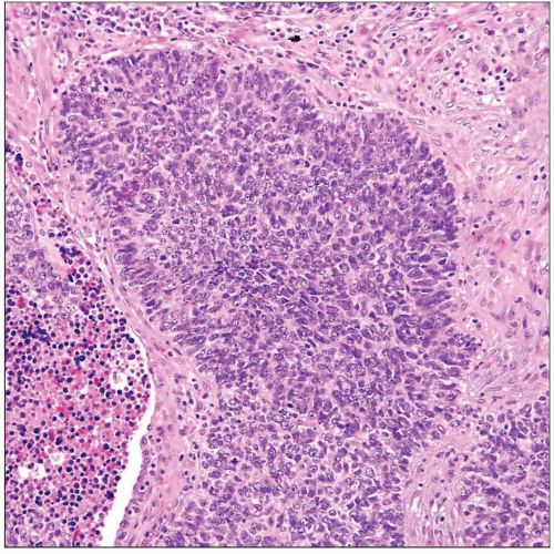 Scanning magnification of small cell variant of poorly differentiated squamous cell carcinoma of the lung shows an island of small tumor cells with peripheral palisading of nuclei. |
 High magnification of small cell variant of poorly differentiated squamous cell carcinoma of the lung shows monotonous round tumor cells with hyperchromatic nuclei and a scant rim of cytoplasm. |
TERMINOLOGY
Definitions
Poorly differentiated squamous cell carcinoma exhibiting small cell morphology and lacking overt features of squamous differentiation
CLINICAL ISSUES
Presentation
Cough
Chest pain
Weight loss
History of cigarette smoking
Treatment
Options, risks, complications
Treatment by surgery, chemotherapy, or radiation depends on stage and grade at presentation
Surgical approaches
Lobectomy and lymph node dissection
Adjuvant therapy
Combination chemotherapy for advanced stages
Prognosis
Depends on grade and stage at presentation; generally poor
IMAGE FINDINGS
General Features
Best diagnostic clue
Peripheral spiculated pulmonary nodule in a smoker
Hilar and mediastinal lymphadenopathy
Bronchial stenosis and associated atelectasis
MACROSCOPIC FEATURES
General Features
Well-circumscribed, tan-white mass
Foci of hemorrhage and necrosis
Intraparenchymatous; unrelated to major bronchi
Sections to Be Submitted
1 section per cm of largest tumor diameter
Size
4-5 cm
MICROSCOPIC PATHOLOGY
Histologic Features
Islands, sheets, and cords of monotonous small, hyperchromatic tumor cells
Tumor cell islands may display prominent basaloid peripheral palisading of nuclei
Tumor cell islands may show prominent central, comedo-like areas of necrosis
Tumor cell cords and islands may be separated by prominent desmoplastic stroma with lymphoid cell infiltrates
Tumor cell islands may be separated by geographic, irregular areas of necrosis
Tumors can show evidence of vascular and perineural invasion
Cytologic Features
Cells have large, hyperchromatic nuclei with dense chromatin pattern and prominent nucleoli
Cells can be round, polygonal, or ovoid and display a conspicuous rim of cytoplasm with distinct cell borders
Tumors may show scattered single-cell keratinization or abortive foci of squamous differentiation
Tumors can display high mitotic activity (> 10 mitoses per 10 HPF)
ANCILLARY TESTS
Immunohistochemistry
Tumor cells
Positive for broad-spectrum cytokeratin and MOC31
Weakly and focally positive for CK7
Uniformly negative for neuroendocrine markers including chromogranin-A, synaptophysin, CD56, and NSE
Negative for TTF-1, CK20, and CEA
May display p63 nuclear staining, which is usually restricted to periphery of basaloid tumor islands
Electron Microscopy
Tumor cells contain desmosomes and tonofilaments
Tumor cells do not display cytoplasmic dense-core neurosecretory granules
DIFFERENTIAL DIAGNOSIS
Small Cell Neuroendocrine Carcinoma
Nuclei show “smudged” nuclear chromatin pattern with little nuclear detail and small or inconspicuous nucleoli
Only scant rim of cytoplasm surrounding nuclei
Stains positive for neuroendocrine markers (chromogranin, synaptophysin, CD56, and NSE)
Shows strong, dot-like paranuclear positivity for CK7
Stay updated, free articles. Join our Telegram channel

Full access? Get Clinical Tree


