Plexiform Fibrohistiocytic Tumor
Cyril Fisher, MD, DSc, FRCPath
Key Facts
Terminology
Fibrohistiocytic tumor of intermediate malignant potential Microscopic Pathology
Tumor based at dermal-subcutaneous junction
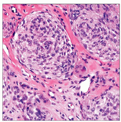
Infiltrative, plexiform, or multinodular growth pattern
3 histological patterns: Histiocyte-like with osteoclast type giant cells, fibroblastic and spindled, mixed
SMA(+) fibroblasts, CD68/CD163(+) histiocyte-like cells
Top Differential Diagnoses
Giant cell tumor of soft parts
May be on spectrum with cellular neurothekeoma
PFHT involves subcutis, not only dermis
PFHT usually presents on extremities and trunk rather than face
PFHT less often myxoid or atypical
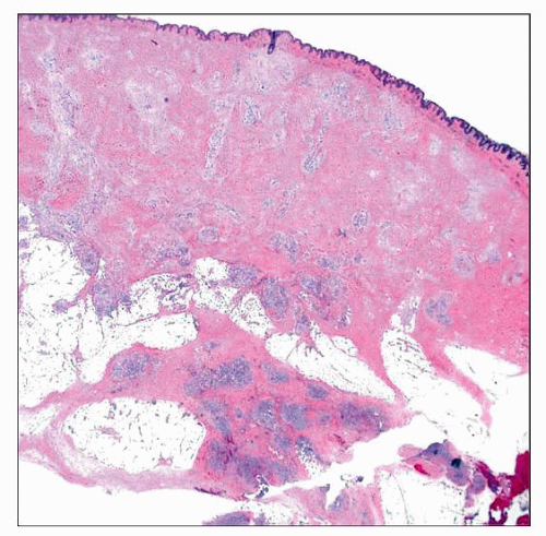
 Low magnification shows plexiform fibrohistiocytic tumor (PFHT) at the dermal-subcutaneous interface with a plexiform appearance. Note the extensions into the dermis and subcutis. |
TERMINOLOGY
Abbreviations
Plexiform fibrohistiocytic tumor (PFHT)
Synonyms
Possibly represents deep form of cellular neurothekeoma
Definitions
Fibrohistiocytic tumor of intermediate malignant potential (rarely metastasizing)
Plexiform pattern
Cellular nodules; interlacing bands of fibroblasts
CLINICAL ISSUES
Epidemiology
Incidence
Rare
Age
Children and young adults
Range: 1-77 years; median: 20 years
Gender
Slight male predominance
Site
Arises at dermal-subcutaneous junction
Extends into dermis and subcutis
Upper extremity is most common site
Followed by lower extremities and trunk
Rare in head and neck region
Presentation
Subcutaneous mass
Treatment
Wide excision with follow-up
Examination of regional lymph nodes, possibly chest imaging
Stay updated, free articles. Join our Telegram channel

Full access? Get Clinical Tree


