Plasmacytoma
Key Facts
Clinical Issues
Incidence
Primary plasmacytomas of lung are rare
May represent approximately 5% of all extramedullary plasmacytomas
Tumor has been described essentially in adults
Symptoms
Cough
Hemoptysis
Dyspnea
Multiple myeloma
Asymptomatic
Prognosis
2-year survival rate approximately 66%
5-year survival rate approximately 40%
Macroscopic Features
Hilar or parenchymal mass
Tumors vary from 2-8 cm in diameter
It is important to submit hilar lymph nodes as they may also be involved
Microscopic Pathology
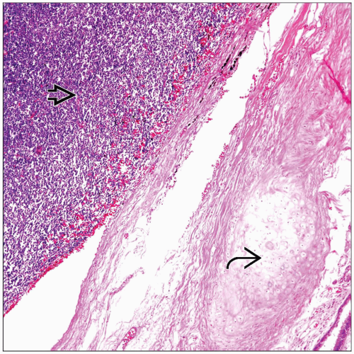
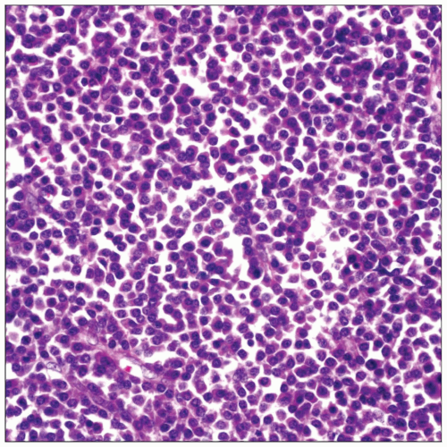
Diffuse plasma cell proliferation
Vague nested pattern
Focal angiectatic areas
Monoclonal gammopathy
Plasma cell with Dutcher bodies
Top Differential Diagnoses
Inflammatory pseudotumor, plasma cell type
Carcinoma
Malignant lymphoma with plasmacytoid features
TERMINOLOGY
Definitions
Malignant neoplasm composed of proliferation of monoclonal plasma cells
ETIOLOGY/PATHOGENESIS
Etiology
Plasma cell dyscrasia
CLINICAL ISSUES
Epidemiology
Incidence
Primary plasmacytomas in lung are rare
Represent approximately 5% of all extramedullary plasmacytomas
Age
Described essentially in adults
Usually from 50-80 years old
Gender
No gender predilection for these tumors
Presentation
Cough
Shortness of breath
Hemoptysis
Multiple myeloma
Asymptomatic
Treatment
Surgical approaches
Pneumonectomy
Lobectomy
Prognosis
2-year survival rate approximately 66%
5-year survival rate approximately 40%
Good possibility for long-term survival
MACROSCOPIC FEATURES
General Features
Hilar or parenchymal mass
Well circumscribed and firm
Yellowish to gray in color
Sections to Be Submitted
It is important to submit hilar lymph nodes as they may also be involved
Size
Tumors vary from 2-8 cm in diameter
MICROSCOPIC PATHOLOGY
Histologic Features
Diffuse plasma cell proliferation
Plasma cell with Dutcher bodies
Vague nested pattern
Focal angiectatic areas
Monoclonal gammopathy
Predominant Pattern/Injury Type
Diffuse
Predominant Cell/Compartment Type


