Pituitary and Sellar Region


Pituitary and Sellar Region


Bernd W. Scheithauer

M. Beatriz

S. Lopes


† Deceased.


Of the wide variety of lesions that affect the sellar region, pituitary adenomas are by far the most common. With advances of histotechnology including immunohistochemistry, the pathologist can engage in clinical correlation with endocrinologists and neurosurgeons and plays an integral role in the management of patients with sellar lesions. In this chapter, these processes are put into perspective for the surgical pathologist. The complex anatomy of the sellar region brings a wide variety of lesions into the clinicopathologic differential diagnosis (Table 12.1). Only those unique or common to this location will be discussed in this chapter.


THE NORMAL PITUITARY


ANTERIOR LOBE

The anterior pituitary or adenohypophysis constitutes the largest portion of the pituitary gland, about 75% to 80%, and it is formed by the pars distalis, the pars intermedia, and the pars tuberalis. The adenohypophysis is epithelial in origin and arises from the Rathke pouch, an invagination in the oral ectoderm. The anterior pituitary has a great variety of cell types and functions (Table 12.2). A number of recognized transcription factors seem to be major participants in anterior pituitary organogenesis in a multistep, highly controlled process (1,2).


TABLE 12.1 Most Common Tumors and Tumorlike Lesions of the Pituitary Gland and Sellar Region


Neuroendocrine neoplasms


Pituitary adenoma


Atypical adenoma


Pituitary carcinoma


Nonneuroendocrine neoplasms


Spindle cell oncocytoma


Pituicytoma


Granular cell tumor


Gangliocytoma


Tumors of nonpituitary origin


Craniopharyngioma


Meningioma


Chordoma


Langerhans cell histiocytosis


Metastases


Cystic lesions


Rathke cleft cyst


Arachnoid cyst


Epidermoid and dermoid cyst


Inflammatory lesions


Lymphocytic hypophysitis


Granulomatous hypophysitis


Xanthomatous hypophysitis


Sarcoidosis


The pars distalis, corresponding to the largest portion of the adenophypophysis, is roughly divided into a central mucoid wedge and lateral components termed acidophilic wings (Fig. 12.1). The term mucoid refers to the abundance of basophilic, periodic acid-Schiff (PAS)-positive cells concentrated and often clustered in this mid portion of the gland. These cells are engaged in the production of adrenocorticotropic hormone (ACTH) and related molecules. The lateral wings contain the majority of growth hormone (GH) and prolactin (PRL) cells, both of which are variably granulated and eosinophilic. Follicle-stimulating hormone (FSH)- and luteinizing hormone (LH)-producing cells are widely and individually distributed throughout the gland. Thyrotrophic hormone (TSH)-producing cells are far smaller in number and more anteriorly situated. The morphologic features of anterior pituitary cells, as well as the biochemical characteristics of their hormone products, are summarized in Table 12.2. The region of the intermediate lobe (pars intermedia), a welldeveloped structure in lower animals, is vestigial in humans and consists primarily of a distinctive form of corticotropic cell and glandular spaces. The latter are remnants of the Rathke cleft and are lined by cuboidal to columnar, ciliated, or mucin-producing cells and only occasionally by granulated adenohypophyseal cells. A sleeve of anterior pituitary tissue extends upward along the anterior aspect of the pituitary stalk (pars tuberalis) and consists mainly of LH/FSH and ACTH cells, all of which are prone to squamous metaplasia with age (3). Salivary gland rests, serous in type, are encountered at the base of the pituitary stalk.

On hematoxylin and eosin (H&E) stain, anterior pituitary cells vary greatly in appearance (Fig. 12.2). Arranged in acini surrounded by a rich network of capillaries (Fig. 12.3), they were once grouped on the basis of tinctorial characteristics as acidophilic, basophilic, or chromophobic. The current cell type recognition is based on hormone content as visualized by immunohistochemistry associated with specific ultrastructural features (Figs. 12.4 and 12.5; Table 12.2). The same is true of pituitary adenomas (Tables 12.3 and 12.4) (4). Pituitary adenoma classification based solely on histochemical stains is no longer recommended because it provides little insight into functional differentiation, which can only be obtained by correlative immunohistochemical and, where needed, ultrastructural studies. These methods have shown GH and PRL cells to be histogenetically related, as are cells engaged in glycoprotein hormone (LH, FSH, and TSH) production. The cells of the anterior pituitary are not limited to the production of these hormones; they have been shown to contain various other peptides (5), as well as growth factors (6).

Electron microscopy has played a pivotal role in categorizing pituitary cells and their tumors. Ultrastructurally, anterior pituitary cells are epithelial in nature and possess the full complement of organelles required for hormone production


and export. Identification of normal and neoplastic pituitary cell types requires attention to such features as cell shape; the content and disposition of organelles; the presence or absence and arrangement of filaments; and of course, the number, size, electron density, and shape of secretory granules (Table 12.4). The mechanisms of hormone secretion vary from transmembrane diffusion to the actual expulsion of secretory granules, a characteristic of PRL cells (7).


TABLE 12.2 Normal Anterior Pituitary Gland: Cellular and Hormonal Features


Cell


Product


Percentage of Cells


Location


Hormonal Characteristics


Histochemical Staininga


Immunohistochemical Staining


Somatotroph


Growth hormone (GH)


50%


Lateral wings


Polypeptide; 191 amino acids; molecular weight (MW), 22,000


Acidophilic


GHb


Lactotroph


Prolactin (PRL)


10%-30% (pregnancy, 50%)


Lateral wings


Polypeptide; 198 amino acids; MW, 23,500


Slightly acidophilic


PRLb


Corticotroph


Adrenocorticotropic hormone (ACTH) and related hormones


10%-20%


Mucoid wedge; often clustered


Polypeptide; 39 amino acids; MW, 4507


Basophilic; periodic acid-Schiff (PAS) positive


ACTH, β-lipotropin (LPH), endorphins, melanocyte-stimulating hormones


Gonadotroph


Luteinizing hormone (LH)c and folliclestimulating hormone (FSH)c


10%


Generalized


βLHc: glycoprotein; 115 amino acids; MW, 28,260


Basophilic to chromophobic


βLH, βFSH, ± α-subunit


βFSHc: glycoprotein; 111 amino acids; MW, 35,100


Thyrotroph


Thyrotropic hormone (TSH)c


5%


Mucoid wedge


βTSHc: glycoprotein; 110 amino acids; MW, 28,000


Basophilic to chromophobic


βTSH ± α-subunit


a Tintorial characteristic of normal pituitary cells are mostly related to the secretory granule density.

b Rare acidophilic stem cells, presumed to be precursor cells producing both GH and PRL, are present in the normal pituitary.

c FSH, LH, and TSH have a common 92-amino acid, 14,000-molecular weight glycoprotein α-subunit in addition to the β-subunit.


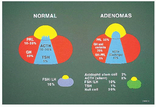
FIGURE 12.1 Schematic of the normal pituitary in horizontal section. Note the proportions and distribution of normal cells and of adenomas. To a significant extent, the locations of growth hormone (GH), prolactin (PRL), and adrenocorticotropic hormone (ACTH) adenomas correspond to the locations of their normal cellular counterparts. Clinically “silent” tumors without endocrine function are usually macroadenomas without specific localization (lower right).


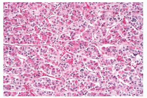
FIGURE 12.2 Normal anterior pituitary. Note the variation in cellular granularity. The staining ranges from acidophilic to chromophobic; several dark-staining basophils are also present.


TABLE 12.3 Pituitary Adenomas: Clinical and Pathologic Characteristics


Adenoma Type


Incidence


Clinical Presentation


Transcription Factor


Hormone


Blood


IHC


Size


Lactotropic adenomas


Sparsely granulated


25%


Amenorrhea and/or galactorrhea; impotence


PIT1


PRL


+


PRL


33% micro/67% macro


Densely granulated


1%


Amenorrhea and/or galactorrhea; impotence


PIT1


PRL


+


PRL


Somatotropic adenomas


Sparsely granulated


5%


Acromegaly or gigantism


PIT1


GH


+


GH, PRL, αSU


14% micro/86% macro


Densely granulated


5%


Acromegaly or gigantism


PIT1


GH


+


GH, PRL, αSU


Adenomas with combined lactotropic and somatotropic features


Mixed GH cell/PRL cell


5%


Acromegaly or gigantism ± hyperprolactinemia


PIT1, ER


GH/PRL


+/+


GH, PRL, αSU


26% micro/74% macro


Mammosomatotroph


3%


Acromegaly or gigantism ± hyperprolactinemia


PIT1, ER


GH/PRL


+/+


GH, PRL, αSU


50% micro/50% macro


Acidophil stem cell


1%


Hyperprolactinemia or “nonfunctional”; only occasional acromegaly


PIT1, ER


GH/PRL


±/+


PRL, GH (focal)


Usually invasive macro


Corticotropic adenomas


Cushing


10%


Hypercortisolism


TPIT


ACTH


+


ACTH


87% micro/13% macro


Nelson


2%


Pigmentation; mass symptoms


TPIT


ACTH


+


ACTH


100% macro


Crooke cell


<1%


Hypercortisolism


TPIT


ACTH


+


ACTH


25% micro/75% macro


Silent corticotropic


3%


Mass symptoms; hypopituitarism


N/A


ACTH


ACTH (diffuse or focal)


100% macro


Glycoprotein adenomas


Gonadotropic


7%-15%


Setting of hypogonadism; functionally silent; mass effects


SF1, ER, GATA2


FSH/LH


15%


βFSH, βLH, αSU


5% micro/95% macro


Thyrotropic


1%


Setting of hypothyroidism or hyperthyroidism


PIT1, GATA2


TSH


+


βTSH, αSU


Usually invasive macro


Silent, subtype 3


3%


Mass effects; hyperprolactinemia or GH effects


PIT1, ER


No specific hormone


Scant to variable GH (10%), PRL (10%), or TSH; rare ACTH


Usually macro


Null cell adenomas


20%


Visual symptoms; hypopituitarism; headaches


Absent


None ± mild elevated PRL as a result of pituitary stalk compression


5% micro/95% macro


αSU, alpha subunit; ACTH, adrenocorticotrophic hormone; ER, estrogen receptor; FSH, follicle-stimulating hormone; GATA2, GATA binding protein 2; GH, growth hormone; IHC, immunohistochemistry; LH, luteinizing hormone; N/A, not applicable; macro, macroadenoma; micro, microadenoma; PIT1, pituitary-specific transcription factor-1 (POU1F1); PRL, prolactin; SF1, steroidogenic factor-1 (NR5A1); TPIT, T-box factor, pituitary (TBX19); TSH, thyrotrophic hormone.


TABLE 12.4 Pituitary Adenomas: Ultrastructural Features


Ultrastructural Type


Cell


Nucleus


Rough Endoplasmic Reticulum


Golgi


Granule Morphology


Miscellaneous


Prolactin cell adenoma


Densely granulated adenoma


Round to oval


Eccentric, oval to irregular


Peripheral parallel stacks


Prominent, ring-shaped


400-1200 nm (600 nm average), electron dense, round to irregular


Sparsely granulated adenoma


Polyhedral


Oval to irregular


“Nebenkern” (concentric whorl) formation


Abundant, ring-shaped or convoluted


150-500 nm (250 nm average), electron dense, round to irregular


Misplaced exocytosis


Growth hormone cell adenoma


Densely granulated adenoma


Round to oval


Central, round to oval, prominent nucleolus


Moderate, peripheral parallel arrays


Prominent, spherical, numerous vesicles


300-600 nm, electron dense, spherical, apposed limiting membrane


Sparsely granulated adenoma


Pleomorphic


Single to multiple, eccentric, irregular


Prominent, peripheral parallel rows or scattered


Abundant, ring-shaped


400-450 nm, electron dense, occasionally 100-250 nm


Paranuclear fibrous bodies, multiple centrioles, smooth endoplasmic reticulum, tubular aggregates in endothelial cells


Adenoma with combined prolactin and growth hormone cell features


Mixed GH-PRL cell adenoma


Variable proportions of sparsely and densely granulated growth hormones and prolactin cells with morphologic features noted above


Mammosomatotroph cell adenoma


Polyhedral


Oval to irregular


Well developed


Prominent, numerous immature secretory vesicles


Two populations: 150-450 nm, electron dense, round to oval, apposed limiting membrane; 350-2000 nm, variably electron dense, elongated, loose limiting membrane (intracellular and extracellular); abundant


Acidophil stem cell adenoma


Elongated


Irregular


Poorly to moderately developed


Moderate; few associated secretory granules


50-300 nm, electron dense; sparse


Paranuclear fibrous bodies, multiple centrioles, misplaced exocytosis, frequent abnormal or giant mitochondria, variable oncocytic transformation


Corticotroph cell adenoma


Densely or sparsely granulated adenoma


Round, angular, or elongate


Round to oval


Abundant, short profiles


Prominent


250-700 nm, round to heart- or teardropshaped, slightly irregular, variably electron dense, often peripheral location


Perinuclear and cytoplasmic 70-Å microfi filaments; enigmatic body (large paranuclear lysosome)


Nelson syndrome


As above but with little or no intermediate filaments


Crooke cell adenoma


As above but with marked to massive accumulation of intermediate filaments


Silent corticotroph cell adenoma


Silent, subtype 1


Resembles densely or sparsely granulated ACTH adenoma


Silent, subtype 2


Small polyhedral


Centrally placed


Numerous


Prominent


150-300 nm, irregular drop-shaped; sparse


No perinuclear and cytoplasmic microfilaments


Glycoprotein adenoma


Gonadotroph cell adenoma


Small to medium, angular to elongated


Oval


Sparse


Moderate, occasionally dilated Vacuolar (“honeycomb”) transformation in females


<150 nm, electron dense ± lucent halo, often peripheral location or in processes abutting capillaries; sparse


Scattered cytoplasmic microtubules


Thyrotroph cell adenoma


Small; angular cells with elongated processes


Irregular to oval


Poorly developed profiles


Moderate


50-250 nm, electron dense with lucent halo, often peripheral location or in processes abutting capillaries; sparse


Scattered cytoplasmic microtubules, occasional large lysosomes


Plurihormonal adenomas


This heterogeneous group of adenomas varies greatly in ultrastructural appearance; some tumors are monomorphous, whereas others consist of two or three distinct cell types


Silent, subtype 3


Irregular polar


Pleomorphic with spheridia


Moderate


Prominent, multifocal


200 nm, cytoplasm and particularly in processes


Abundant smooth endoplasmic reticulum


Null cell adenoma


Small polyhedral


Irregular, indented


Sparse stacks, some ribosomal clusters


Moderate


100-250 nm, electron dense ± lucent halo; sparse


Microtubules, annulate lamellae; variable to abundant mitochondria, microtubules


See references 38 and 39.


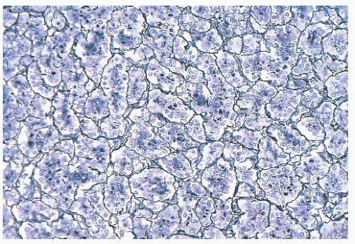
FIGURE 12.3 Normal anterior pituitary. Acini and cords of cells are demonstrated (reticulin stain).


FIGURE 12.4 Normal growth hormone (GH) cells. The high density of these cells in the lateral wing of the anterior lobe, which here are seen immunostained for GH, is sufficient to mimic adenomas, particularly on frozen section.


FIGURE 12.5 Normal adrenocorticotropic hormone (ACTH) cells. The nodularity normally exhibited by some ACTH cells may be mistaken for pituitary hyperplasia, particularly in the setting of Cushing disease when no adenoma is identified.

Folliculostellate cells akin to sustentacular cells in the paraganglia are also encountered in the anterior pituitary (5,8). Small in number, their immunoreactivity for S-100 protein, glial fibrillary acidic protein (GFAP), galectin 3, and annexin 1, and their lack of neuroendocrine marker staining distinguish them from the hormone-producing cells. The function of folliculostellate cells is still controversial, but it appears to be diverse and related to phagocytosis, secretion of growth factors, and intercellular communication (9). It has even been suggested that they represent stem cells (8).


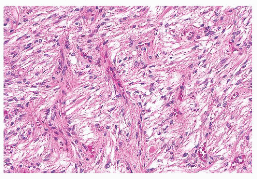
FIGURE 12.6 Posterior pituitary. The posterior lobe consists of the axonal processes and terminations of the vasopressin and oxytocin-producing supraoptic and paraventricular nuclei. In addition, endothelial cells and pituicytes, modified astrocytes, contribute to its cellularity.


POSTERIOR LOBE

The pituitary stalk and the posterior lobe or neurohypophysis represent part of a neurosecretory unit that begins in the magnocellular neurons of the supraoptic and paraventricular nuclei. Coursing via the stalk to the posterior lobe, their unmyelinated axons and terminations carry and store the hormones vasopressin and oxytocin. In their course, the 1-nm diameter axons often show the formation of swellings (Herring bodies) in which neurosecretory materials accumulate. Pituicytes, modified glial cells of primarily astrocytic type, are found throughout the posterior lobe (Fig. 12.6). At the ultrastructural level, axons and their swellings (Herring bodies) and terminations contain secretory granules of varying electron density, as well as multilamellar bodies and electron-lucent vesicles (10). In intimate association with the pituicyte processes, axonal terminations are seen to abut or lie within perivascular spaces. Lastly, scattered corticotroph cells are a normal feature of the posterior lobe (Fig. 12.7). Derived from the intermediate lobe, they appear to be physiologically distinct from anterior lobe corticotrophs
(ACTH-secreting cells). Their accumulation with age, a process termed basophil invasion of the posterior lobe, is of unknown clinical significance. Such cells are believed by some to give rise to so-called silent corticotroph cell adenomas (5,11).


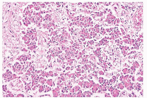
FIGURE 12.7 Basophil invasion. A normal feature of the posterior lobe is the presence of adrenocorticotropic hormone cells at its interface with the anterior lobe (periodic acid-Schiff).


BASIC CONCEPTS ON PITUITARY ADENOMAS

In neurosurgical series, pituitary adenomas represent 10% to 20% of intracranial neoplasms (12). A higher proportion of clinically functioning adenomas (65%) occurs in surgical series (13,14). Incidental or subclinical adenomas are encountered in nearly 25% of autopsies (15,16). The majority are either null cell adenomas (50%) or prolactinomas (45%). For an in-depth discussion of the endocrinologic aspects of pituitary tumors, the reader is referred to authoritative texts (17,18).

Pituitary adenomas show somewhat of a predilection for women in the third to sixth decades of life. However, no age group is exempt, including childhood, a period wherein 2% of all adenomas occur (19,20). ACTH-producing tumors are those most often encountered in pediatric patients (19).

The vast majority of pituitary adenomas are sporadic, but a small percentage of adenomas are associated with hereditary syndromes, including multiple endocrine neoplasia type 1 (MEN1), Carney complex, McCune-Albright syndrome, and a few other rare familial syndromes described as familial isolated pituitary adenoma group (21). The majority of adenomas arising in these hereditary syndromes are GH-secreting adenomas (22), but somatotroph adenomas linked to either MEN1 or Carney complex correspond to only 3% of all GH-secreting tumors (23).

Multiple adenomas are encountered in less than 1% of surgical specimens (24), although a higher incidence is seen as incidental findings in autopsy (16,25). Coexistence of ACTH and PRL cell adenomas is most common (26), which is not surprising given the high incidence of incidental PRL cell adenomas in autopsy series. The distinction of two tumors in a single biopsy specimen is often difficult and may require both immunohistochemical and ultrastructural confirmation (24).

Endocrinologically functional tumors are often small, whereas silent or nonfunctioning tumors are large, coming to attention only as a result of mass effects. An accessible distinction of microadenomas (Fig. 12.8), which are defined by neuroimaging as tumors 1 cm in size or smaller, from macroadenomas (Fig. 12.9), which are tumors exceeding 1 cm, has long been in use (27). Giant adenomas, which are presently defined as adenomas greater than 4 cm in maximal dimension, are rare (28). Diffuse adenomas are ones that fill and expand the sella, often compressing the residual gland into a thin membrane. Larger or massive adenomas often efface the sellar floor, displace surrounding structures, and undergo suprasellar extension.


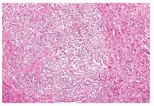
FIGURE 12.8 Microadenoma. Relative circumscription and early compression of surrounding parenchyma are seen. The acinar architecture is effaced.

Suprasellar extension with elevation of the sellar diaphragm is common, as is the extension of tumor through its frequently incompetent diaphragmatic opening. The result of significant suprasellar extension is chiasmal compression with visual disturbance, typically bitemporal hemianopsia. Compression of the pituitary stalk may lead to disruption of the hypothalamicpituitary trafficking of hormones leading to hypopituitarism and increased PRL release due to negative feedback, the socalled “pituitary stalk effect” (17,18). It is of note that pituitary adenomas, regardless of their size, are rarely associated with diabetes insipidus.

Massive suprasellar extension may result in the deep indentation of the brain in the region of the third ventricle. Extension into the middle, anterior, or, less often, posterior fossa may also be seen. Gross, operatively or radiographically apparent invasion of adjacent bony and soft tissues of the sellar region is not infrequently seen. In any case, it predisposes patients to tumor recurrence (29). Microscopic dural invasion, when systematically sought, is common, ranging from 65% to 95%, depending on tumor size (30,31). Documenting the presence of dural or bone invasion by microscopy is recommended for follow-up of the patient. Lateral growth into the cavernous sinus may occur through infrequent naturally occurring discontinuities in the fibrous membrane separating the sella from the sinus (32) or by way of invasion.

Pituitary apoplexy, which is defined as rapid enlargement of an adenoma by intratumoral hemorrhage and variable infarction, may be a surgical emergency (Fig. 12.10). Alternatively, the process may undergo subclinical evolution. Seen in approximately 10% of operated adenomas, it takes the form of hemorrhagic, necrotic, or cystic foci (33,34). Apoplexy affects all types of adenoma, but large, nonfunctioning tumors are particularly
prone. The specimens usually consist of blood and necrotic tumor. Identification of the underlying tumor is aided by reticulin stains, which highlight the abnormal stromal pattern of the adenoma (Fig. 12.11).


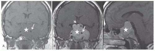
FIGURE 12.9 Macroadenoma. Coronal pre- and postcontrast T1 (A,B) and sagittal postcontrast T1 (C)-weighted sequences demonstrate a large sellar mass lesion (white star), which expands the sella and completely replaces the normal pituitary gland. The infundibular stalk is not identified. The lesion has significant suprasellar extension with left cavernous sinus invasion with encasement of the left internal carotid artery (long white arrow in A and B). Note the mass effect on the optic chiasma (short white arrow in B), which is stretched and thinned over the superior aspect of the lesion. (Courtesy of Dr. Sugoto Mukherjee, Department of Radiology, University of Virginia Health System, Charlottesville, VA).


THE ROLE OF THE PATHOLOGIST

The assessment of pituitary adenomas begins at the time of surgery. Although smears or frozen sections may be performed to identify the tumor, systematic assessment of resection margins is an unrealistic endeavor. Total surgical excision may be complicated by factors such as irregular tumor shape, lack of cleavage planes and a capsule, and grossly inapparent foci of dural invasion (Fig. 12.12). Furthermore, because of lack of stroma, the soft consistency of adenomas obscures landmarks and may even promote contamination of the operative field.

Although pituitary adenomas are the most common tumors in the sellar region, a spectrum of lesions enters into their differential diagnosis. The number of diagnostic possibilities is markedly reduced by attention to such factors as the presence or absence of sellar enlargement and clinical and biochemical data regarding hormone secretion and hyperfunction. A working relationship between the pathologist and surgeon is essential, not only to ensure the procurement of an adequate specimen but also to maximize clinicopathologic correlation.


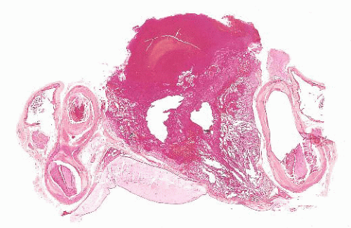
FIGURE 12.10 Pituitary apoplexy. The sellar region in coronal sections shows a massive hemorrhage within a macroadenoma. (Courtesy of Dr. K. Kovacs, St. Michael’s Hospital, Toronto, Ontario, Canada.)


SPECIMEN HANDLING

Fresh tissues should be promptly transported from the operating room on a moist Telfa pad; delayed fixation and drying artifact must be avoided. Smears and touch preparations are preferable to frozen sections in the assessment of adenomas. Smears best demonstrate cytologic detail (Fig. 12.13). Cytologic methods obviate the mechanical and freezing artifacts that affect permanent sections and immunohistochemical preparations. Once the tissues are frozen, they often show nonspecific or reduced immunoreactivity. Furthermore, they are useless for ultrastructural study.

The specimen should be formalin fixed only after a minute portion, at minimum a single 1-mm fragment, has been placed in glutaraldehyde for possible ultrastructural study. With an adequate sample, consideration can be given to freezing a portion for biochemical or genetic studies. When diagnostic tissue is scant or no lesion is identified on cytologic or frozen section assessment, the entire specimen should be step sectioned to obtain H&E and unstained slides.

In addition to the preparation of an H&E-stained slide, consecutive microsections should be cut for reticulin stains (Fig. 12.14), as well as immunostains for pituitary hormones. Performance of a full hormone battery, including GH, PRL, ACTH, LH, FSH, TSH, and α-subunit of glycoproteins, is preferable for better classification of the tumors, but immunostains can be selectively applied, depending on the clinical setting and size of the specimen. This is particularly true in the setting of Cushing disease, in which serial immunostains for ACTH may be more useful in confirming the presence of a small, often fragmented pituitary adenoma rather than the entire hormonal panel. In large institutions with advanced endocrine and neurosurgical departments, the full spectrum of antibodies for pituitary hormones is usually applied to all but the adenomas of Cushing disease. As stated earlier, electron microscopy plays an important, albeit defined, role in the assessment of adenomas.


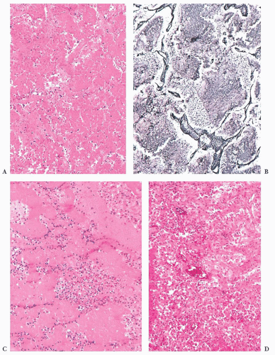
FIGURE 12.11 Pituitary apoplexy. (A) Hematoxylin and eosin-stained sections of such specimens often show only hemorrhage and extensive necrosis. (B) The underlying pattern of adenoma is highlighted by reticulin stain. (C) In the subacute phase, aggregates of polymorphonuclear leukocytes should not be mistaken for infection. (D) In chronic phases, ingrowth of granulation tissue may be conspicuous.


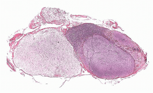
FIGURE 12.12 Microinvasive adenoma. This whole-mount sagittal section of the pituitary demonstrates the adenoma. Note the irregular outlines of the tumor, as well as the early invasion of the dural capsule, on the anterior aspect of the gland.


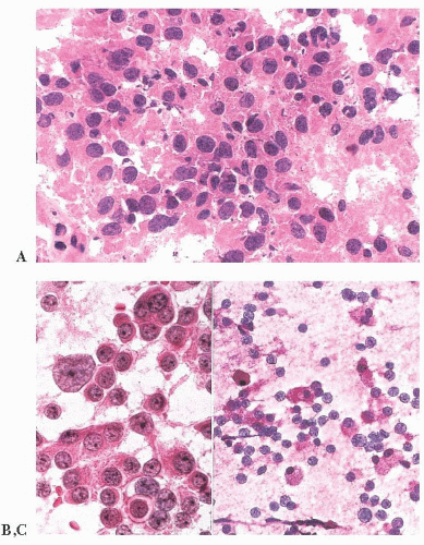
FIGURE 12.13 Pituitary adenoma. This is a comparison of a hematoxylin and eosin-stained frozen section (A) and a touch preparation (B). The latter shows excellent cytologic detail, including prominence of nucleoli, binucleation, nuclear atypia, the presence of a mitosis, and cytoplasmic uniformity. A touch preparation from normal pituitary tissue is far less cellular; it shows variation in cytoplasmic staining; and it lacks both nuclear abnormalities, as well as mitoses (C).


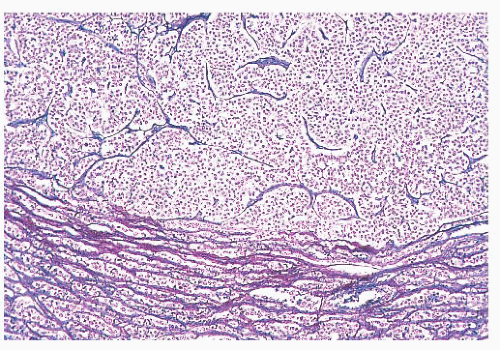
FIGURE 12.14 Pituitary adenoma. The lack of reticulin content and the compression of surrounding parenchyma are demonstrated (reticulin stain).


CLASSIFICATION OF PITUITARY ADENOMAS

In broad terms, the World Health Organization (WHO) and Systematized Nomenclature of Medicine (SNOMED) classify adenohypophyseal tumors as typical and atypical adenomas (WHO 8272/0 and 8272/1), as well as carcinomas (WHO 8272/3) (35).

Morphologically, adenomas may show a variety of growth patterns including diffuse, papillary, and trabecular arrangements similar to other neuroendocrine tumors, which can be present in any adenoma type (Fig. 12.15). Their recognition, although of no prognostic significance, is worthwhile because a spectrum of lesions enters into the differential diagnosis of sellar region pathology. Cytologically, adenomatous cells may be acidophilic, basophilic, or chromophobic; however, these tintorial characteristics do not identify specific adenoma types. Note that all adenomas are synaptophysin immunoreactive but not all stain for chromogranin. Adenomas are focally immunoreactive for cytokeratin, in particular CAM5.2, and epithelial membrane antigen (EMA) (36). Electron microscopy has revealed much about the nature of normal pituitary cells (37) and their respective tumors. At present, although less used in clinical practice (38), ultrastructural analysis continues to play a valuable role in the diagnosis of unusual adenoma types and the expanding spectrum of rare sellar region tumors (Table 12.4) (37,38 and 39).

The classification of adenomas and their clinicopathologic and immunohistochemical features are summarized in Table 12.3. The major adenoma subgroups are discussed in turn in the following sections.


PITUITARY ADENOMA SUBTYPES


PROLACTIN CELL ADENOMA

Nearly half of the newly clinically diagnosed pituitary adenomas are PRL-producing tumors (40). The so-called prolactinomas or lactotropic tumors are composed entirely of PRL
cells. Although the majority of prolactinomas are medically treated by dopamine agonists, a significant number of the patients undergo surgical resection due to several clinical issues (17,18).


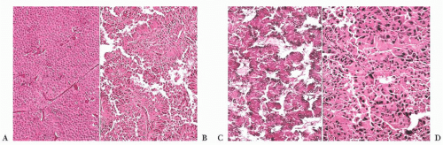
FIGURE 12.15 Pituitary adenomas. The hematoxylin and eosin-stained appearance of these adenomas, which include diffuse (A), papillary (B), ribbon (C), and pleomorphic (D) patterns, illustrates their broad morphologic spectrum and highlights the diagnostic use of immunohistochemistry.

Prolactinomas are the second most frequently occurring adenoma in MEN1 after somatotroph adenomas (22,41). On the other hand, isolated familial prolactinomas are rare (42). Whereas estrogens cause prolactinomas in experimental animals and pregnancy may be associated with enlargement of some, these agents alone are generally incapable of inducing prolactinomas in humans (43,44 and 45).

Microadenomas generally occur in reproductive-age women who exhibit exquisite sensitivity to PRL excess by manifesting with amenorrhea, galactorrhea, or both. In men and postmenopausal women, prolactinomas may appear to be clinically nonfunctional, growing to macroadenoma dimensions and exhibiting invasion (46,47 and 48). The basis for this relative aggressiveness appears to be related to differences in tumor invasion and proliferative activity (47,48). Approximately 50% of prolactinomas treated by surgery are macroadenomas and a third of the cases are grossly or radiographically invasive at initial surgery (48). Not surprisingly, the frequency of invasion increases with tumor size. Serum PRL levels are uniformly elevated in patients with prolactinomas, although the levels range from little more than normal (˜20 ng/mL) to extremely high (>2000 ng/mL). In general, PRL levels correlate with the tumor size (17,18). As noted earlier, mild increases of PRL (<150 ng/mL) may be a result of “stalk section effect” and are not diagnostic of adenoma (17,18).


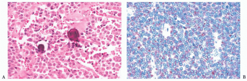
FIGURE 12.16 Prolactin cell adenomas. These are nearly all chromophobic, and they often contain spherical microcalcifications (A). Immunoreactivity for prolactin shows a characteristic globular reaction in the paranuclear Golgi zone (B).

Nearly all prolactinomas are sparsely granulated and thus chromophobic (Fig. 12.16). Densely granulated (eosinophilic) examples are rare. Approximately 10% to 20% of prolactinomas feature psammomatous microcalcification (49). The latter is usually scant but may be so abundant that it forms a “pituitary stone.” The presence of microcalcifications in an adenoma strongly suggests the diagnosis of prolactinoma, as
does the rare finding of spherical amyloid bodies (Fig. 12.17) (50). Immunoreactivity for PRL is typically strong but is paranuclear in location (Fig. 12.16). The ultrastructural features of PRL cell adenoma are distinctive (Table 12.4) and include abundant rough endoplasmic reticulum, as well as “misplaced exocytosis” or granule extrusion between neoplastic cells (37,38).


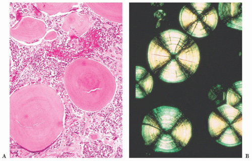
FIGURE 12.17 Prolactinoma with amyloid deposition. Such spherical bodies are virtually diagnostic of a prolactin cell adenoma (B, polarization).

Given the efficacy of dopamine agonist therapy, the frequency of prolactinomas in surgical series has dramatically decreased from 30% to 10%. These agents produce atrophy of tumoral cells with resultant tumor shrinkage or arrest; the effect is reversible with cessation of therapy and thus not curative. The morphologic effects of such dopamine agonists as bromocriptine have been well characterized (51,52). They include diminution of cell size, condensation of nuclei, reduction in synthetic and secretory organelles, cessation of granule extrusion, and interstitial collagen deposition. Necrosis plays no significant part. Perhaps because of uneven D2 receptor loss, these changes may not affect all tumor cells. When administered long-term, they may induce dense tumoral fibrosis (Fig. 12.18).

Small tumor size and normalization of PRL levels after surgical or medical therapy of prolactinoma are favorable prognostic features. Increased age, male sex, large tumors, and invasion are negative predictors for surgical outcomes (48). Recurrence has been associated with invasion (48,53). The main differential diagnosis of prolactinoma is the acidophil stem cell adenoma (39) (see “Acidophil Stem Cell Adenoma” section). Key distinguishing features include its often large size in the presence of low PRL levels, minor immunoreactivity for both PRL and GH, and distinctive ultrastructural features.


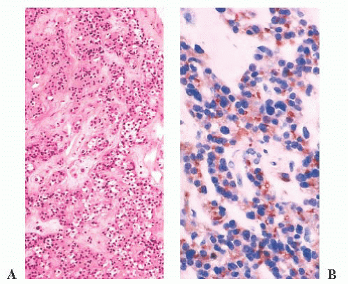
FIGURE 12.18 Treated prolactin (PRL) cell adenoma. Microscopically, these adenomas show perivascular fibrosis, which here is seen on hematoxylin and eosin stain (A) and on immunostain for PRL (B), the reactivity of which persists.


GROWTH HORMONE-PRODUCING ADENOMAS


Growth Hormone Cell Adenoma

Two clinical disorders are associated with GH-producing adenomas: gigantism, which begins in childhood or adolescence, and acromegaly, a far more common disease affecting adults. These effects are largely mediated by insulin-like growth factor-1
(IGF-1) produced by the liver and more reliably elevated than GH levels.

Although GH cell adenomas are associated with clinical or immunohistologic evidence of GH production, only a minority of adenomas produces GH alone (54,55). The vast majority of adenomas produce both GH and PRL or are plurihormonal, also expressing TSH and/or α-subunit. Acromegaly can also be the result of somatotroph hyperplasia caused by ectopic production of growth hormone-releasing hormone (GHRH) by a variety of endocrine tumors and in the McCune-Albright syndrome (21,22). As previously commented, GH adenomas also occur in Carney complex and familial isolated pituitary adenoma (22).

The pathology of GH-producing or somatotroph adenomas has been extensively studied (56,57 and 58). Two variants of GH cell adenomas are described: the densely granulated (eosinophilic) and the sparsely granulated (chromophobic) adenomas. Densely granulated adenomas are more frequent than sparsely granulated GH cell adenoma (59). The histologic patterns of GH cell adenomas are often nonspecific and diffuse, but nuclear pleomorphism and multinucleation are most often seen in sparsely granulated (chromophobic) tumors. The latter often show only scant GH immunoreactivity (Fig. 12.19). In contrast, densely granulated eosinophilic tumors stain strongly (Fig. 12.20). A characteristic of sparsely granulated adenomas is the presence of paranuclear eosinophilic, low-molecular-weight keratin containing “fibrous bodies” (Fig. 12.19C). At the ultrastructural level, these consist of intermediate filament whorls, often enmeshing organelles (Table 12.4). Sparsely granulated GH cells do not occur in the normal pituitary. In contrast, the cells of densely granulated tumors resemble normal GH cells, their eosinophilia being a result of abundant larger secretory granules. Such tumors lack fibrous bodies. Although the endocrinologic features and secretory activity of densely granulated and sparsely granulated variants of GH cell adenoma are similar, sparsely granulated tumors are more aggressive (59). These tumors are mostly macroadenomas, tend to arise in younger patients than densely granulated adenomas (44 vs. 50 years), and have higher invasive incidence (55,59). On rare occasion, GH adenomas are associated with gangliocytomas (60). Clinically nonfunctional or “silent GH cell adenomas” are rare (61).

Medical treatment of GH-producing adenomas involves the use of long-acting somatostatin analogs, such as octreotide. Morphologically, their effect varies but includes mild to moderate interstitial fibrosis (62). Tumor shrinkage is minor despite reduction in GH levels (62).


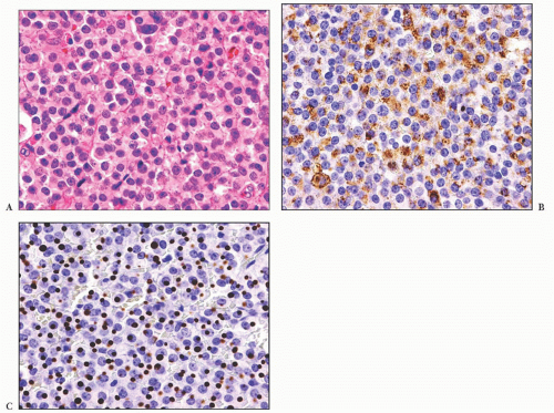
FIGURE 12.19 (A) Growth hormone cell adenoma, sparsely granulated (chromophobic) type. Note the presence of paranuclear hyaline, fibrous bodies. (B) Immunoreactivity for growth hormone may be weak and present in only a portion of cells. (C) Immunoreactivity for cytokeratin (CAM 5.2) highlights the fibrous bodies.


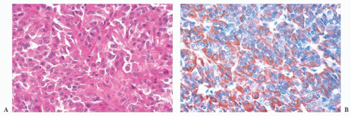
FIGURE 12.20 (A) Growth hormone (GH) cell adenoma, densely granulated (eosinophilic) type. Note the prominent acidophilia and the lack of fibrous bodies. (B) GH immunoreactivity is strong.


Mixed Growth Hormone Cell-Prolactin Cell Adenoma

As has already been noted, most GH-producing adenomas also secrete PRL. Indeed, nearly 40% of acromegalic patients have hyperprolactinemia (55). Because any number of lesions in the sellar region may be associated with hyperprolactinemia as a result of stalk section effect, a diagnosis of GH adenoma with a PRL-producing component cannot be made without immunohistochemical confirmation.

The predominant clinical feature of the mixed GH cell-PRL cell adenoma is acromegaly. The effects of concomitant GH and PRL elevation are not always apparent. Such tumors, which are variably acidophilic, are of interest because they are composed of two distinct, albeit related, cell types—somatotrophs and lactotrophs (Fig. 12.21) (37,38). They represent approximately 5% of pituitary adenomas. Both GH and PRL immunoreactivity are strong. Distinguishing them ultrastructurally from the acidophil stem cell adenoma is important but generally easy. The latter, an aggressive form of GH- and PRL-producing adenoma, shows much less hormone reactivity and masquerades as a prolactinoma, with GH elevation and acromegaly being infrequent (see the following section) (39).


Mammosomatotroph Cell Adenoma

This unusual GH- and PRL-producing tumor represents only 1% of pituitary adenomas and is the most common tumor underlying gigantism (63). It is of interest because this well-differentiated, eosinophilic tumor is composed of a single cell type sharing ultrastructural features of both GH and PRL cells. Key features include densely granulated cells resembling somatotrophs but exhibiting both large (up to 1500 nm) granules and misplaced exocytosis, a feature of lactotrophs (37,38,64). Immunoreactivity for both hormones is strong. Again, making a distinction from acidophil stem cell adenoma is essential (Table 12.4).


Acidophil Stem Cell Adenoma

This unique form of pituitary adenoma is uncommon and comprises less than 1% of all adenomas and 5% of GH-producing tumors. Most patients present with hyperprolactinemia with minor symptomatology related to GH hypersecretion (39). The adenoma is composed of immature cells, perhaps stem cells, of the acidophil (GH and PRL cell) line. Small numbers of acidophil stem cells are found in the normal pituitary. On light microscopy, acidophil stem cell adenomas are chromophobic or somewhat oncocytic in appearance (Fig. 12.22). In general, acidophil stem cell adenoma shows greater immunoreactivity for PRL than for GH, which may be very focal or lacking. Scattered fibrous bodies, immunoreactive for cytokeratin, may be seen. Ultrastructurally, the tumor consists of a single, rather poorly differentiated cell type showing features intermediate between GH cells (fibrous bodies) and PRL cells (misplaced exocytosis) (Table 12.4) (39). Secretory granules are sparse and small. An often conspicuous feature is the presence of giant mitochondria, which may be apparent on light microscopy as cytoplasmic vacuoles, some approaching the size of the nucleus (Fig. 12.22). Electron microscopy is required to establish a firm diagnosis of acidophil stem cell adenoma (Table 12.4).

Only gold members can continue reading. Log In or Register to continue

Stay updated, free articles. Join our Telegram channel

Sep 22, 2016 | Posted by in PATHOLOGY & LABORATORY MEDICINE | Comments Off on Pituitary and Sellar Region

Full access? Get Clinical Tree

Get Clinical Tree app for offline access