Pilar (Tricholemmal) Cyst
Steven D. Billings, MD
Key Facts
Clinical Issues
90% of pilar cysts present on scalp
Microscopic Pathology
Circumscribed simple cyst
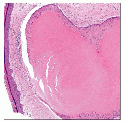
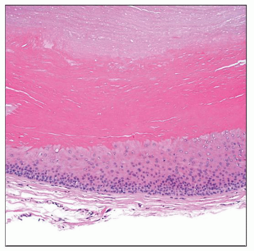
Filled with dense eosinophilic keratin
Stratified squamous epithelium
Abrupt keratinization from large polygonal keratinocytes
Granular layer generally absent
Rupture with associated granulomatous response common
Diagnostic Checklist
Abrupt tricholemmal keratinization is a key diagnostic feature
Simple unilocular cyst
If architectural complexity is present, consider proliferating pilar cyst or malignant pilar cyst
 Scanning magnification shows a dermal-based benign cystic lesion with dense eosinophilic tricholemmal keratinization, characteristic of a pilar cyst. |
TERMINOLOGY
Synonyms
Pilar cyst
Trichilemmal cyst
Isthmus-catagen cyst
Definitions
Benign unilocular cyst lined by squamous epithelium lacking a granular layer and containing abundant dense keratin material
CLINICAL ISSUES
Epidemiology
Incidence
2nd most common type of cutaneous cyst
Age
Most common in adults
Gender
More common in women than men
Site
90% of pilar cysts present on scalp
Presentation
Autosomal dominant inheritance common
Stay updated, free articles. Join our Telegram channel

Full access? Get Clinical Tree


