Penile and Vulvar Intraepithelial Neoplasia
Elsa F. Velazquez, MD
Key Facts
Terminology
Most warty, basaloid, and mixed VIN and PeIN replace 2/3 to entire thickness of epithelium (VIN III and PeIN III) and represent carcinoma in situ
VIN/PeIN I-II is rare
Differentiated VIN/PeIN is considered high-grade lesion
Microscopic Pathology
Differentiated (simplex) PeIN and VIN (HPV-unrelated)
Elongated and anastomosing rete ridges
Atypical basal cells with hyperchromatic nuclei
Subtle abnormal maturation (large eosinophilic keratinocytes)
Whorling and keratin pearl formation
Usually associated with LS&A
Preferential association with HPV-unrelated variants of invasive SCC (keratinizing SCC, verrucous carcinoma)
Basaloid, warty, and mixed (warty-basaloid) PeIN and VIN (a.k.a. usual VIN) (HPV-related)
Basaloid VIN/PeIN: Basaloid cells replace most to full thickness of epithelium
Warty VIN/PeIN: Pleomorphic cells with koilocytic changes replace most to full thickness of epithelium
Warty-basaloid VIN/PeIN: Pleomorphic cells with koilocytic changes seen on upper epithelium and basaloid cells replace lower epithelium
Warty, basaloid, and mixed VIN/PeIN is usually seen adjacent to HPV-related variants of invasive SCC (basaloid and warty types)
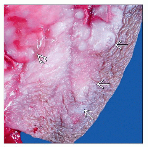 Vulvar intraepithelial neoplasia may appear as a raised, thickened white plaque  . This lesion is associated with an invasive component . This lesion is associated with an invasive component  that is exophytic. (Courtesy T. Quinn, MD.) that is exophytic. (Courtesy T. Quinn, MD.) |
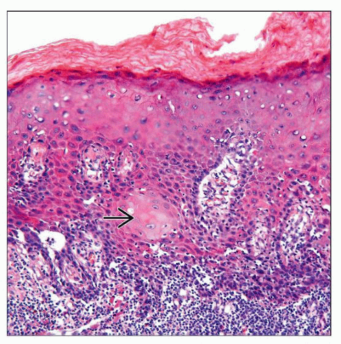 Enlarged keratinocytes with abundant eosinophilic cytoplasm throughout most of the epithelium are seen in differentiated PeIN/VIN. Characteristic keratin pearl formation is present  . . |
TERMINOLOGY
Abbreviations
Penile intraepithelial neoplasia (PeIN)
Vulvar intraepithelial neoplasia (VIN)
Synonyms
Erythroplasia of Queyrat, Bowen disease, squamous cell carcinoma in situ (SCCis)
Definitions
VIN and PeIN are considered intraepithelial (in situ) precursor lesions of invasive SCC
ETIOLOGY/PATHOGENESIS
Pathogenesis
Bimodal pathway of tumor progression in vulvar and penile SCC (HPV-related and HPV-unrelated)
Basaloid, warty, and warty-basaloid (mixed) VIN and PeIN are HPV-related (especially HPV 16)
Differentiated (simplex) VIN and PeIN are HPV-unrelated
May be related to lichen sclerosus et atrophicus (LS&A)
May be associated with P53 mutations
CLINICAL ISSUES
Epidemiology
Incidence
Real incidence is unknown
2/3 associated with invasive SCC
Age
5th and 6th decades
About 1/2 of patients with VIN are < 40 years old
Presentation
Differentiated PeIN and VIN
Older patients
Usually arises in setting of chronic scarring, inflammatory dermatosis, especially lichen sclerosus et atrophicus (LS&A)
Warty, basaloid, and mixed PeIN and VIN (a.k.a. VIN of usual type in vulvar pathology)
Younger patients
Patients may have history of condyloma
Treatment
Surgery, locally destructive treatments
Prognosis
Most studies are retrospective and real prognosis remains unknown
MACROSCOPIC FEATURES
General Features
VIN and PeIN have heterogeneous gross appearance
Solitary or multifocal
Flat to slightly elevated hyperkeratotic or even condylomatous lesions
Pearly white, moist, erythematous, dark brown/black macules, papules, or plaques
MICROSCOPIC PATHOLOGY
Histologic Features
Differentiated (simplex) PeIN and VIN
Stay updated, free articles. Join our Telegram channel

Full access? Get Clinical Tree


