Chapter 50 Penetrating Neck Injury (Case 33)
Case: A 44-year-old female presents to the ED with a stab wound to the left neck.
Differential Diagnosis
| Carotid artery injury | Tracheal injury/laryngeal injury |
| Jugular vein injury | Esophageal injury |
PATIENT CARE
Clinical Thinking
• Consider review of the primary and secondary surveys in Chapter 47, Introduction to the Trauma Patient: Using the Primary and Secondary Surveys.
• Airway: Assess whether the airway is patent and if the patient can protect his or her airway. If not, you must protect the airway by endotracheal intubation. C-spine injury must be considered and the C-spine protected until injury is definitely excluded.
• In a patient with a stab wound to the neck, early control of the airway is key. If there are doubts about whether the patient can protect his or her airway, intubate the patient.
• Breathing: Assess for breath sounds over each lung field. Assess for the presence of crepitus, subcutaneous emphysema, or air bubbling from the wound.
• Circulation: Place two large-bore IV lines in the antecubital fossa and begin fluid resuscitation with normal saline. Look for active bleeding or an expanding hematoma. Place direct pressure over any active bleeding sites. Assess the patient for hemodynamic instability (hypotension, tachycardia).
• Disability: Evaluate the pupils and calculate the GCS score. Make a note of any neurological deficits.
• Injury to a major venous structure in the neck can lead to death from air embolus. Cover all open wounds, apply pressure as necessary, and place the patient in the Trendelenberg position.
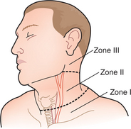
• When the platysma muscle has been violated, there is the potential for injury to underlying structures. Use the location of the injury (zone I, II, or III; Fig. 50-1) to guide your diagnostic workup.
• Zone I: From the thoracic outlet as defined by the clavicles inferiorly to the cricoid cartilage superiorly. Structures at risk: major vascular structures (great vessels, carotid and vertebral arteries, internal jugular veins), the apices of the lungs, esophagus, thoracic duct, and cervical nerve trunks.
• Zone II: From the cricoid cartilage inferiorly to the angle of the mandible superiorly. Structures at risk: major vascular structures (carotid and vertebral arteries, jugular veins), the esophagus, and major airway structures (pharynx, trachea, larynx).
• Classically, all patients with a zone II injury that violated the platysma underwent mandatory neck exploration; this led to many operations in which no injury was identified. Currently, patients with zone II injuries who are stable and have no hard definitive signs of underlying injury undergo further evaluation (CT angiography of the neck) and selective management based on the injuries identified. Zones I and III are bounded by bony structures, and injuries to these areas can be more difficult to access. Stable patients with an injury to zones I or III should undergo diagnostic studies (CT angiography of the neck) to identify injuries and provide a “road map” for the appropriate operative intervention.


