INTRODUCTION
The pancreas contains two distinctive types of glandular tissue, an exocrine component and an endocrine component, which allow the organ to perform two different functions. Exocrine glands secrete their products into a duct system. The ducts, in turn, transport these secretions to a specific target. Acini are the functional units of the exocrine pancreas. Endocrine glands secrete their products directly into the blood, rather than into a duct system. Islets of Langerhans are the functional units of the endocrine pancreas.
NORMAL ANATOMY AND HISTOLOGY OF THE PANCREAS
The pancreas is a retroperitoneal organ located in the upper abdomen and surrounded by a number of important organs and blood vessels. Although the pancreas is one continuous organ, it is thought of as having three anatomic regions based on its spatial relationship to surrounding structures. There are also minor functional differences between the anatomic regions, and certain diseases have a predilection for one region versus another.
The head of the pancreas is nestled within the curvature of the duodenum that abuts the right lateral and right inferior borders of the pancreas. The right borders of the superior mesenteric vessels, which lie directly posterior to the pancreas, mark the transition to the body of the pancreas. The body of the pancreas encompasses the majority of the pancreas and transitions distally into a tapered tail of the pancreas. In addition to the superior mesenteric vessels, the aorta and inferior vena cava lie posterior to the pancreas. Other organs that lie in close proximity to the pancreas include the stomach (anterior to the pancreas), spleen (at the distal tip of the pancreas), and left kidney (posterior to the tail of the pancreas) (Figure 11-1).
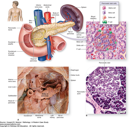
Before discussing the anatomy of the pancreatic ductal system, it is helpful to first develop a working knowledge of the microscopic anatomy of the pancreas. Low-power examination of the pancreas reveals an organ composed of lobules of tissue (Figure 11-2).
Exocrine tissue makes up the vast majority of the lobule. The functional unit of the exocrine pancreas is the acinus (pl. acini). Acini are organized collections of secretory cells that surround a central lumen. Acinar cells have peripherally located nuclei and abundant granular cytoplasm and secrete a variety of active enzymes and inactive proenzymes into the acinar lumen (Figure 11-3). These secretion products are carried from the acini to the duodenum via the ductal system. Proenzymes are converted to active enzymes in the duodenum. A summary of some of the important pancreatic enzymes is included in Table 11-1.

Dotting a sea of exocrine tissue are the islets of Langerhans, the functional units of the endocrine pancreas. In hematoxylin and eosin-stained sections, islets appear paler than their more abundant exocrine counterparts (Figure 11-4). A summary of some of the important islet produced hormones is included in Table 11-2.
| Hormone | Cell of Origin | Function |
|---|---|---|
| Insulin | Beta cells |
|
| Glucagon | Alpha cells |
|
| Somatostatin | Delta cells (a.k.a. D cells) |
|
| Pancreatic polypeptide | PP cells |
|

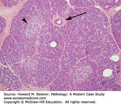
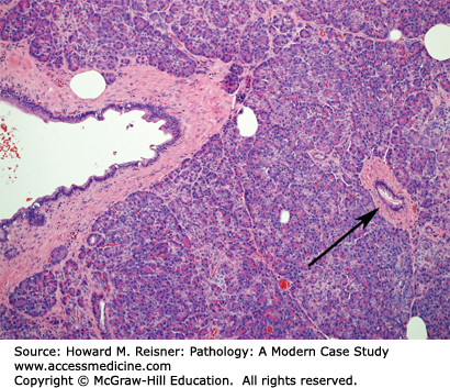
Acinar cell secretions are collected in the acinar lumen and are transported to the duodenum through a series of progressively enlarging pancreatic ducts. Intralobular ducts are small ducts present within pancreatic lobules. Interlobular ducts are larger ducts present between lobules. Interlobular ducts drain into the main pancreatic duct, which usually merges with the common bile duct just proximal to the ampulla of Vater, the site where bile and pancreatic secretions enter the duodenum. In some people, a separate accessory pancreatic duct is present and empties directly into the duodenum, separate from the ampulla of Vater. Pancreatic duct epithelial cells secrete bicarbonate, which helps to neutralize the highly acidic material entering the duodenum from the stomach (Figures 11-5 and 11-6).

QUICK REVIEW
Anatomically, the pancreas is subdivided into a head, body, and tail.
The pancreas contains two distinctive types of glandular tissue: an exocrine component and an endocrine component.
Exocrine pancreas: Acini secrete digestive enzymes into the pancreatic ducts for transport to their site of function in the duodenum.
Endocrine pancreas: Islets of Langerhans secrete hormones (e.g., insulin and glucagon) directly into the blood.
PRINCIPAL DISEASES OF THE PANCREAS
A diverse collection of neoplastic and non-neoplastic diseases can affect the pancreas. This chapter will focus on the more common diseases, including pancreatitis, diabetes mellitus, and neoplasia.
Stay updated, free articles. Join our Telegram channel

Full access? Get Clinical Tree


