Papilloma and Related Benign Lesions


Papilloma and Related Benign Lesions


FREDERICK C. KOERNER


This chapter discusses a heterogeneous group of lesions. Some are fundamentally papillary (intraductal papilloma and subareolar sclerosing papilloma); others are prominently papillary (florid papillomatosis and radial sclerosing lesion [RSL]) or partly papillary (cystic and papillary apocrine metaplasia). Syringomatous adenoma is discussed because it is clinically and histologically part of the differential diagnosis of florid papillomatosis. Collagenous spherulosis, also included here, is a stromal alteration seen in various papillary lesions including papilloma, ductal hyperplasia, and, rarely, adenosis.


PAPILLOMA

Papillomas are discrete benign tumors of the epithelium of mammary ducts. They arise most often from lactiferous ducts in the central part of the breast, but they can occur peripherally in any quadrant. Intracystic papilloma is the designation applied to a papilloma in a cystically dilated duct. Large, complex papillomas that have a cystic component have sometimes been referred to as papillary cystadenomas, whereas solid, noncystic papillomas have been variously classified as ductal adenoma, solid papilloma, or adenomyoepithelioma (AME). A solitary papilloma is a single discrete papillary tumor in one duct. Multiple papillomas usually occur in contiguous branches of the ductal system. Solitary papillomas are more common than multiple papillomas. A distinction must be made between intraductal papilloma and papillomatosis (epitheliosis). The latter terms are used to describe usual (conventional) ductal hyperplasia (UDH), the common form of ductal hyperplasia, which may coexist with solitary or multiple papillomas.


Clinical Presentation

In 1951, Haagensen et al.1 reviewed a series of 367 patients with benign intraductal papillary breast lesions who had been treated at Presbyterian Hospital in New York between 1916 and 1941. After excluding 243 patients with “microscopic papillomas,” 14 with incomplete data, and 2 with a papilloma and unrelated nonpapillary carcinoma, the authors concentrated on describing 108 patients treated for gross, benign intraductal papillomas. Microscopic papillomas were characterized as “small multiple papillary projections, with or without fibrous cores, which project into the ducts and cysts of chronic cystic mastitis.” By contrast, gross papilloma was defined as a “definite disease entity in which one or more papillomas grow within a relatively localized portion of a duct, or in several adjacent ducts, and attain sufficient size to fill up the duct and become evident grossly.”

The study revealed some now well-established features of duct papilloma. The majority (75%) were located in the central part of the breast. Discharge, bloody or nonbloody, was the primary symptom in 72% of cases, but it was less commonly seen with peripheral lesions (29%) than with those in a central duct (86%). Lesions that caused nipple discharge, whether central or peripheral, tended to have a frond-forming papillary configuration, whereas a more solid growth pattern was observed in tumors not associated with a discharge.

Solitary papillomas may occur at any age from infancy to the ninth decade, but they are most frequent in the sixth and seventh decades of life. The tumors usually occur in the subareolar region and come to attention because of a discharge from the nipple. Women with multiple papillomas tend to be younger than women with solitary papillomas and most often present in their 40s and early 50s. Multiple papillomas develop peripherally more often than centrally and typically present as a palpable lesion. Papillomas may occur more frequently in African American women than in women of other ethnic origins.2

Papillomas have developed in unusual settings. One report3 documented the growth of a papilloma in residual breast tissue following a transverse rectus abdominus muscle (TRAM) flap reconstruction. Two publications4,5 describe the presence of papillomas in ectopic breast tissue in axillary lymph nodes (ALNs) of women with mammary papillomas.

Although papillomas afflict women almost exclusively, the literature contains reports of 26 papillomas in men. Volmer6 lists the cases described prior to 1985. In 2006, Yamamoto et al.7 tabulated clinical details of 10 additional cases, and five other examples8,9,10,11,12 have also been published. The patients range from 7 months13 to 82 years14 of age, the range seen in women. Two patients presented during their early teenage years,8,15 and the author has examined a papilloma from a 15-year-old boy. The presence of several cases in adolescent boys seems a curious coincidence, which
might merit further attention. Symptomatic intervals as long as 6 years have been noted.16 Two patients reported longterm use of phenothiazines.7,17 One of these patients had an elevated serum prolactin level,7 and the papillary epithelium in the other case17 exhibited secretory changes.

Nipple discharge occurs in most patients with a central papilloma. Bloody discharge, more commonly associated with papillary carcinoma, can develop in a papilloma as a result of degenerative changes. A subareolar mass may be palpable in patients with a central solitary papilloma, and a palpable tumor may be the first clinical manifestation of a peripheral papilloma. A papilloma in a 44-year-old man produced a hard, lobulated mass fixed to the chest wall.10


Imaging Studies

Cystic, solitary papillary tumors may appear well circumscribed on mammography, but the presence of a cystic component is best appreciated by ultrasonography, which seems to be more sensitive than mammography for the detection of papillomas. Francis et al.18 reported that 44% of mammograms in their study of 36 women with papillomas were described as normal, whereas only 5% of sonograms appeared normal. These authors also cited similar findings presented in earlier publications. Ductography can also demonstrate the presence of a central papilloma in patients who experience a nipple discharge.19 Magnetic resonance imaging (MRI) in women with solitary intraductal papillomas and nipple discharge typically reveals duct dilation and the presence of small, oval, smoothly contoured, enhancing intraductal masses,18,20 but papillomas do not always display these characteristic features. In the study of Kurz et al.,21 4 of 20 papillomas had irregular shapes, 3 had irregular or spiculated margins, 8 displayed heterogeneous enhancement, and the characteristics of the time-intensity curves varied. After reviewing images from 15 papillomas, Daniel et al.22 reported that 7 displayed suspicious morphologic findings such as irregular margins or rim enhancement and that 4 were not evident on the MRI studies.


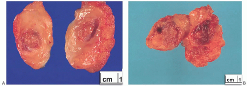
FIG. 5.1. Papilloma. Gross appearance of three lesions. A: Intracystic papilloma. B: Solid intracystic papilloma virtually filling the cyst lumen. C: An everted cystic multinodular papilloma from a 17-year-old girl. D: A histologic whole mount of a tumor similar to the one in (B).

Cardenosa and Eklund19 compared the clinical and mammographic findings in patients with solitary and multiple papillomas. The latter group was subdivided into peripheral and central lesions. Most multiple peripheral papillomas were asymptomatic and were detected as a result of mammographic abnormalities, including calcifications, nodules, masses, or various opacities.

After a detailed mammographic and sonographic study of 40 papillary tumors, which were later excised and examined histologically, Lam et al.23 concluded that “radiologic features are not sufficiently sensitive or specific to differentiate benign from malignant papillary lesions.” By using a combination of both imaging modalities, the authors achieved a sensitivity of 61%, a specificity of 33%, a positive predictive value (PPV) of 85%, and a negative predictive value (NPV) of 13%.


Gross Pathology

A part of the mass associated with some solitary papillomas is a cyst formed by the dilated duct in which the papilloma grew (Fig. 5.1). The cyst may contain clear fluid, bloody fluid, or clotted blood. The papilloma usually forms a single mural nodule protruding into the lumen, but occasionally multiple, separate, or aggregated nodules are present (Fig. 5.2). Some papillomas grow as soft, solid friable masses that obliterate the cystic space.

Solitary papillomas are typically bosselated, soft-to-firm tumors that appear gray to reddish brown when examined grossly in the fresh state. The lesions are well circumscribed and appear to be enclosed by a capsule formed by the duct wall and accompanying reactive changes. Multiple associated papillomas can sometimes be appreciated grossly when they form nodules in contiguous dilated ducts (Fig. 5.3).


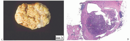
FIG. 5.1. (Continued)


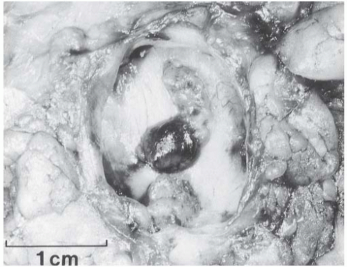
FIG. 5.2. Papilloma. Gross appearance of an intracystic papilloma consisting of two mural nodules. The cyst wall is a distinct fibrous membrane with a smooth inner surface.


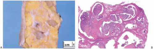
FIG. 5.3. Multiple intraductal papillomas. A: Gross specimen showing multiple papillomas. The two largest lesions are solid, tan tumors. A small cystic papilloma is visible in the center. B: A cluster of small intraductal papillomas.

Solitary papillomas may form clinically symptomatic tumors 1 cm or less in diameter in a major lactiferous duct (Fig. 5.4), but the average size is 2 to 3 cm. Benign cystic papillomas may be larger than 10 cm. Tumors formed by multiple papillomas are typically larger than 2 cm.1,24


Microscopic Pathology

The basic microscopic structure of a papilloma consists of the proliferation of ductal epithelium supported by frondforming fibrovascular stroma. The most orderly form of papilloma consists of branching fronds of stroma supporting a layer of epithelium composed of epithelial and myoepithelial cells (Fig. 5.5). The epithelial cells are cuboidal to columnar, and they display little pleomorphism, nuclear hyperchromasia, or mitotic activity. The supporting stroma may arise from a single base or from several foci in the duct wall. Epithelium lining the nonpapillary portion of the duct usually exhibits little or no hyperplasia.

Some papillomas have a more complex structure caused by stromal overgrowth, hyperplasia of the epithelium, or
both processes, resulting in fusion of the papillary fronds (Fig. 5.6). The most exaggerated form of this process is the solid intraductal papilloma, in which virtually all the space between fibrovascular stalks is filled by solid sheets of proliferative ductal epithelial cells. More often, secondary lumens are formed within the hyperplastic epithelium, resulting in irregular microlumens, micropapillary fronds, focal solid areas, or heterogeneous combinations of these patterns (Fig. 5.7).


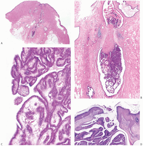
FIG. 5.4. Papilloma of lactiferous duct. A,B: A dumbbell-shaped papilloma with a central zone of fibrosis. C: The papillary fronds are composed of somewhat edematous fibrovascular stroma with a thin layer of epithelium on the surface. Similar epithelium lines the lumen of the dilated duct. D: A papilloma at the level of a lactiferous duct orifice. E: Florid epithelial hyperplasia in a lactiferous duct papilloma.

Foci of apocrine metaplasia are not uncommon in papillomas (Fig. 5.8), and rarely most or all of the epithelium is of the apocrine type. When present in the conventional
papillary configuration, apocrine metaplasia is usually cytologically bland, but apocrine atypia manifested by nuclear pleomorphism and cytoplasmic clearing can be seen in sclerosing papillary tumors. This is especially common when sclerosing adenosis (SA) is incorporated into a papilloma.


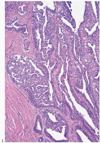
FIG. 5.4. (Continued)


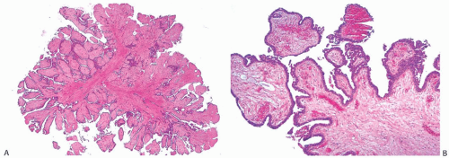
FIG. 5.5. Papilloma. A: This papilloma has dense collagenous stroma. B: Epithelium on the surfaces of the fronds is thin. C: A thin layer of cuboidal and columnar epithelial cells overlies a continuous layer of myoepithelial cells (arrows). D: Slight epithelial hyperplasia at the surface of one papillary frond.

The appearance of the fibrovascular stroma varies considerably among papillomas. In some lesions, the stroma is limited to slender inconspicuous strands consisting of thin-walled capillaries and sparse fibroblasts, collagen, and mononuclear cells and forming a network that supports the voluminous epithelium. The distribution of this stroma stands out especially clearly in sections stained for reticulin, vimentin, basement membrane proteins, or vascular markers such as CD34 and CD31. Expansion of the fibrovascular stroma by accumulated histiocytes occurs in papillomas and is an exceedingly rare finding in papillary carcinoma (Fig. 5.9).

Collagenization of the fibrovascular stroma occurs in some papillomas. The papillary architecture is accentuated when this process is limited to the intrinsic papillary structure. If myofibroblastic proliferation accompanies collagenization of the stroma, the papillary arrangement is likely to become distorted. Epithelial elements entrapped in this stroma may simulate invasive carcinoma within or at the periphery of the lesion (Figs. 5.10 and 5.11). In the most extreme situations, fibrous sclerosis is so severe as to virtually obliterate the papilloma, reducing it to a nodular scar containing sparse benign glandular elements (Fig. 5.12). Such a lesion may be difficult to distinguish from a fibroadenoma (FA). This problem is typified by a case report purported to represent an infarcted FA in which the illustrations are highly suggestive of a fibrotic and partially infarcted cystic papilloma.25

The epithelium of intraductal papillomas contains a myoepithelial cell layer.26 Inconspicuous elongated myoepithelial cells with nuclei are flattened along the basement membrane. Hyperplastic myoepithelial cells typically form a prominent layer of cuboidal cells that tend to have relatively clear cytoplasm (Fig. 5.13). Myoepithelial cells in papillomas can be demonstrated with a variety of immunostains.26,27 When immunostaining is done to detect myoepithelial cells, it is necessary to differentiate stromal and myoepithelial reactivity. Myoepithelial cells are immunoreactive for
actin, calponin, myosin heavy chain, CD10, p63, and CK5/6 (Fig. 5.13). All of these proteins except p63 are cytoplasmic markers, which vary in their proclivity to react with stromal cells. The p63 immunostain localizes in myoepithelial cell nuclei and rarely in the nuclei of epithelial cells in a papillary lesion.28 Cross-reactivity in stromal cells is not observed. For this reason, it is preferable to include a stain for p63 among those chosen to detect myoepithelial cells. The myoepithelium may become noticeably attenuated and focally undetectable by immunostains in regions of sclerosis in a papilloma. The focal absence of immunoreactive myoepithelium, by itself, is not diagnostic of carcinoma in this setting. When there is marked hyperplasia of myoepithelial cells in a benign papillary tumor, the differential diagnosis includes AME.


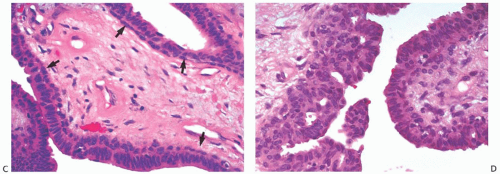
FIG. 5.5. (Continued)


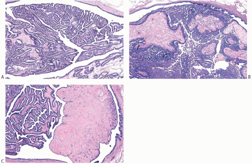
FIG. 5.6. Papillomas. Histologic appearances of three different tumors. A: Papilloma with simple epithelium and complex glands. B: Papilloma with prominent epithelial hyperplasia. C: Papilloma with complex glands and a discrete nodular area of sclerosis that distorts the underlying papillary architecture.

Infarction occurs in solitary and in multiple papillomas (Figs. 5.14 and 5.15). No specific cause for the necrosis is evident in most cases. The presence of chronic inflammation and hemosiderin in and around many papillomas suggests that these lesions are prone to transient bleeding secondary to ischemia or incidental trauma. Needling procedures can contribute to infarction and cause fresh hemorrhage,
sometimes associated with displaced epithelium. Spontaneous infarction usually involves superficial portions of a papilloma. Rarely, the entire lesion is infarcted either spontaneously or as a consequence of a biopsy. The underlying structure of a fully infarcted papilloma can be demonstrated with a reticulin stain. In some infarcted lesions in which degeneration is not advanced, the structure of the epithelium can be displayed more clearly with immunostains such as cytokeratin (CK) and p63.29 There is no procedure for reliably distinguishing between the completely infarcted epithelium of a papilloma and that of a papillary carcinoma. However, if myoepithelium can be demonstrated in the infarcted tissue, the tumor is more likely to be a papilloma than a papillary carcinoma. Cytologic atypia is commonly found in the partially degenerated epithelium of a papilloma in the vicinity of infarcts. The atypia is usually manifested by nuclear hyperchromasia and pleomorphism. These cytologic abnormalities may lead to an erroneous diagnosis of carcinoma in the fine-needle aspiration (FNA) specimen from an infarcted papilloma30,31 or in a needle core biopsy specimen.


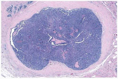
FIG. 5.7. Papilloma. Florid ductal hyperplasia obscures the papillary architecture of this solid papilloma.


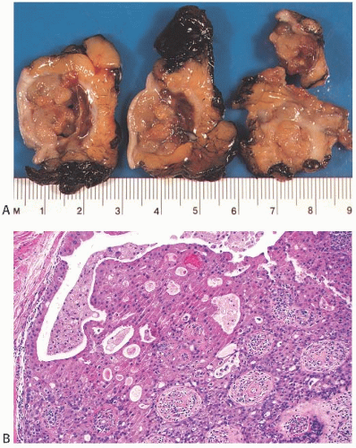
FIG. 5.8. Papilloma with apocrine metaplasia. A: The light brown color of this cystic papillary lesion is typical of tumors with extensive apocrine metaplasia. B: Hyperplastic apocrine epithelium on the surface of a papillary frond.


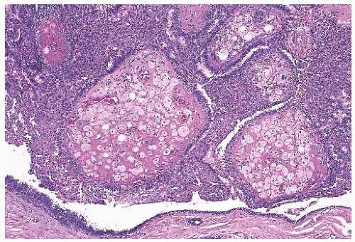
FIG. 5.9. Papilloma with stromal histiocytes. The fibrovascular stroma is prominent as a result of accumulated histiocytes in a papilloma, which nearly fills a cystically dilated duct. Part of the duct wall is seen at the bottom of the image.

Squamous metaplasia can occur in the epithelium of a papilloma (Fig. 5.16). It is more likely to be found when there is infarction and probably represents a reactive or reparative process.32 Rarely, squamous metaplasia constitutes a conspicuous component of the papilloma or of the epithelium lining the cystic portion of the lesion. Extension of squamous metaplasia to the epithelium of adjacent ducts is an uncommon finding.33 Entrapped metaplastic epithelium in the stromal reaction may simulate metaplastic or squamous carcinoma, and in some instances the distinction between these processes is very difficult.32 Electron microscopy and immunohistochemistry (IHC) suggest that some examples of squamous metaplasia derive from myoepithelial cells.34 Sebaceous metaplasia of luminal cells of a papilloma has also been reported.35

Intraductal papillomas can be recognized by frozen section (FS), but in most circumstances, it is preferable to rely upon paraffin sections for the diagnosis of papillary tumors. It is sometimes difficult to prepare satisfactory FSs from these fragile lesions or to identify foci of carcinoma that have developed in a papilloma.


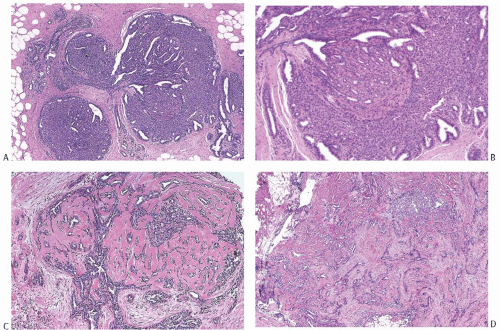
FIG. 5.10. Papilloma with stromal sclerosis. A: A trilobed solid papilloma showing early central sclerosis. B: Florid duct hyperplasia and adenosis in the lesion shown in (A). C: Well-developed sclerosis is present throughout this papilloma. D: Sclerotic stroma is uniformly distributed in this lesion.


Cytology

The diagnosis of an intraductal papilloma may be suggested by the cytologic findings in an FNA specimen.36,37 The smear typically has cohesive three-dimensional groups of cytologically benign-appearing cells accompanied by variable amounts of stromal cells, apocrine cells, inflammatory cells, and histiocytes. Two diagnostic problems can arise in this setting.

First, a significant number of lesions classified as papillary on the basis of findings in needle aspiration specimens prove not to exhibit a papillary architecture when excised. Of the 70 cases studied by Simsir et al.38 in which needle aspiration specimens were classified as papillary, only 31 (44%) showed a papillary architecture when examined histologically. FAs, fibrocystic changes (FCCs), a phyllodes tumor, and carcinomas accounted for the remaining 39 cases. Although usually correctly classified as benign, FAs and FCCs account for the most common discrepancies. Apocrine cells, foamy histiocytes, and bipolar nuclei associated with myoepithelial cells are characteristic of a papilloma. Although one can also observe these cells in aspirates from FAs, the epithelium from the latter lesion tends to be distributed in flat sheets rather than in three-dimensional groups, and it usually has a honeycomb appearance. Furthermore, the epithelial groups from FAs typically lack fibrovascular stroma, and one usually observes fragments of stroma devoid of blood vessels and unassociated with epithelial cells. Most aspirates of FCCs appear less cellular than those from papillomas, and they do not contain branched, three-dimensional cell clusters supported by fibrovascular stroma. These generalizations notwithstanding, many smears do not demonstrate the findings sufficiently clearly that one can make a secure diagnosis of papilloma. Even in retrospect, Simsir et al.38 could not distinguish the smears of three FAs from those of papillomas. Mak and Field39 recounted similar observations. The authors studied 56 cases in which smears were interpreted as papillomas. The excision specimens revealed papillomas in 42 (75%) of the cases yielding a PPV of 0.74. FAs, radial scars, and other benign proliferative lesions accounted for 10 discrepancies, atypical ductal hyperplasia (ADH) 2 discrepancies, and tubular carcinomas the remaining two discordant cases.

Second, the cytologic detection of malignancy in a papilloma and the recognition of a papillary carcinoma often prove especially difficult. For example, in the study of FNA
specimens of papillary lesions by Masood et al.,40 3 of 21 tumors interpreted as benign papillary lesions proved to be micropapillary ductal carcinoma in situ (DCIS) upon study of an excision specimen. Immunostaining may help in the diagnosis of FNA specimens of papillary breast tumors. Chang et al.41 reported that the percentage of Ki67-positive cells was significantly higher in papillary carcinomas (21.0 ± 19.23%) than in papillomas (6.23 ± 7.25%). These investigators did not find cyclin-D1 reactivity to be a useful differential feature. Staining of cell-block preparations for calponin to reveal myoepithelial cells can provide diagnostic information in certain cases.42 For further discussion, see Chapter 14.


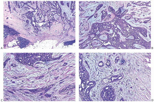
FIG. 5.11. Papilloma with stromal sclerosis. Images are from one tumor. A: The border of the lesion is circumscribed. Note the collections of lymphocytes. B: An area near the center of the lesion showing residual papilloma with fenestrated epithelium. C: Attenuated epithelium is distributed between layers of myofibroblasts and collagen within the tumor. D: This image from the periphery of the lesion shows rounded groups of cells and isolated cells, a characteristic finding at the edges of a papilloma with sclerosis.


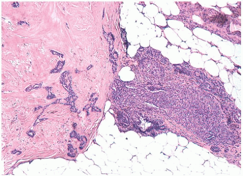
FIG. 5.12. Papilloma with extreme stromal sclerosis. The main tumor is almost entirely effaced by collagenized stroma. An intraductal papilloma persists in a peripheral duct.


Core Biopsy

The diagnosis of papillary tumors of the breast by needle core biopsy has been the subject of extensive investigation, and the literature contains more than 40 reports dealing with this topic. Publications by Skandarajah et al.,43 Bernik et al.,44 Jung et al.,45 and Brennan et al.46 tabulate data from many investigations, and Table 5.1 lists findings from those comprising more than 50 cases.2,43,45,47,48,49,50,51,52,53,54 Interpretation of this body of information is hampered by several factors. First, most studies represent retrospective analyses of cases in which a core biopsy reportedly demonstrated a papilloma. In the early studies, only a few patients underwent excisions, so selection bias may have influenced the results. Second, the designs of the studies have varied widely. Certain ones included retrospective review of histological or radiologic studies, whereas others did not. The histologic classification of the papillary lesions differed, the expertise of the radiologists and pathologists varied, and most studies do not describe in detail the extent of the study of the core biopsy specimens. Statements about the concordance of the radiologic studies and the pathology studies were not included in every report. Finally, during the more than two decades spanned by these studies, the practices of both breast imaging and breast pathology have changed in ways such as the use of vacuum-assisted biopsy methods and immunohistochemical staining, and these advances could have influenced the findings of the studies.


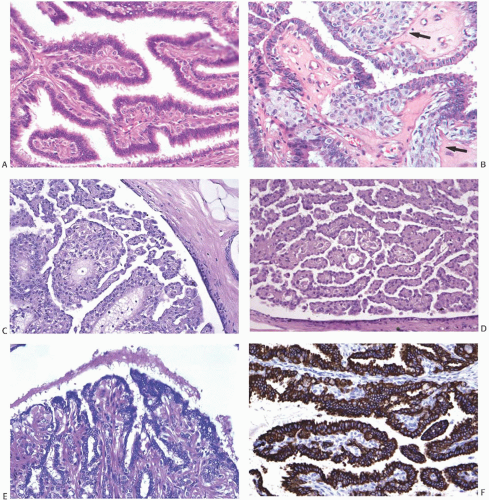
FIG. 5.13. Myoepithelial cell hyperplasia. A: Hyperplastic myoepithelial cells forming an expanded zone beneath the thin epithelium. B: The myoepithelial cells have an epithelioid phenotype (arrows). C,D: Myoepithelial cells with an epithelial phenotype are markedly hyperplastic in these papillomas. The epithelium is reduced to a thin layer of flat cells overlying the multilayered myoepithelium. E: A papilloma in which the myoepithelial cells have a striking myoid phenotype and resemble smooth muscle. F: The AE1/AE3 immunostain is reactive in the epithelium but not in the myoepithelium. G: The myoepithelium is immunoreactive for myosin heavy chain, which shows no reactivity in the epithelium. H: Clusters of epithelioid myoepithelial cells resemble lobular carcinoma in the stroma of this papilloma. I: Reactivity for E-cadherin in the epithelial and myoepithelial cells is shown. Lobular carcinoma would be E-cadherin negative.


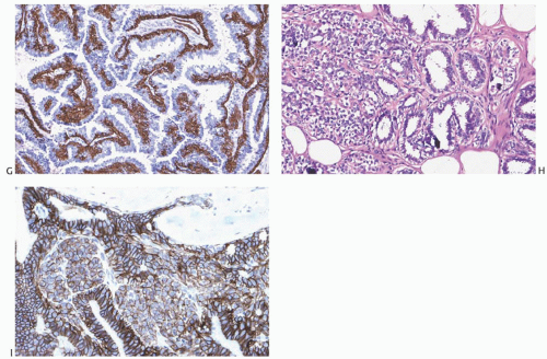
FIG. 5.13. (Continued)


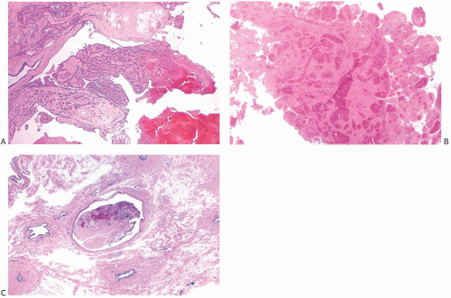
FIG. 5.14. Infarcted papilloma. A: Low-magnification view of infarction in the apical portion of a papilloma. B: This papillary lesion is entirely infarcted. C: A partly infarcted and sclerotic intraductal papilloma.


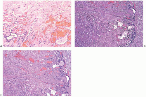
FIG. 5.15. Infarcted papillary tumors. A: Hemorrhage is present at the border of the infarcted zone (above) in a sclerosing papilloma. B,C: The ghost architecture of this papillary lesion is visible (left), and a thin zone of residual viable epithelium is present at the periphery (right). This was probably a solid papilloma.


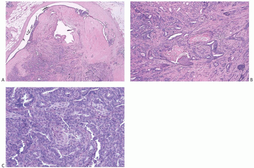
FIG. 5.16. Papilloma with squamous metaplasia. A,B: An intracystic sclerosing papilloma with focal squamous metaplasia. C: A small cluster of cells exhibiting squamous differentiation is present in the epithelium of this florid papilloma.


TABLE 5.1 Core Biopsy and Excision of Benign Intraductal Papillomaa: A Selected Literature Review


Excision Findings (%)


References


Cases


Benign


Atypical


Malignant


Rizzo et al.2


101


72


19


9


Skandarajah et al.43


80


67


14


19


Jung et al.45


160


88


6


6


Chang et al.47


100


83


13


4


Chang et al.48


64


86


11


3


Jaffer et al.49


104


84


8


8


Jakate et al.50


90


78


13


9


Kil et al.51


76


92


0


8


Lu et al.52


66


82


12


6


Rizzo et al.53


234


73


18


9


Shin54


86


79


7


14


Total


1161


80


12


8


aBenign intraductal papilloma refers to the diagnosis made on the core biopsy sample.


Despite these significant differences in design, a coherent picture has emerged, and two generalizations seem established. First, an excision is usually indicated when a core biopsy specimen of a papillary tumor contains atypical ductal or lobular cells (ADH, atypical lobular hyperplasia [ALH], or lobular carcinoma in situ [LCIS]). Between one-third and one-half of such patients will prove to have a carcinoma in an excision specimen. This conclusion mirrors the approach advocated for atypical epithelial proliferations detected in other histologic settings. Second, patients in whom a core biopsy specimen discloses a benign papilloma have a significant likelihood of harboring an atypical epithelial proliferation in an excision specimen. The literature contains data from more than 2,000 core biopsy specimens showing only benign papillomas. Subsequent excision specimens demonstrated benign findings in approximately 80%, atypical ductal or lobular cells (ADH, ALH, or LCIS) in approximately 10%, and DCIS or invasive carcinoma in approximately 10%.

Researchers have attempted to improve the predictive values of the findings present in core biopsy specimens demonstrating benign papillomas by incorporating ancillary clinical, radiologic, or histologic features in their analysis. Scattered reports have identified findings that seem to indicate an increased risk for the presence of atypical cells in an excision specimen; however, other investigations have not borne out the predictive value of these findings in most instances. The literature does suggest that two features may identify patients at elevated risk for atypia: age and size of the lesion. Arora et al.55 observed an increasing likelihood of an upgrade to malignancy with the increasing age of the patient, and five other studies found that older patients are more likely to have atypical cells discovered on excision.46,53,56,57,58 This association notwithstanding, investigators have not discovered an age threshold below which an excision would seem unnecessary.

The size of a papilloma may also influence the likelihood of discovering atypical cells in an excision specimen. In the study of Jung et al.,45 the presence of a palpable mass indicated an increased likelihood of the detection of carcinoma and so did the presence of a mass on the mammogram. Among studies examining imaging features, two reported that the presence of a mass greater than 1.5 cm detected by sonography47 or by either sonography or mammography51 indicated an increased likelihood of carcinoma in a subsequent sample. Another investigation50 observed that benign papillomas associated with atypia in excision specimens were larger than those that were not. Three other studies did not confirm the predictive nature of the size of the lesion.49,57,58


Additional features such as clinical information, radiologic findings, details of the biopsy procedure, and the experience of the radiologists have not improved the predictive values consistently. Many authors stress the need for careful correlation of the radiologic and pathologic findings and point out that lack of correlation accounts for many discrepancies. Nevertheless, in the study by Bernik et al.,44 9 of 17 benign papillomas with concordant radiologic studies proved to have ADH in the excision specimen, and 2 of the 17 contained carcinomas. Other authors report the same experience.

Pathologists have searched for way to improve their diagnostic abilities, and both the use of immunohistochemical stains and the expertise of pathologists have come under study. Shah et al.59 found that the use of immunohistochemical staining for calponin, CK5/6, and p63 allowed pathologists to recognize foci of ADH associated with papillomas. This technique improved the accuracy of all four participating pathologists to the point that they concluded that papillomas lacking atypia “do not require excision in the absence of suspicious clinical/radiological findings.” Tse et al.60 employed staining for estrogen receptor (ER), CK14, and p63 in an attempt to resolve discrepancies encountered in 15 core biopsy specimens. Although the use of these stains reduced the rate of discordance by 69%, the staining results did not eliminate either false-positive or false-negative cases.

The expertise of the pathologist influences the correlation between findings of core biopsy and excision specimens, but even experienced breast pathologists cannot exclude the presence of atypical cells in an excision specimen. In the study of Jakate et al.,50 diagnoses made by pathologists with fellowship training in breast pathology on core biopsy specimens were less likely to differ from those of the subsequent excision specimen than were diagnoses made by general pathologists; nevertheless, a substantial number of discrepancies remained. An “upgrade” rate of 26.3% was observed for diagnoses made by general pathologists, whereas a rate of 16.3% was observed for breast pathologists. Of the 86 cases in which breast pathologists made a diagnosis of benign papilloma on a core biopsy specimen, 10 excision specimens displayed atypia and 3 showed malignancy.

Several investigations include patients who did not undergo an excision following the diagnosis of benign papilloma on a core biopsy. The authors reported that 7 of the 437 patients (1.6%) included in these studies developed carcinoma. Followup periods ranged from 2 to 5 years, and the nature of the follow-up varied. These variations, among many other aspects of the studies, make it impossible to draw secure conclusions about the safety of careful clinical surveillance as an alternative to excision of benign papillomas diagnosed by means of a core biopsy. A conservative approach may prove safe, but investigators have not formulated the criteria to identify either the appropriate patients or the details of the program of surveillance.

Data from the foregoing studies do not provide definitive guidelines for the management of a papilloma without atypia diagnosed by needle core biopsy. A decision as to whether surgical excision should be performed will be influenced by factors such as the size of the lesion, evidence of residual tumor following the biopsy, the ease of mammographic follow-up, family history of breast cancer, and patient concerns. Among women recommended for follow-up without an initial surgical excision, some will require surgical biopsy at a later date. In a series studied by Sexton et al.,61 59 of 78 patients (i.e., 75%) with a papilloma diagnosed by needle core biopsy did not undergo surgical biopsy and were followed for 3 to 5 years. Subsequent interval mammographic changes necessitated surgical excision in 10 of the 59 (17%), and 2 (3%) had a subsequent needle core biopsy. All subsequent biopsies were reportedly “benign.” This study suggests that up to 20% of patients enrolled in follow-up after a needle core biopsy diagnosis of papilloma will undergo another biopsy within 5 years of the initial procedure. The long-term risk for the development of carcinoma at the site of an incompletely excised papilloma that was sampled by core biopsy has not been determined.


Immunohistochemistry

Immunohistochemical staining of papillomas reveals the results expected for mammary epithelial, myoepithelial, and stromal cells.26 The epithelial cells in papillomas exhibit nuclear immunoreactivity for ER, which can appear either scattered or diffuse (Fig. 5.17). Most investigations of papillomas using immunohistochemical staining have centered on the differentiation of benign papilloma from papilloma with atypical hyperplasia (atypical papilloma)59,60 and papillary carcinoma (see Chapter 14).


Electron Microscopy

Electron microscopy reveals the expected ultrastructural features of the cells that compose papillomas.16,62,63 The epithelial cells have well-developed microvilli on their luminal surfaces and numerous terminal bars and desmosomes between adjacent cells. The nuclear contours appear irregular and invaginated, and the chromatin shows peripheral condensation. The nucleoli sit in the center of the nuclei, they appear round or oval, and they often have irregular borders.
Cytoplasmic organelles include rough endoplasmic reticulum, free ribosomes, Golgi apparatus, and mitochondria. The cytoplasm contains variable numbers of intermediate filament, lipid droplets, lysosomes, and dense secretory granules. Myoepithelial cells sit between the epithelial cells and the basement membrane. They exhibit polygonal or spindly shapes and possess fusiform nuclei. Myofilaments with dense bodies run parallel to the long axis of the cell, and tonofilaments arranged in thick bundles are arranged in a curvilinear pattern. The basal aspects of the myoepithelial cells often display scalloping. Rare cells display features of both epithelial and myoepithelial cells. A thick basement membrane, which often exhibits splitting and reduplication, separates the epithelium from the stroma.


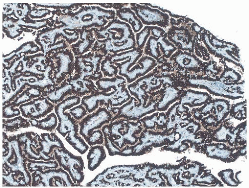
FIG. 5.17. Estrogen receptors. Immunoreactivity for ER is present in virtually all the epithelial cells.


Other Studies

Clonal analysis based on restriction fragment length polymorphism of the X-linked phosphoglycerate kinase (PGK) gene of nine solitary papillomas demonstrated monoclonality in all nine.64 Furthermore, analysis of widely separated sites of two papillomas showed that the same allele of the PGK gene was inactivated at each site. A few studies have contrasted genetic alterations in papillomas with those in papillary carcinoma. Chapter 14 summarizes the findings.


RADIAL SCLEROSING LESIONS Terminology

RSLs are proliferative abnormalities that have a stellate configuration radiologically and histologically. Clinical interest in RSLs derives from the realization that these abnormalities may be difficult to distinguish from carcinoma by mammography and the concern that they are precursors of the development of carcinoma.

The existence of these lesions has been recognized throughout most of the 20th century. In 1928, Semb83 referred to RSLs as rosettes or proliferation centers that might give rise to carcinoma. Bloodgood84 drew attention to RSLs in a study of “borderline breast tumors,” emphasizing diagnostic problems that they present and uncertainty about their precancerous potential.

RSLs have been described by a variety of names introduced since the 1970s. Fenoglio and Lattes85 described 30 examples, which they termed “sclerosing papillary proliferations” because the lesions had a prominent papillary component. This name has not been met with general acceptance because some of the lesions in this category have little or no papillary element. Fisher et al.86 suggested the term “nonencapsulated sclerosing lesion,” which has the advantage of avoiding issues related to histogenesis. “Infiltrating epitheliosis” has not been widely accepted because it could be misconstrued as indicating an invasive malignant neoplasm.87 The unpopularity of “indurative mastopathy” is probably attributed to the fact that most of the lesions are too small to be palpable as indurated foci and the vague meaning of the term “mastopathy.”88

“Radial scar,” a widely used name for these lesions, is a translation of “strahlige Narben,” the term Hamperl89 introduced in 1975. This designation refers to the stellate configuration of most of these lesions; it is short, and it avoids terminology that suggests association with particular proliferative ductal lesions. However, use of the word “scar” implies that there is a reparative process in the stroma. Although the stellate configuration has a cicatrix-like appearance, it is possible that the stromal change is an integral part of the proliferative lesion, perhaps enhanced by paracrine growth
factors, rather than a reparative process. In this regard, Jacobs et al.90 compared the “expression of factors involved in vascular stroma formation” in radial scars with invasive carcinomas. When compared with normal breast tissue, the stroma of both radial scars and invasive carcinomas displayed increased vascularity as well as “focally increased expression” of mRNA for collagen type IV, total fibronectin, vascular permeability factor/vascular endothelial growth factor, and other markers. These results provided molecular confirmation of the presence of a vasoproliferative process in both radial scars and invasive carcinomas; however, they did not identify the mechanism by which this process occurs, nor did they necessarily indicate that the same mechanism is involved in both lesions. The term radial sclerosing lesion, used here, is preferable because it describes the mammographic and histopathologic appearance of the process without implying histogenesis, and it is sufficiently nonspecific to encompass the many histologic variants included in this category. RSLs are discussed in this chapter devoted to benign papillary tumors because a substantial proportion has a component of papillary ductal proliferation.


Clinical Presentation

Most RSLs are microscopic lesions not detectable by palpation or mammography. They are usually discovered during examination of specimens resected for unrelated indications. Consequently, the distribution of the ages of women with RSLs parallels that of women undergoing breast surgery. RSLs are uncommon before the age of 30 years and most frequent between the ages of 40 and 60 years. The reported frequency of these subclinical lesions varies depending upon the groups of patients studied and the diagnostic criteria. RSLs have been detected in 1.7%,91 1.8%,92 5.3%,89 7.1%,93 14%,94 and 28%95 of benign breast specimens and in 4%,86 16%,96 and 26%94 of mastectomy specimens from patients with carcinomas. The broad and overlapping ranges of the frequency of RSLs associated with benign and carcinomatous breasts suggest that RSLs occur with similar frequency in both circumstances. The amount of tissue available for study and the thoroughness with which it is examined are important factors affecting these results. Multiple microscopic RSLs are not uncommon in one breast,96 and both breasts can be affected.95 A paper by Anderson and Battersby97 describes a woman with 80 RSLs in her right breast and 46 in her left breast. RSLs virtually never affect men. One report98 mentions in passing the presence of a RSL in the breast of a man with breast carcinoma.


Imaging Studies

Most RSLs are smaller than 2 cm when detected radiologically. Typical lesions are characterized by a lucent or dense center, radiating slender strands of tissue, and changes in appearance in different imaging projections99 (Fig. 5.18A). The ability to visualize a RSL mammographically is enhanced if the lesion is situated in predominantly fatty tissue that provides contrast for the core and radial strands of fibrous stroma containing proliferating epithelium and cysts. In one study of RSLs subjected to stereotactic aspiration biopsy, the average size was 8 mm,100 and in another report, the size averaged 1.3 cm.101 Microcalcifications are detected in some but not in all RSLs.100,102,103 When evident using sonography, RSLs form an irregular hypoechogenic mass with ill-defined borders and diminished posterior acoustic transmission.104 Certain radiologic features favor the radiologic diagnosis of a RSL over a stellate carcinoma, but these are not sufficiently distinctive to serve as the basis for a specific diagnosis.101,102,103,105,106 MRI may offer valuable diagnostic information. In one study,107 18 RSLs did not demonstrate enhancement, whereas 11 of the 12 carcinomas showed irregular areas of focal enhancement with ill-defined borders. These findings led to a specificity of 89%, a sensitivity of 82%, an overall accuracy of 87%, and a K coefficient of 0.97 for the differentiation of RSLs and invasive carcinomas.


Gross Pathology

Most RSLs excised after mammographic localization have gross appearances similar to those of small invasive carcinomas. The nodule is firm and when bisected reveals a pale, retracted center in which there may be white streaks (Fig. 5.18B). Slender bands of pale stroma extend radially into the fat from the core. Small cysts can be appreciated in some lesions. A minority of RSLs lack a distinct stellate gross configuration; instead, they present as ill-defined firm areas or circumscribed nodules.


Microscopic Pathology

Anderson et al.108 defined the histologic appearance of the RSL or, in their terminology, the “radial scar” as “a distinct histologic structure, characterized by a sclerotic center with a central core containing obliterated duct(s), elastin deposits, and mostly infiltrating tubules and the center is surrounded by a corona of contracted ducts and lobules, which may show different types of proliferative lesions” (Figs. 5.18C-F and 5.19).

The proliferative components that most commonly contribute in differing proportions to RSLs are duct hyperplasia, SA, and cysts (Figs. 5.20 and 5.21). There is a common architectural or structural configuration to RSLs despite the various components. The central nidus is a relatively sclerotic zone composed of fibrosis and elastosis. Abundant elastin in the walls of ducts and throughout the stroma appears as a dense, sometimes granular, eosinophilic or weakly basophilic deposit that can be highlighted by an elastic tissue stain such as Verhoeff or van Gieson. One or more ductal structures within the core appear to be partially or completely obliterated. Sections of a RSL in a relatively early phase of development reveal branching and budding ductal structures in the core. At this stage, the stroma appears relatively cellular with spindle cells distributed around the ductal units and extending along radiating fibrous bands toward the periphery. A light, scattered infiltrate of lymphocytes and plasma cells is found in the stroma. Conspicuous lymphocytic aggregates are uncommon in the typical RSL, and their presence may indicate a low-grade adenosquamous carcinoma (LGASC) (see Chapter 16). In later stages, the stromal cells are less abundant as the tissue becomes collagenized, and elastosis is more pronounced. Electron microscopy reveals that many of the stromal cells are myofibroblasts,109 an observation that can be confirmed by IHC.


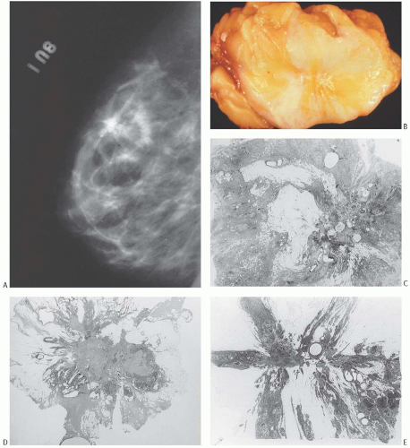
FIG. 5.18. Radial sclerosing lesion. A: Mammogram showing a RSL with a large calcification. B: Gross appearance of the lesion seen in (A). The RSL is the stellate whitish lesion near the center of the tissue. C-F: Whole-mount histologic sections showing different structural patterns. Relatively ill-defined lesion with multiple cysts (C). Asymmetric, oval lesion with dense central fibrosis. A proliferative component and cysts are present around half of the circumference (D). Lesion with a stellate pattern (E). Ovoid tumor with a contour that is partly smooth and partly stellate (F).


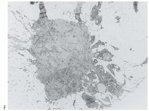
FIG. 5.18. (Continued)

Small ductules and distorted lobules are distributed between bands of sclerotic tissue radiating from the core into the surrounding stroma. A “corona” of ducts, lobules, and cysts, variably present at the periphery of the lesion, is created by incorporation of these structures from the surrounding tissue (Fig. 5.20). This peripheral zone is not evident around every RSL, and it may appear incomplete or asymmetric owing to intrinsic differences among lesions or as a consequence of asymmetric sectioning. The peripheral zone can also include nonproliferative ducts and lobules, which appear to be drawn toward the core. In some lesions, the “corona” consists mainly or entirely of cysts (Fig. 5.20C). RSLs usually occur as isolated, separate lesions, but on occasion, contiguous foci may be joined to form a larger complex and palpable proliferative lesion in a fashion analogous to the formation of an adenosis tumor.


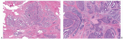
FIG. 5.19. Radial sclerosing lesions, elastosis. A: Foci of SA radiate from the elastotic core. B: Florid duct hyperplasia around an elastotic core.

The fibrous reaction associated with RSLs typically entraps small ductules and, less commonly, nerves. Like nests of epithelium trapped in the stroma at the periphery of a sclerosing papilloma (Fig. 5.11), those in a RSL simulate invasive carcinoma (Figs. 5.21 and 5.22). This is an important consideration when examining needle core biopsy samples (Fig. 5.23). The presence of a myoepithelial cell layer demonstrated with the p63, calponin, or actin immunostains characterizes epithelial entrapment within a RSL and thereby helps to avoid an erroneous diagnosis of invasive carcinoma. Somewhat less than 5% of RSLs demonstrate entrapment of small nerves.110 They are probably incorporated into RSLs by the same mechanism that is responsible for this phenomenon in other sclerosing lesions111 (Figs. 5.20C and 5.24).

The epithelium within a RSL can exhibit a range of changes. Apocrine metaplasia frequently occurs in the cystic component of RSLs, and it may occasionally be present more widely in the proliferative component, especially in areas of SA. Clear cell change and nuclear atypia are not uncommon in this apocrine epithelium. Squamous metaplasia is relatively infrequent in RSLs (Fig. 5.25), and those uncommon examples with squamous metaplasia may resemble metaplastic carcinoma, especially the low-grade adenosquamous variant.


Hyperplasia and Carcinoma in RSLs

Ductal proliferations in RSLs can take the form of florid and atypical hyperplasia (Figs. 5.26 and 5.27) as well as DCIS. Duct hyperplasia in a RSL may be solid, cribriform, micropapillary, or any combination of these structural patterns. Focal necrosis occurs in the hyperplastic duct epithelium of about 10% of RSLs (Fig. 5.26). The epithelium with these comedo-like foci is usually indistinguishable from the epithelium in hyperplastic ducts lacking necrosis in the same RSL. The presence of mitoses or necrosis is evidence of atypical hyperplasia in a RSL. Foci of ADH or ALH have been observed in 21%112 to 51%113 of RSLs. Often, the significant
proliferative foci are distributed in multiple tissue fragments in a needle core biopsy specimen. Care should be taken to avoid an erroneous diagnosis of DCIS or invasive carcinoma in this setting. Myoepithelial cells can be demonstrated around the perimeter of most hyperplastic ducts by using immunostains for p63, CD10, smooth muscle myosin-heavy chain (SMM-HC), or actin. However, in the central part of the lesion, myoepithelium may be substantially attenuated and even undetectable around hyperplastic ducts (Fig. 5.23).


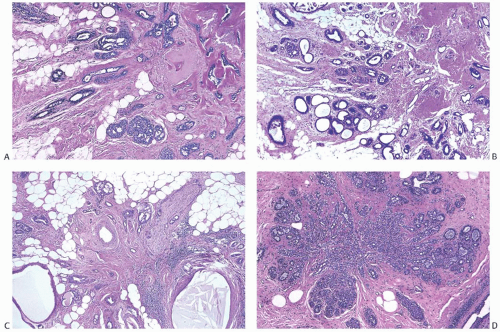
FIG. 5.20. Radial sclerosing lesions. A: The elastotic center is in the upper right corner. Mild ductal hyperplasia is present at the periphery. B: The elastotic center is in the upper right corner. The lesion features adenosis with microcystic dilation of glands. C: A RSL with peripheral apocrine cysts and an entrapped nerve (upper right). D: A RSL composed of SA.


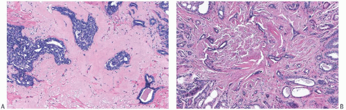
FIG. 5.21. Radial sclerosing lesion. A: Ductal hyperplasia and fibroelastotic stroma. B: Adenosis with a pattern that simulates tubular carcinoma.


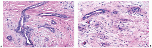
FIG. 5.22. Radial sclerosing lesion simulating invasive carcinoma. A: Irregular ductules in desmoplastic stroma. B: The distinction between epithelial and stromal cells is obscured.


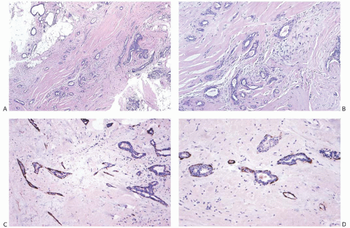
FIG. 5.23. Radial sclerosing lesion, needle core biopsy. A: A low-magnification view of the biopsy sample shows small angular glands in the fibroelastotic stroma. A focus of ductal hyperplasia is present near the right border. This specimen was misinterpreted as tubular carcinoma. B: Angular glands in the myofibroblastic stromal proliferation. C: Actin-positive myoepithelial cells surround these glands in a RSL. D: Incomplete and focally absent SMM-HC immunoreactivity in a RSL.


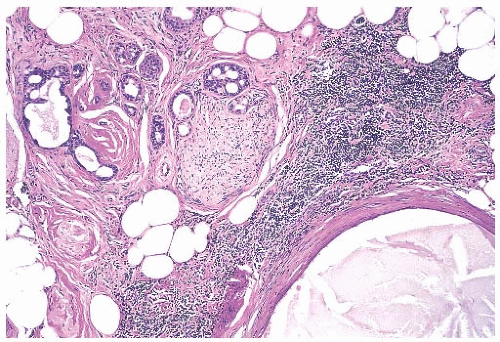
FIG. 5.24. Nerve entrapment in a radial sclerosing lesion. A nerve is shown in the upper center at the periphery of a RSL. Small glands at the perimeter of the perineurium indent the nerve.


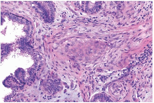
FIG. 5.25. Squamous metaplasia in a radial sclerosing lesion.

The presence of carcinoma, including tubular carcinoma, in RSLs has been well documented,114 but the literature does not provide a coherent set of estimates of the frequency of this association. Values as high as 31% and 32% have been reported,112,115 and in one series, 28% of mammographically detected RSLs larger than 1 cm had foci of carcinoma.116 In this context, it is important to distinguish between carcinomas with a stellate configuration and RSLs with foci of carcinoma. In the absence of residual RSL, it is not possible to prove that an entirely stellate carcinoma arose in a RSL. Carcinoma is most frequently found in RSLs larger than 0.6 cm,117 and it occurs more often in RSLs from women older than 50 years.117,118 Both ductal and lobular carcinomas (Fig. 5.28) occur in RSLs, and the frequency of noninvasive carcinomas outweighs that of invasive carcinomas.110,112,117,118 The carcinomas usually involve only a small region of the RSL, sometimes as little as 5%, and they more frequently occupy the periphery rather than the center.110,117,118

Only gold members can continue reading. Log In or Register to continue

Stay updated, free articles. Join our Telegram channel

Jun 5, 2016 | Posted by in PATHOLOGY & LABORATORY MEDICINE | Comments Off on Papilloma and Related Benign Lesions

Full access? Get Clinical Tree

Get Clinical Tree app for offline access