Papillary Endothelial Hyperplasia (Masson Tumor)
Amitabh Srivastava, MD
Key Facts
Terminology
Benign, reactive, intravascular papillary endothelial proliferation
Clinical Issues
Wide site distribution; located in deep dermis or subcutaneous tissue
Macroscopic Features
Small, cystic lesions with red-purple discoloration
Microscopic Pathology
Circumscribed lesion with pseudocapsule
Papillary structures lined by endothelial cells
Significant nuclear pleomorphism is absent
Top Differential Diagnoses
Angiosarcoma
PEH lacks nuclear atypia, tumor cell necrosis, and mitotic activity present in angiosarcoma
Hemangioma
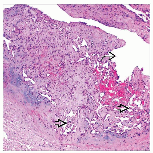 PEH is a well-circumscribed reactive lesion in which papillary fronds  lined by a single layer of endothelial cells proliferate within a vascular lumen lined by a single layer of endothelial cells proliferate within a vascular lumen  . . |
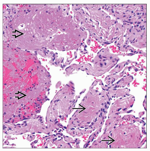 Fibrin thrombi  are apparent in early stages, and with time are replaced by papillary fronds with a fibrous core are apparent in early stages, and with time are replaced by papillary fronds with a fibrous core  characteristic of PEH. characteristic of PEH. |
TERMINOLOGY
Abbreviations
Papillary endothelial hyperplasia (PEH)
Stay updated, free articles. Join our Telegram channel

Full access? Get Clinical Tree


