Other Unusual and Rare Variants of Melanoma
David Cassarino, MD, PhD
Key Facts
Terminology
Rare variants of melanoma including chondroid and osseous melanoma, myxoid melanoma, rhabdoid melanoma, and balloon cell melanoma
Etiology/Pathogenesis
Related to UV exposure in most cases
Clinical Issues
Very rare tumors
Microscopic Pathology
Most variants often show areas of more conventionalappearing melanoma &/or overlying melanoma in situ
Balloon cell/clear cell melanoma
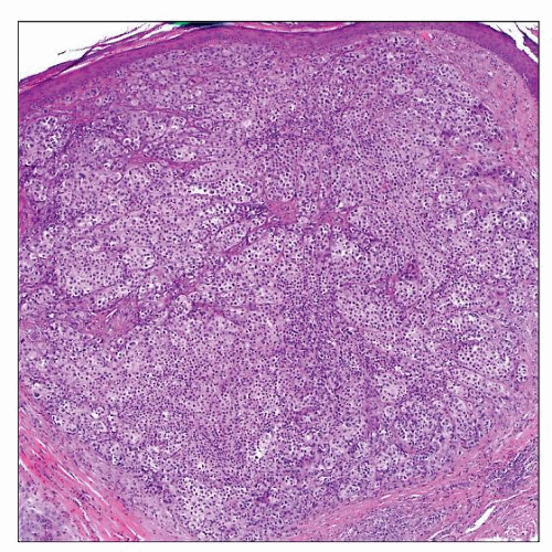
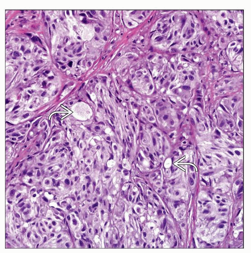
Composed of enlarged cells with abundant clear to vacuolated-appearing cytoplasm
Chondroid melanoma
Malignant epithelioid cells associated with prominent myxochondroid-appearing matrix
Osteoid melanoma
Malignant epithelioid cells associated with prominent osteoid-appearing material
Myxoid melanoma
Atypical epithelioid cells scattered in prominent myxoid matrix
Rhabdoid melanoma
Proliferation of markedly enlarged and atypical-appearing melanocytes with abundant eosinophilic-staining cytoplasm
Small cell melanoma
Nests and sheets of small, hyperchromatic-staining cells with scant cytoplasm (high N:C ratio)
 Clear cell melanoma shows a dense nodular to sheet-like dermal proliferation of pale-staining cells. These tumors can mimic clear cell sarcoma, but the superficial location would be highly unusual. |
TERMINOLOGY
Definitions
Rare variants of melanoma including clear cell/balloon cell, chondroid, osteoid, myxoid, and rhabdoid melanoma
ETIOLOGY/PATHOGENESIS
Environmental Exposure
Related to UV exposure in most cases
CLINICAL ISSUES
Epidemiology
Incidence
Very rare tumors
Age
Typically elderly patients
Site
Sun-damaged skin, typically head and neck region, upper trunk, forearms
Presentation
Usually presents as papule or nodule with irregular, asymmetric borders
Treatment
Surgical approaches
Complete and wide excision, with clinical margins appropriate for conventional melanoma of similar
Breslow depth
Prognosis
Depends on typical melanoma prognostic features such as Breslow depth, ulceration, mitotic count, and presence of perineural or angiolymphatic invasion
Rhabdoid melanoma reportedly shows aggressive course in most cases
MICROSCOPIC PATHOLOGY
Histologic Features
Depends on histologic variant
Most cases often show areas of conventional-appearing melanoma &/or overlying melanoma in situ
Clear cell/balloon cell melanoma
Enlarged cells with abundant clear to vacuolatedappearing cytoplasm
May be due to glycogen
May appear deceptively bland in some cases
Chondroid melanoma
Malignant epithelioid cells associated with myxochondroid-appearing matrix
Often shows areas of overlying melanoma in situ or more conventional melanoma with nesting
Osteoid melanoma
Malignant epithelioid cells associated with prominent osteoid-appearing material
Myxoid melanoma
Atypical epithelioid to spindle-shaped cells scattered in prominent myxoid matrix
Rhabdoid melanoma
Proliferation of markedly enlarged and atypicalappearing cells with abundant eosinophilic-staining cytoplasm
Peripheral displacement of nucleus by cytoplasmic material (intermediate filaments)
Signet ring cell melanoma
Composed of atypical epithelioid cells with pale to clear-staining cytoplasm
Likely due to degenerative changes or glycogen
Peripheral displacement of nucleus leads to signet ring cell appearance
Small cell melanoma
Nests and sheets of small, hyperchromatic-staining cells with scant cytoplasm (high N:C ratio)
Mimics basal cell carcinoma (BCC), Merkel cell carcinoma, or other neuroendocrine carcinomas
Stay updated, free articles. Join our Telegram channel

Full access? Get Clinical Tree


