Osteonecrosis


Osteonecrosis


Although risk factors and clinical associations have been identified, the pathogenesis of osteonecrosis is still unknown (1). Theories such as fat emboli, fatty induced intraosseous pressure, intravascular coagulation, microfractures, apoptosis of osteoblasts and osteocytes, and others continue to be explored.

Coagulation disorders such as seen in thrombophilia (increased predisposition to clots) and hypofibrinolysis (decreased ability to lyse clots) have been increasingly associated with osteonecrosis.

Mankin (2) has classified osteonecrosis as the result of mechanical vascular interruption, thrombosis, and embolism, injury to or pressure on a vessel wall, or that due to venous occlusion (Table 14.1). The most common cause of osteonecrosis is that which follows trauma, especially the osteonecrosis that follows femoral neck fractures in the elderly. Fractures at this site are thought to compromise an already tenuous blood supply, the deep branch of the medial circumflex artery (3). Osteonecrosis is known to complicate developmental problems such as slipped capital femoral epiphysis and the femoral head deformities seen in Legg-Calvé-Perthes disease in childhood. Osteochondritis dissecans is a well-described osteonecrotic entity, most commonly seen in the medial aspect of the lateral femoral condyle. Numerous anatomically consistent sites exist where osteonecrosis may be observed either idiopathically or following trauma (Table 14.2).

Hall (4) has articulated the preference of these osteonecrotic syndromes for convex as opposed to concave subarticular areas of the bone. He attributed the predilection for these sites to the larger cartilaginous (and thus avascular) covering of convex subarticular surfaces, which may result in a more tenuous blood supply or more concentrated biomechanical forces transmitted across this type of surface. Although trauma is generally thought to be the etiology in these syndromes, the etiology in males, in weight-bearing areas, and particularly in some clinical syndromes related to stress, such as the capitellum in adolescent baseball pitchers or tennis players, suggests a multifactorial etiology (4).

To the list of orthopaedic osteonecrotic syndromes, one may add the list of transient ischemia phenomena that may explain bizarre puzzling spectra of illnesses, including transient osteoporosis, Sudeck atrophy, and reflex sympathetic dystrophy (RSD) syndrome (5).

In considering osteonecrosis, arterial disruption is the most explicable pathogenesis, the most common example being that of osteonecrosis following femoral neck fractures. Elegant studies by Crock (6) and others have clearly demonstrated the fine but tenuous arterial supplies to the femoral head (Fig. 14.1). Although there is some arterial supply from vessels from the ligamentum teres, the medial circumflex branch of the femoral artery supplies most of the femoral head. This arterial vasculature courses along the femoral neck and is quite susceptible to disruption with subcapital fractures of the neck, particularly displaced subcapital fractures of the neck. The blood supply to the femoral head is primarily from the medial circumflex artery, with its deep branch the most important. The deep branch terminates as the posterior superior nutrient arteries that enter the head of the femur via the vascular foramina in the posterior-superior and anterior-superior quadrants of the femoral head and neck (7). The incidence of osteonecrosis following displaced femoral neck fractures has been reported to be as high as 84 percent in the literature. Given the tenuousness of the circulation, it is remarkable that not all femoral neck fractures eventuate in bone necrosis.

A contributing factor to osteonecrosis following hip fracture may be a tamponade effect caused by hemarthrosis-induced increased intracapsular pressure.

Pathologically, the events following arterial disruption have been clearly described (8,9). The classic features of osteonecrosis are characterized by loss of cells—hematopoietic, fatty, and bone cells (Table 14.3). Interestingly, emptying of osteocyte lacunae are thought to be an age-related change, with focal loss typically seen in interstitial lamellae, outer rings of cortical bone, and subchondral
cortical bone (9). In ischemic necrosis, initially fat necrosis and death of hematopoietic cells are noted, followed in several weeks by loss of osteocytes. Characteristically, the bone looks acellular, with empty ghostlike osteocytic lacunae and no evidence of osteoblast surface activity (Fig. 14.2). Osteonecrotic tissue often shows evidence of past remodeling such as creeping substitution or juxtaposition of new bone, suggesting that following the initial ischemia the bone-forming potential is stimulated. Most probably, the length of the ischemic episode dictates the extent to which, if possible, remodeling and new formation can occur. Obviously, terminal ischemia, that is, complete disruption of the only arterial supply, results in irreversible death. It has been estimated that, with ischemia, in less than 2 hours the changes that occur in osteocytes are probably reversible, but most likely irreversible damage occurs after 4 hours (10). Correlation with light microscopic features has been made (Table 14.3).


TABLE 14.1 Classification of Osteonecrosis


Disorder


Mechanisma


Traumatic osteonecrosis (fracture)


MVI, VVO


Septic osteonecrosis


MVI, T&E, I/P-VW, VVO (?)


Nontraumatic osteonecrosis (adults)


Stress or fatigue fractures


MVI, VVO


Alcohol abuse


T&E, I/P-VW, VVO (?)


Dysbarism (caisson’s disease, decompression sickness)


T&E, I/P-VW


Gaucher disease


I/P-VW


Connective-tissue disorders (e.g., rheumatoid arthritis or systemic lupus erythematosus)


MVI, I/P-VW


Arteritis or vasculitis


I/P-VW


Hemoglobinopathy (sickle cell disease)


T&E


Coagulopathy


T&E, I/P-VW


Radiation injury


I/P-VW


Corticosteroid administration


MVI, T&E, I/P-VW, VVO (?)


“Aging”-related lesions of the distal femur


I/P-VW


Pregnancy


VVO (?)


Gout


Secondary event


Pancreatitis


?


“Idiopathic” or spontaneous osteonecrosis


?


Childhood diseases (e.g., Legg-Calvé-Perthes, Sever, Kohler, Larsen, Blount, or Panner disease)


?


Other


Myeloproliferative disorders (polycythemia)


Fat emboli


a Putative pathogenetic mechanism: MVI, mechanical vascular interruption; VVO, venous and venular occlusion (Chandler disease); T&E, thrombosis and embolism; I/P-VW, injury to or external pressure on vessel wall.


Modified from Mankin HJ. Nontraumatic necrosis of bone (osteonecrosis) [Review]. N Engl J Med. 1992;326:1473-1479.)


Changes thought to be reversible are electron microscopically detectable condensation of nuclear chromatin and irregularities of the cytoskeleton. Irreversible changes seen later include coarse aggregation of chromatin, broadening of the interchromatin
space, and aggregation of interchromatin granules and the development of large interruptions on the plasma membrane and actual extrusion of cytoplasm. These changes are not detectable by light microscopy. However, Catto (8,9) has studied osteonecrosis following femoral neck fractures. Within 24 hours of injury, fibrin and hemorrhage are the most obvious changes. With ensuing days, there is a proliferation of capillaries, with foamy macrophages appearing about day 4. New bone formation is not usually evident at the fracture site in less than 5 days. However, active osteoblast formation and juxtaposed new bone formation are noticeable by the second week. The hematopoietic tissue shows loss of nuclear detail within days after death has occurred and complete loss of nuclei with cloudy-like appearance 2 weeks afterward. In humans, osteocytic nuclei begin to disappear after 4 to 5 days, but are more obvious 2 weeks later. In a rabbit model of bone ischemia, James and Steijn-Myagkaya (11) showed irreversible damage at 24 hours and empty lacunae after 24 hours. Despite the slow loss of osteocytic nuclei, traditionally a hallmark of osteonecrosis, the bone is most likely dead after the vascular injury and long before all cellular detail is lost. Therefore, the pathologist interpreting histopathologic sections, relying solely on cellular criteria of osteocytic empty lacuna spaces, may underdiagnose the presence of osteonecrosis.


TABLE 14.2 Clinicopathologic Sites and Syndromes Associated with Osteonecrosis


  1. Posttraumatic—femoral neck fractures, proximal half of scaphoid, posterior half of talus


  2. Avascular necrosis of the hip—(N.B., convex surfaces) knee, talus (especially after dislocation), carpal (especially after dislocation), other: humerus, tibia, radius


  3. Slipped capital femoral epiphysis


  4. Legg-Calvé-Perthes disease (femoral head osteonecrosis)


  5. Osteochondritis dissecans (knee)


  6. ? Reflex sympathetic dystrophy


  7. Freiberg disease (osteonecrosis of the distal metatarsal bone)


  8. Kienbock disease (osteonecrosis of the carpal lunate)


  9. Kohler disease (osteonecrosis of the tarsal navicular)


  10. Panner disease (osteonecrosis of the capitellum)


  11. Other: talar dome, femoral condyle, humeral head


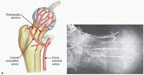
FIGURE 14.1. Vascularity of the femoral head. Specimen radiograph (A) and gross femoral head (Continued)

To summarize, the changes seen histopathologically in bone undergoing necrosis are sequential and affect hematopoietic cells, marrow fat, and bone cells in a chronological fashion. Hematopoietic cells are the first to undergo anoxic death in approximately 6 to 12 hours. This is followed by the death of bone cells (osteocytes, osteoclasts, and osteoblasts) in 12 to 48 hours and subsequently marrow fat in 48 hours to 5 days.

Variability in the sensitivity of the various bone cells and marrow cells to anoxia raises the intriguing question of when the necrosis of one element is irreversible in leading to necrosis of the entire tissue. Intuitively, some reversibility is possible, perhaps explaining the enigmatic entities of reflex sympathetic dystrophy, bone bruise, bone marrow edema syndrome, and transient osteoporosis, the etiologies of which may, in part, be due to transient ischemia or anoxia rather than irreversible full necrosis.

The identification of dead bone is initially best assessed by necrosis of hematopoietic cells and fat cells. Although the loss of nuclei in lacuna spaces may be seen within a few days, it is usually not complete until several weeks and, in the nontraumatized sections of bone adjacent to fracture, may not be evident for several more weeks. In addition, empty bone lacunae may be a technical artifact due to inadequate dehydration or fixation or decalcification. The reparative response is characterized by a fibrovascular proliferation, including macrophages, and the subsequent juxtaposition of new bone on top of necrotic cancellous bone by active osteoblast proliferation. Concomitant areas of active osteoclastic resorption of dead bone may also be obvious.


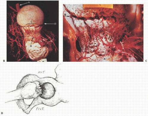
FIGURE 14.1. (Continued) (B) (A, medial femoral artery; B, cervical arteries; C, subcapital zone; D, site of subcapital fracture) and (C) (A, cervical branches; B, site of subcapital fractures; C, lateral circulation) with arterial injection showing fine tenuous arterial circulation to the femoral head, a site at great risk for disruption at the time of subcapital fractures (D). (After Crock HV. A revision of the anatomy of the arteries supplying the upper end of the human femur. J Anat. 1965;99:77-88.)


TABLE 14.3 Timetable of Microscopic Bone Changes in Osteonecrosis


>2 h


Irreversible damage rabbit model (10,11)


<6 h


Loss of osteocyte nuclear DNA; irreversible cell damage


>6 h


Histologic deterioration of bone marrow cells and osteoblasts


˜24 h


Pyknosis of osteocyte nuclei; initial losses noted


2nd d


Ghostlike foggy appearance of marrow


3 d


Decreased affinity of osteocyte nuclei for H&E and Feulgen stains


2-4 wk


Definitive loss of osteocytes in light microscopic sections (9)


h = hours, d = days, wk = weeks


H&E, hematoxylin and eosin.


Modified from Catto M. Pathology of aseptic bone necrosis. In: Davidson JK, ed. Aseptic Necrosis of Bone. Excepta Medica, 1976:3-100 and James J, Steijn-Myagkaya GL. Death of osteocytes. Electron microscopy after in vitro ischaemia. J Bone Joint Surg Br. 1986;68:620-624.


Generally, necrotic bone is identified histologically by poorly staining cortical and trabecular bone with the loss of nuclear detail as mentioned previously. The marrow itself usually undergoes change, which may be identified in early reversible stages as replacement by foamy macrophages (lipophages). A fuzzy disruption of the normal distinct clear fatty lobules is often evident as well as the appearance of small degenerating fat cells. Saponification of fat in necrotic marrow is identified as fine granular and flocculent, faintly eosinophilic, or deeply staining granules. More obvious calcification similar to that seen in dystrophic calcification states is eventually noted. Calcification of marrow is the end stage of marrow necrosis, the result of free fatty acids from dead marrow fat cells reacting with available released calcium. These calcifying sites lie dormant and usually are nonprogressive. Often asymptomatic, they appear only clinically as a clue that previous infarction has occurred.

Bone necrosis confined to the medullary cavity is often irreversible. However, osteonecrosis involving both cortical and cancellous bone appears more resilient in its reparative potential (2). Postinjury hyperemia gives way to a vascular fibrous repair revascularizing the dead bone. Both osteoclastic resorption of dead bone and osteoblast production of new bone ensue often juxtaposed on previous necrotic spicules.

Clinically, osteonecrosis is usually associated with pain.


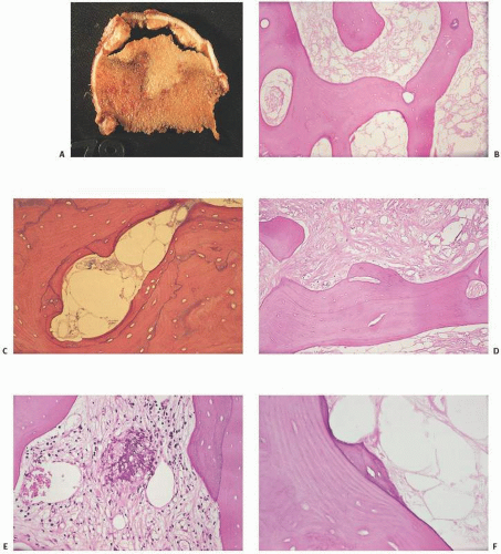
FIGURE 14.2. Dead bone. Dead bone has a gross (A) and microscopic pallor (B) (lack of staining intensity). The bone characteristically lacks nuclei in osteocyte lacunar spaces (B, C) and, until phagocytic activity ensues, also lacks surface remodeling by osteoblasts and osteoclasts. The marrow simultaneously undergoes necrosis with loss of nuclei of fat or marrow cells, and a foggy appearance ensues followed by fibrosis of marrow (D) or even saponification (calcification of the fat) (E). Attempts at repair include new bone apposition. This creeping substitution may be jagged (F) or smooth (Continued)


FIGURE 14.2. (Continued) (G). Eventually, the bone is resorbed with large osteoclast-type resorption spaces (H). Thrombi may be observed in osteonecrosis, giving credibility to thrombophilia as a significant etiologic event (I). In some cases, ischemic marrow and microfractures support a link between subchondral insufficiency fractures and osteonecrosis (J). Infarcted bone and fat may be associated with a bluish discoloration of the mineralized tissue (K, L).


Roentgenographically, osteonecrosis may be identified by a decreased uptake on bone scan in its early phase. Several weeks later, intense uptake can be noted owing to several factors: the hyperemia of the reparative response and mineralization of new bone to replace old bone. When saponification of dead marrow fat occurs, x-ray densities can be noted (12) (Fig. 14.3). Magnetic resonance imaging (MRI) classically shows a reduced signal in both T1- and T2-weighted images. Hypervascular reparative tissue may vary signal intensity (2).


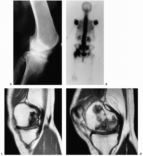
FIGURE 14.3. Roentgenographic, bone scan, and magnetic resonance imaging (MRI) findings in osteonecrosis. Fuzzy, irregular radiopacities are typical of late-stage osteonecrosis on routine x-rays, distal femoral condyle (A). Bone scan is hot, due to both hypervascularity and new bone production in hip (B). (C-F) Areas of necrosis in the medial femoral condyle as demonstrated on MRI images; all images are fast-spin echo. C and D are proton density (TR 4,000; TE 30/Ef). (Continued)

Practical applications of MRI in the imaging of osteonecrosis of the femoral head include detection of early or small lesions, to differentiation of osteonecrosis from other processes, and to
prediction of the likelihood of collapse (13). The classic pattern of osteonecrosis of the femoral head is a circumscribed ovoid or crescent-shaped low signal in subchondral bone, the rim representing the reparative zone between normal and ischemic damaged bone. A second inner rim, the “double lesion sign,” may be seen as a high signal on T2 and is thought to be pathognomonic of osteonecrosis by some (13).


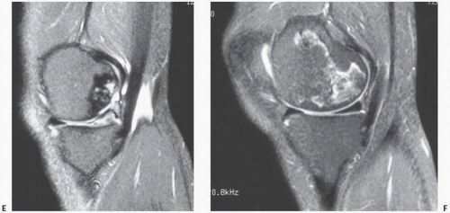
FIGURE 14.3. (Continued) E and F are fat-suppressed T2-weighted (TR 3,500; TE 39 Ef). Images C and E demonstrate damage of the articular cartilage superficial to the area of necrosis.

Multifocal osteonecrosis, defined as disease involving three or more anatomical sites, is most often associated with corticosteroid therapy, especially in the treatment of lupus. Femoral head involvement is common, as is bilaterality. Knee, shoulder, and ankle joints are also commonly involved.


TABLE 14.4 Causes of Osteonecrosis of the Femoral Head in One Large Series


No. of Patients


Type


Total


Male


Female


No. of Resected Femoral Heads


Osteonecrosis, all cases


345


337


Postfracture osteonecrosis


113


33 (mean age 58 y)


80 (mean age 66 y)


113


Idiopathic osteonecrosis, all cases


232


112 (mean age 49 y)


120 (mean age 55 y)


264


Idiopathic osteonecrosis, excluding lesions developing in osteoarthritis or rheumatoid arthritis


187


95 (mean age 46 y)


92 (mean age 53 y)


211


Idiopathic osteonecrosis, developing in osteoarthritis


37


15 (mean age 67 y)


22 (mean age 66 y)


40


Idiopathic osteonecrosis, developing in rheumatoid arthritis


8


1 (age 44 y)


7 (mean age 54 y)


13


After Sissons HA, Nuovo MA, Steiner GC. Pathology of osteonecrosis of the femoral head. A review of experience at the Hospital for Joint Diseases, New York. Skeletal Radiol. 1992;21:229-238. Copyright © 1992. Reprinted with permission of Springer-Verlag New York, Inc.


A large group of clinical syndromes associated with osteonecrosis have been documented (Table 14.2). Idiopathic (“avascular” or “aseptic”) osteonecrosis of the femoral head is the most completely studied.


Idiopathic Necrosis

Avascular necrosis of the bone is a significant cause of arthritis of the hip (12), the knee (14,15), and other major joints (Table 14.4).


Spontaneous Osteonecrosis of the Knee

A link between osteonecrosis and (insufficiency) fractures has been proposed in the condition referred to as spontaneous osteonecrosis of the knee (SPONK or SONK).These patients, usually elderly, develop pain predilected to the medial femoral condyle. Histopathologic findings consistent with fracture remodeling have led to the suggestion that the primary event leading to SPONK is a subchondral insufficiency fracture and that localized osteonecrosis seen in association with the disorder is the result of a fracture (14). Distinctions between SPONK and secondary causes of osteonecrosis of the knee have been drawn (15). Unlike the osteonecrosis secondarily associated with corticosteroids, ischemia, or atraumatic necrosis, SPONK is typically noted in older patients with no known risk factors and is usually unilateral with one condyle involved. Other joint involvement is rare. SPONK is located usually in a subchondral or epiphyseal region. In secondary osteonecrosis, patients are typically younger (>55 years) with bilateral disease often involving multiple condyles and multiple joints. Lesions may be diaphyseal, metaphyseal, or epiphyseal (15).


Avascular/Aseptic Necrosis

In the femoral head, avascular necrosis (AVN) has been estimated to develop in 10,000 to 20,000 new patients per year in the United States, and accounts for 5 to 12 percent of total hip replacements (1). It is termed avascular because there is no definitively identified vascular disruption, and aseptic because it is not related to infection.

AVN of the femoral head is more common in males, and its peak incidence is in the fourth decade. Conservative treatment options appear to delay but not halt the progression of the disorder (16).


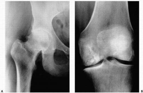
FIGURE 14.4. Avascular necrosis of the femoral head (A) and knee (B). Irregular radiodense subarticular portions of the femoral head and knee indicate dead (and partially reparative) bone.

Areas of osteonecrosis that are immediately adjacent to an articular joint may result in arthritis owing to a fracture of the necrotic bone and subsequent collapse of the overlying articular surface (Fig. 14.4).

The clinical onset of avascular AVN is usually sudden, although local pain may be present from months to years. Furthermore, the duration of symptoms is shorter than that in either rheumatoid arthritis or osteoarthritis. The hip seems to be the joint most commonly affected by an infarct. It occurs in the femoral head rather than the acetabulum. In this regard, it is generally true that the convex surface of any joint is most often affected. Avascular necrosis of the femoral head occurs in the subchondral bone of the superolateral or weight-bearing area of the femoral head. It may involve more than one skeletal site. Multiple lesions are often symmetrical, often varying in clinicopathologic significance.

The characteristic radiologic features of AVN include joint space preservation and subchondral fracture with changed contour and increased density. Bone scan may show hot uptake before roentgenographic changes are evident. The increased density results from reparative new bone, collapse and condensation of dead bone, marrow fat calcification, and subsequent trabecular thickening surrounding the infarct. Suggested staging based on x-ray, scan, and MRI changes have been proposed (1,17) (Table 14.5).

Most staging or grading systems used to assess the extent of damage use MRI to assess the extent of involvement of the femoral head (<15 percent, 15 to 30 percent, >30 percent) and the extent of sclerosis and cystic change, the degree of head depression and/or collapse, and the degree of acetabular changes (17).

The changes in the contour of the joint result from the failure of the reparative tissues to support the articular surface, with subsequent collapse of the infarcted area. This classically results in a subarticular lucent zone, the crescent sign (18) (Figs. 14.5 and 14.6).


TABLE 14.5 Stages of Osteonecrosis Based on Roentgenographic Studies


Stage


Criteria


0


Normal or nondiagnostic radiograph, bone scan, and MRI


Ia


Normal radiograph, abnormal bone scan, and/or MRI


IIa


Abnormal radiograph showing “cystic” and sclerotic changes in the femoral head


IIIa


Subchondral collapse producing a crescent sign


IVa


Flattening of the femoral head


Va


Joint narrowing with or without acetabular involvement


VIa


Advanced degenerative changes


a The extent or grade of involvement should also be indicated as A, mild; B, moderate; or C, severe.


MRI, magnetic resonance imaging.


Modified after Steinberg ME, Hayken GD, Steinberg DR. A quantitative system for staging avascular necrosis. J Bone Joint Surg Br. 1995;77:34-41 and Mont MA, Hungerford DS. Non-traumatic avascular necrosis of the femoral head [Review]. J Bone Joint Surg Am. 1995;77:459-474.


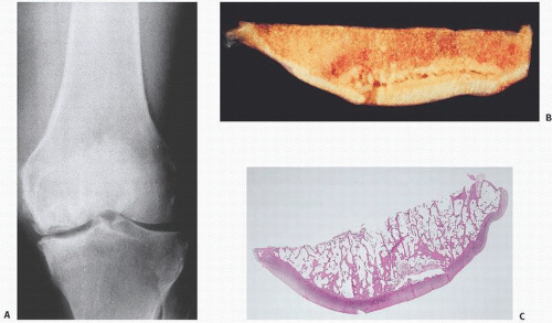
FIGURE 14.5. Avascular necrosis of the knee. Anteroposterior x-ray of the knee in a middle-aged woman with sudden onset of knee pain. Sclerosis and disruption of the medial femoral condyle is seen (A). A coronal section [(B) gross; (C) microscopic] taken through the medial condyle of a patient with osteonecrosis of the knee shows a zone of bone necrosis immediately under the articular surface characterized by an opaque-yellow appearance. Immediately beyond the necrotic zone is a band of hyperemia. (Continued)

It should be emphasized that the necrosis involves only bone and bone marrow and not, except in rare cases, the articular cartilage, which receives its nutrition from the synovial fluid. Therefore, the joint space on roentgenograms remains intact. At least in the initial stages of the disease, this radiologic feature clearly distinguishes early AVN from other forms of joint disease, in which the first radiologically evident change is a loss of articular cartilage and joint space narrowing.

Gross examination of a joint surface resected in a patient with early-stage clinical osteonecrosis is likely to reveal fairly intact articular cartilage, although some wrinkling of the surface that marks the edge of the necrotic area will probably be evident. On coronal sectioning, the infarcted zone exhibits a characteristic bright yellow opaque appearance. The infarcted area is usually large, involving 3 cm of subchondral bone and penetrating 2 cm deep (12). If the infarct is recent, a hyperemic zone is present at its margin, to be replaced later by a zone of fibrous scarring.

An infarct heals from the periphery by invading the necrotic marrow with granulation tissue and ensheathing the necrotic trabeculae by a layer of new bone (so-called creeping substitution) identical to that seen in bone repair from other injuries. Some infarcts heal without complication, and such instances are unlikely to be detected clinically because the process is generally asymptomatic. However, some cases are complicated by collapse, perhaps resulting from accumulated “fatigue” microfractures of the necrotic bone trabeculae.


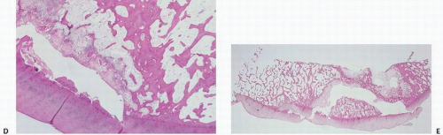
FIGURE 14.5. (Continued) Separating the necrosis bone from the overlying cartilage is a gap created by the collapse of the bone trabeculae in the necrotic segment. Microscopically, there is collapse and compression of the dead subchondral bone. A radiolucent space ensues (C, D). Dead bone may adhere to residual viable cartilage and eventually dislodge into the joint, manifesting itself as a loose body (E).


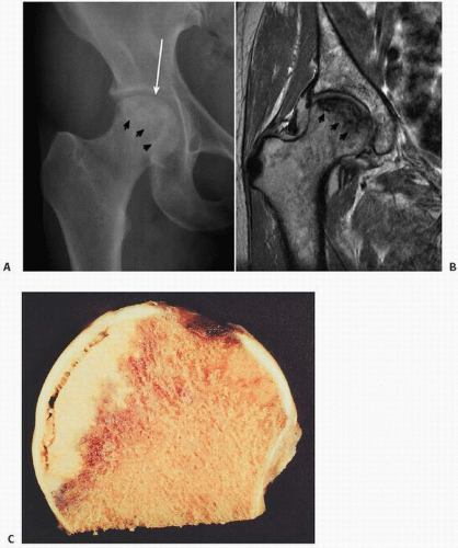
FIGURE 14.6. Avascular necrosis of the femoral head. Roentgenogram (A) and coronal MRI image (B) demonstrate avascular necrosis of the femoral head seen as density sclerosis in A and low signal in B (black arrows). Note the crescent sign (long arrow in A indicating subchondral collapse, corresponding to the lack of perfect sphericity of the femoral head in B. (Continued)


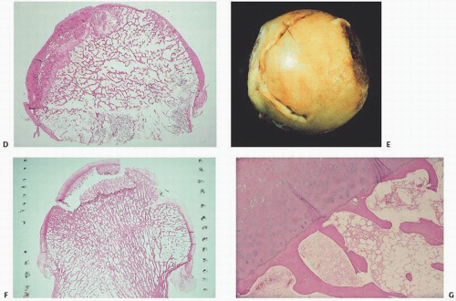
FIGURE 14.6. (Continued) Coronal section showing crescenteric zone of opaque yellow dead bone with radiolucent subchondral zone [(C) gross; (D) microscopic]. The zone of dead bone may or may not be evident on gross examination, depending on whether the cartilage has collapsed and broken off. The femoral head cartilage is dimpled (E) and fractured (F). Microscopically, there is a dislodged fragment of viable cartilage and adherent dead bone (G).

With the progression of AVN, the collapse of the necrotic segment and flattening of the joint surface ensues. The articular cartilage detaches from the underlying bone, which in turn gradually fragments and erodes, eventuating in secondary osteoarthritis.

The lack of a definitive identification of vascular disruption in AVN does not preclude a vascular etiology. Atsumi and Kuroki (19) have postulated repetitive vascular ischemia.

Deformation and incomplete revascularization of the bone in cases of AVN may, in fact, be due to repeated episodes of infarction.


Idiopathic Avascular, Aseptic Necrosis of the Hip

Mont et al. (17) have recently summarized key points regarding AVN of the femoral head:


  • Magnetic resonance imaging is the most sensitive and specific diagnostic method.


  • Symptomatology, although important for planning treatment strategies for individual patients, is not a reliable indicator of disease severity because it is quite variable; thus, it should not be part of the classification system.


  • A major determinant of prognosis is whether the femoral head is at the precollapse or postcollapse stage.


  • Size of lesion is important for prognosis; however, the best method of assessing the size of the lesion has yet to be fully elucidated or defined.


  • Acetabular articular cartilage involvement is an important determinant of disease severity in classification systems. Again, the method to determine this, whether preoperatively or with intraoperative inspection, has not been fully elucidated.


  • The amount of femoral head depression is a major prognostic indicator of disease progression.


  • Measurement of the amount of the depressed femoral head that is involved as well as the length of the crescent may not be reproducible.


  • The location of the lesion may not be as important as size when dealing with large lesions, because lateral lesions are usually large. Medial lesions, although quite rare, are typically small and are associated with the best prognosis.


  • While small, medially located lesions have a low rate of progression, the natural history of asymptomatic medium-sized, and especially large, osteonecrotic lesions is progressive in most patients.

Both nonoperative (lipid-lowering agents, anticoagulants, shock-wave therapy, pulsed electromagnetic fields) and operative treatments (core decompression, bone grafts, stem cell grafts, cementation) have been applied to this puzzling entity (1).


Osteochondrosis/Osteonecrosis Clinical Syndromes


Osteochondritis Dissecans

Osteochondritis dissecans (OCD) is a lesion of the subchondral bone that may involve partial or total separation of a fragment of the articular cartilage and subchondral bone from the articular surface (20). It can present in skeletally immature children and adolescents (juvenile form) or in skeletally mature adults (adult form).

Classically, the lateral aspect of the medial femoral condyle is affected (Fig. 14.7). Pathologically, this is usually associated with osteonecrosis localized to the subchondral bone fragment. Although the knee is the most common site, this type of subchondral osteonecrosis can occur in other joints, including the capitellum of the elbow, hip, talus, humerus, and patella. Bilaterality has been well documented in the femur. Although it is generally believed to follow a traumatic event with subsequent loss of the blood supply, this is not clearly established. It may be, as previously described, a multifactorial etiology including vascular injury, with biomechanical factors as well.

The occurrence of OCD in twins and in families supports the idea that there may be a genetic predisposition to the disorder (21).

The zone of osteonecrosis may be roentgenographically detectable by an area of sclerosis and a radiolucent linear line, not dissimilar to the crescent sign of AVN of the knee or hip (Figs. 14.5 and 14.6). In osteochondritis dissecans of the knee, this subchondral zone of osteonecrosis may remain anatomically confined to the femoral condyle or in fact be dislodged into the joint, forming a loose body. In fact, the loose subchondral osteonecrotic bone may be the nidus for the formation of an even larger loose body.


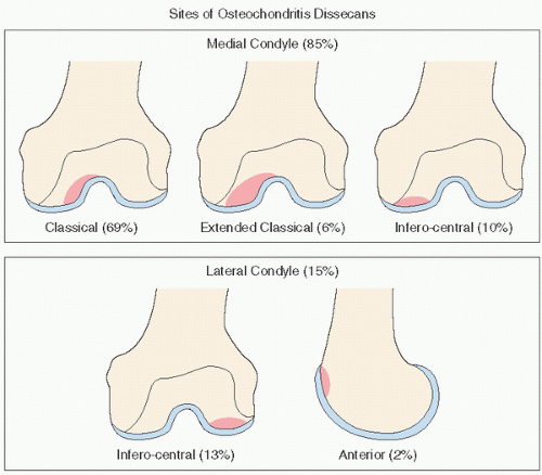
FIGURE 14.7. Osteochondritis dissecans: sites.

Symptoms reflect the various stages of development (Fig. 14.8) of this phenomenon. Sometimes vague, symptoms may include pain, or if the loose body is dislodged, mechanical locking of the joint. Osteochondritis dissecans is most common in men in the second and third decades of life.


Legg-Calvé-Perthes Disease

Legg-Calvé-Perthes disease occurs during childhood and is thought to be due to isolated or multiple episodes of infarction, most likely due to interruption of the medial femoral circumflex artery, absence or occlusion of its lateral epiphyseal branches, and even absence of anastomosis between the circumflex vessels and branches of the obturator artery (22) (Fig. 14.9). The resulting injury results in partial or total necrosis of the epiphysis, with boys being affected four times more frequently than girls. Others have postulated a coagulopathy etiology.

The incidence of Legg-Calvé-Perthes disease has been shown to be increased in the presence of factor V Leiden mutation, in the presence of prothrombin G20210A mutation, in association with elevated levels of factor VIII and in association with protein S deficiency (23).

The intermittent pain and limping associated with Legg-Calvé-Perthes injury is now thought to be due to pathologic fracture of tissue and the ensuing subchondral osteonecrosis. The well-described crescent subchondral line in children, an early radiographic feature of Legg-Calvé-Perthes disease, mimics that seen in the adult
population. Although destruction of the growth plate may ensue restricting blood flow to the epiphysis, and although the anatomical configuration of the femoral head may remain normal initially, eventual subchondral collapse may ensue with a flattened mushroom-shaped femoral head. Suggested factors determining the prognosis for any given patient include the patient’s age of onset, extent of involvement of the femoral head, loss of containment of the femoral head by the acetabulum in the weight-bearing position (subluxation), and a loss of motion of the hip joint. In the absence of subchondral fracture, AVN of the femoral head in children may resolve with no deformity. Legg-Calvé-Perthes disease is thought by some to be a disorder characterized by AVN, which, if complicated by pathologic subchondral fracture, may lead to further damage including resorption of bone and collapse of the femoral head.


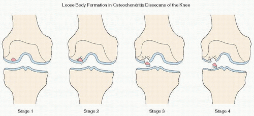
FIGURE 14.8. Osteochondritis dissecans: staging.


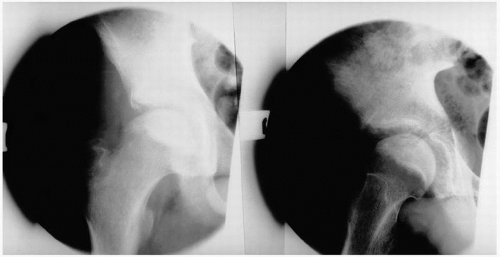
FIGURE 14.9. Legg-Calvé-Perthes disease. Roentgenographic features.

Numerous clinical correlations with bone necrosis have now been established, and multifactorial effects have been proposed (Fig. 14.10).


Fat Embolization

Fat embolism as a cause of osteonecrosis has been suggested clinically for more than 30 years. The support for an etiology of
osteonecrosis from fat embolization derives from biophysical forces. Fat has a greater viscosity than plasma, and its surface tension may facilitate adherence to arterial walls. It has been speculated that increased blood viscosity or increased arterial wall length or decreased vascular diameter as well as decreased intravascular pressure gradients all may reduce blood flow and potentiate intraosseous fat trigger damage. There are at least three proposed etiologies for fat embolization: the hyperlipidemia associated with fatty liver, endogenous production of lipoprotein, and the destruction of marrow fat. The latter may be a contributing factor in posttraumatic osteonecrosis.


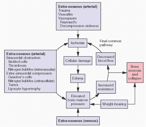
FIGURE 14.10. Proposed pathogenesis of osteonecrosis.

Experimental models have demonstrated that following fat embolism, hypoxia and hypercapnia occur and that chemical mediators such as phospholipase A2, nitrate/nitrite, methylguanidine, and other proinflammatory cytokines are significantly increased (24).

Only gold members can continue reading. Log In or Register to continue

Stay updated, free articles. Join our Telegram channel

Jul 24, 2016 | Posted by in PATHOLOGY & LABORATORY MEDICINE | Comments Off on Osteonecrosis

Full access? Get Clinical Tree

Get Clinical Tree app for offline access