Palpation of supraorbital ridge and frontal bone.
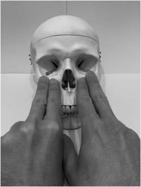
Palpation of infraorbital and lateral orbital ridges.
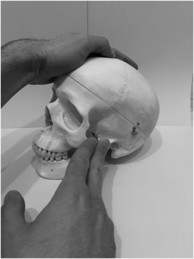
Palpation of zygomatic arch.
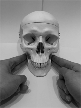
Palpation of mandibular ramus and angle.
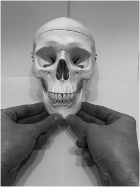
Palpation of mandibular body and mental protuberance.
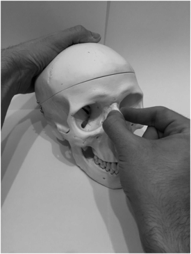
Palpation of nasal bones.
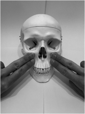
Palpation of malar/zygomatic bones.
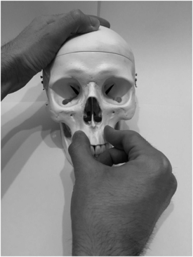
Palpation of maxilla/midface stability.
How is a nasal septal haematoma diagnosed?
A septal haematoma presents as an anterior blue-red septal swelling. This can be confused with septal deviation due to a fracture. Palpation with a Jobson–Horne probe or a gloved little finger will determine whether the swelling is soft and fluctuant (haematoma) or firm (deviated septum).
Are there specific tests for the maxilla?
Maxillary fractures are important, because they can cause both obstruction of the naso-/oropharynx and signify trauma to the base of the skull. The maxilla is held between the thumb and forefinger of one hand while the free hand examines for movement externally with gentle anterior–posterior pressure. The two points of reference are the sides (Le Fort I) and the bridge of the nose (Le Fort II/III). The maxilla should not be manipulated if there are signs of a base of skull fracture, in which case one should proceed straight to CT scanning.
What are the features of an orbital blow-out fracture?
Blow-out fractures are caused by direct blunt trauma to the orbit resulting in fracture of the orbital floor or medial wall. It is characterised by enophthalmos (depression of the eyeball in the orbit) and entrapment of the inferior rectus muscle, which does not allow the eye to look superiorly. Eye movement assessment is performed with a standard ‘H’ pattern, which can be augmented by including a central vertical movement up. This is particularly sensitive for entrapment secondary to an infraorbital fracture. Other features may include exophthalmos/proptosis due to a retrobulbar haematoma.
What are the features of a zygomatic arch fracture?
Zygomatic arch fractures may be associated with facial asymmetry due to a depressed cheekbone. Lateral subconjunctival haemorrhage in the eyes may indicate a potential fracture at the zygomaticofrontal suture.
What are the common fracture patterns of the midface?
These are described by the Le Fort classification:
- Le Fort I
Bilateral detachment of the alveolar complex and palate or low-level subzygomatic fracture
- Le Fort II
Pyramidal, subzygomatic fracture of the maxilla
- Le Fort III
Craniofacial dislocation; high-level suprazygomatic fracture of the central and lateral parts of the face
It is important to appreciate that these are an approximation of potential injury, as high-energy facial traumas can lead to variable and often mixed injury patterns.
Stay updated, free articles. Join our Telegram channel

Full access? Get Clinical Tree


