The vector gets the attention, but a corrective gene can be perfectly engineered and still fail if it never reaches the right cells intact. In retinal gene therapy, that final step belongs to a surgical instrument.
Roughly 57% of retinal gene therapies are delivered straight under the retina by subretinal injection, a procedure carried out in fractions of a millimetre. With ophthalmic gene therapy now approved and in clinical use, its biggest remaining risk is no longer the adeno-associated virus (AAV) construct itself, but whether the dose reaches the target cells intact.
This guide explains what subretinal delivery is, why the instrument matters as much as the vector, how the procedure is performed, and how delivery cannulas are classified and chosen.
What is subretinal delivery in ophthalmic gene therapy?
Subretinal delivery in ophthalmic gene therapy is a surgical procedure that places a therapeutic agent into the potential space between the neurosensory retina and the retinal pigment epithelium (RPE). The surgeon raises a small, controlled pocket of fluid, called a bleb, that lifts the retina just enough to seat the vector against the target cells.
Subretinal delivery is the most direct of the three retinal routes because it puts the vector in contact with the photoreceptors and RPE cells that need it. It is performed in an operating room and usually follows a pars plana vitrectomy.
The instrument at the tip is a fine subretinal cannula, also called a subretinal injection needle or delivery cannula. It is the single point of contact between an expensive biologic and fragile neural tissue. Its bore, tip finish, and handling shape the outcome.

Figure 1. Subretinal injection: the cannula raises a fluid bleb between the retina and the RPE, seating the vector against the target cells.
Why does the delivery instrument matter as much as the vector?
The delivery instrument matters as much as the vector because the complications increasingly trace back to the procedure, not the drug. At the ARVO 2026 Annual Meeting in May 2026, three-year outcome data from retinitis pigmentosa gene-therapy trials reported a telling detail.
The serious adverse events, including cataract, a rhegmatogenous retinal detachment, and persistent subretinal fluid, were attributed to the surgical delivery procedure rather than to the vector itself. That is the essence of the last-millimetre problem.
A vector can be immaculately engineered and still underperform if any of the following happens:
- the injection plane is wrong (intraretinal rather than subretinal);
- shear stress or dead-space in the cannula degrades viable titre;
- reflux or leakage at the retinotomy reduces the delivered dose;
- tissue trauma at the entry site triggers inflammation.
Every item on that list is a function of instrument geometry, bore, material, and handling, not vector design. Precision instrumentation is therefore one of the most direct ways to reduce delivery risk.
How is a subretinal injection performed, step by step?
A subretinal injection is performed as a short sequence of surgical steps, and the technique shows how instrument choice is woven into every stage. A representative transvitreal approach proceeds in four steps.
- Vitrectomy. A standard 23 or 25 gauge three-port pars plana vitrectomy (PPV) removes the vitreous to give controlled access to the posterior segment.
- Retinotomy and bleb creation. A narrow-bore cannula, commonly a 38G outer and 41G inner polyimide or Teflon-tipped tip, makes a self-sealing retinotomy and raises a parafoveal fluid bleb.
- Vector infusion. In a widely used two-step technique, saline first creates the bleb. The viral vector is then infused slowly through the same retinotomy, either by hand or via a microinjector.
- Closure. The self-sealing retinotomy is allowed to close, sometimes with a fluid-air exchange.
Recent technical work has focused on how manual versus microinjector-assisted delivery, injection speed and pressure, and adjuncts such as internal limiting membrane peeling affect efficacy and iatrogenic damage. Robot-assisted delivery under intraoperative OCT guidance is an active area of development.
How are ophthalmic gene-therapy delivery instruments classified?
Ophthalmic gene-therapy delivery instruments are classified by the action they perform, the same way surgical instruments are traditionally grouped. For a retinal gene-therapy procedure, that classification maps cleanly onto the delivery workflow.
- Cutting and opening instruments, such as blades and micro-vitreoretinal (MVR) knives used to create sclerotomy ports.
- Grasping and holding instruments, such as micro-forceps for membrane peeling and tissue manipulation.
- Cannulating and delivering instruments, meaning the subretinal injection cannula itself, plus infusion and extrusion cannulas.
- Manipulating and stabilising instruments, such as picks, spatulas, and manipulators that control the tissue plane during injection.
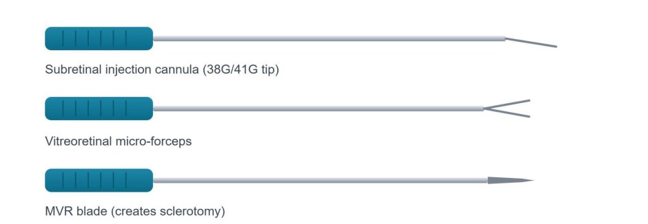
Figure 2. Representative delivery instruments: a cannulating instrument, a grasping instrument, and a cutting instrument.
If you are learning the procedure, map each surgical step to its instrument class first. Then the delivery cannula’s role becomes clear as the one instrument that both accesses and deposits into the subretinal space.
How are retinal gene therapies delivered?
Retinal gene therapies are delivered by three routes, and their share of clinical use is heavily skewed toward the subretinal approach.
| Delivery route | Approximate share |
| Subretinal | ~57% |
| Intravitreal | ~37% |
| Suprachoroidal | ~3% |
Figure 3. Approximate share of retinal gene-therapy delivery by route, based on 2025 to 2026 literature.
The pattern shows where the instrument stakes are highest. Subretinal, the majority route, is the most instrument-dependent. Intravitreal is simpler but non-targeted.
Suprachoroidal delivery, given via a transscleral microneedle and often in an office setting, is emerging quickly but remains a small share.
Which delivery route is used, and when?
The delivery route used depends on how targeted the therapy needs to be, and it determines the setting, the technique, and the instrument set. Use this comparison as a first-pass map.
| Route | Setting | Targeting | Core instruments | Typical use |
| Subretinal | Operating room, post-vitrectomy | Highest, direct RPE and photoreceptor contact | Subretinal injection cannula, micro-forceps, MVR blade | Inherited retinal diseases needing focal, high-precision delivery |
| Intravitreal | Clinic or OR | Broad, non-targeted | Injection needle, lid speculum | Programs prioritising simplicity and repeat dosing |
| Suprachoroidal | In-office | Wide posterior coverage | Transscleral microneedle | Broad chorioretinal coverage without vitrectomy |
Table 1. Retinal gene-therapy delivery routes and their instruments.
If the therapy must reach photoreceptors or RPE cells focally, then the subretinal route and its OR-grade microsurgical instruments are indicated. If broad retinal surface coverage matters more than pinpoint targeting, then suprachoroidal microneedle delivery is the emerging alternative.
How do you choose the right subretinal delivery cannula?
You choose the right subretinal delivery cannula by working through a short set of criteria in order.
- Match the cannula to the route and gauge. Transvitreal subretinal delivery typically uses a fine 38G or 41G tip on a 23 or 25 gauge shaft. Confirm compatibility with the vitrectomy system.
- Minimise dead-space and control bore. Every microlitre of internal dead volume is dose that is not delivered. Treat bore and internal volume as dose-integrity parameters.
- Check tip material and finish. Polyimide and Teflon-tipped cannulas reduce trauma. Burrs and poor tolerances increase reflux and RPE damage.
- Confirm self-sealing behaviour. A clean, self-sealing retinotomy limits reflux of the infused vector.
- Decide manual versus microinjector delivery. A microinjector can standardise injection speed and pressure. Manual delivery relies more on the surgeon’s tactile feedback.
If reproducible injection pressure and volume are the priority, then favour a microinjector-compatible cannula system. If tactile control is preferred, then a well-finished manual cannula with low dead-space is the safer default.
What makes a quality microsurgical instrument?
What makes a quality microsurgical instrument is documented build integrity, because when an instrument sits between an approved biologic and neural tissue, its quality is a clinical variable, not a catalogue detail.
Look for documented manufacturing origin and consistency, single-use sterility where reprocessing risk is unacceptable, verifiable tolerances and tip finish, and lifecycle support such as repair programs that keep reusable instruments performing to the same specification over time.
Reviewing a full catalog of ophthalmic microsurgical instruments shows the range of cannulas, forceps, and blades these steps rely on, and how single-use and reusable options differ in practice.
Conclusion
The hardest science in ophthalmic gene therapy has largely been solved. What now separates a successful outcome from a wasted dose is retinal gene therapy delivery: the cannula, forceps, and blade that carry an AAV vector into the subretinal space, and the technique that guides them.
Treating those instruments as a clinical variable, rather than a consumable, is one of the most practical ways to protect an expensive therapy at the point of care.
Key takeaways
- Subretinal injection is the dominant, most targeted, and most instrument-dependent route for delivering gene therapy for inherited retinal disease.
- Delivery outcomes hinge on cannula bore, tip finish, and vitreoretinal technique. Recent trial complications were linked to the procedure, not the vector.
- Choosing the right gene therapy delivery device means matching gauge to the vitrectomy system, minimising dead-space, and deciding between single-use and reusable instruments.
Final thoughts: As inherited retinal disease programs expand, the teams that respect the delivery instrument as much as the vector will be the ones that most reliably turn a promising therapy into restored sight.
Sources
- Ophthalmology Times, ARVO 2026: gene therapy in retinitis pigmentosa.
- Retina Today, Current Subretinal Gene Therapy Delivery (Jan/Feb 2026).
- PMC, Surgical Approaches to Retinal Gene Therapy: 2025 Update.
- Eye (Nature), Technique of retinal gene therapy: delivery into the subretinal space.
- Retinal Physician, Expanding the Role of Suprachoroidal Injections.
This article is an educational overview of surgical delivery instrumentation and does not constitute clinical or procedural guidance. Clinical figures and techniques should be verified against primary literature and institutional protocols before use.

Stay updated, free articles. Join our Telegram channel

Full access? Get Clinical Tree


