chapter 15 Nutrition and digestion
 Macronutrients make up the bulk of the food consumed, creating the fuel the body runs on and serving as precursors to its synthesis.
 Digestion refers to breaking down food to molecules that are small enough to be absorbed by the body.
 Most nutrient absorption occurs in the small intestine through specialised epithelial cells called enterocytes.
 The macronutrients—protein, carbohydrates and lipids—are digested and absorbed by specialised active transport systems.
 Minerals are either major elements or trace elements and are required for a number of physiological processes.
The need to eat and drink
Nutrition is the process by which food is taken into and used by the body. The food and drink we consume contains nutrients, the chemicals taken into the body that provide energy and serve as the building blocks for new molecules. There are six major classes of nutrients required for a healthy diet: lipids (Ch 4), proteins (Ch 5), carbohydrates (Ch 8), vitamins, minerals and water. However, it is not just a simple matter of taking nutrients from food for use in the body; the nutrients consumed are unlikely to be the appropriate kind, or in the appropriate abundance, for our needs. Food is eaten, broken down through digestion and the products absorbed. Cells can then use these to produce the chemical energy and to build the complex molecules required to make up our cells, organs and tissues. This occurs through the myriad of chemical reactions that occur in living cells—cellular metabolism.
Digestion and absorption
Digestion refers to breaking down food to molecules that are small enough to be absorbed by the body. Digestion is both mechanical, the chewing that takes place in the mouth or the churning of the stomach, and chemical, involving the breaking of covalent bonds by digestive enzymes (Table 15-1).
TABLE 15-1 Digestive enzymes of the gastrointestinal tract
| Source | Enzymes | Substrates |
|---|---|---|
| Saliva | amylase | starch |
| lingual lipase | triacylglycerol | |
| Stomach | pepsin | proteins |
| gastric lipase | triacylglycerol | |
| Pancreas | proteases | proteins |
| amylase | starch | |
| pancreatic lipase | triacylglycerol | |
| Intestinal lining (microvilli) | disaccharidases | disaccharides |
| peptidases | peptides |
Absorption of nutrients begins in the stomach, though the bulk of absorption takes place in the small intestine. The lining of the gut is made up of a layer of epithelial cells known as enterocytes, which are responsible for absorption of nutrients and passing them to the bloodstream or lymphatic system (through capillaries and lacteals respectively) to make them available for the rest of the body. The epithelium of the small intestine is optimised for absorption. The lining is folded and then finger-like projections known as villi (singular: villus) are formed on the folded surface. The outer layer of cells on the villi is made up of enterocytes. To further increase the available surface area the cell membrane of each enterocyte that is exposed to the gut is also folded on itself to form microvilli. These microvilli are so fine and numerous that under the microscope they resemble the bristles of a brush, hence the layer of enterocytes with their microvilli is often referred to as the brush border. This folding increases the surface area of the gut lining an estimated 600-fold compared to a smooth cylindrical gut, thus maximising the ability of enterocytes to absorb nutrients quickly and efficiently.
Macronutrients The need to … eat carbohydrates
Some tissues of the body have an absolute requirement for glucose and it is estimated that a daily intake of >130 g is required before the body has to resort to using protein via gluconeogenesis (see Ch 17) to maintain blood glucose levels, a situation best avoided.
Absorption of carbohydrates
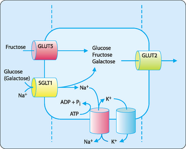
Carbohydrates in the diet mainly consist of starches, cellulose, sucrose and small amounts of fructose and lactose. Digestion of carbohydrates begins in the oral cavity with salivary amylase and is completed in the small intestine by pancreatic amylase. Amylases break down complex carbohydrates into the disaccharide maltose (see Ch 8) and then enzymes in the membrane of the enterocytes that specifically target disaccharides take over. Maltose is broken down to two glucose molecules by the enzyme maltase, lactose to glucose and galactose by lactase and sucrose to glucose and fructose by sucrase. Enterocytes take up the resulting monosaccharides using specific transporters, many of which are members of the glucose transporter (GLUT) protein family that includes GLUT1 to 4. Each of these transporters has different affinities for glucose. Those with a high affinity will transport glucose even when the concentration of glucose is very low (e.g. GLUT1, GLUT3). Some have an intermediate affinity (GLUT4) requiring slightly higher levels of glucose before transport can occur and others have a low affinity for glucose and need a relatively high concentration of glucose before transport will occur.
Glucose is taken into enterocytes using a secondary active transport mechanism. Glucose (and to a lesser degree galactose) is co-transported with Na+ by the symporter SGLT1. ATP is then used to drive a sodium–potassium antiporter to pump the sodium back out of the cell (Fig 15-1). This ensures that the sodium concentration in the cell is less than that in the gut lumen; thus the cell can use this concentration gradient to drive the uptake of glucose. This system is important in the efficient absorption of glucose from the gut as the concentration of glucose in the gut lumen drops rapidly and would result in poor absorption if it were a passive process.
The need to … eat protein
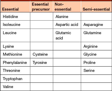
Humans also need to consume all of the essential amino acids, which the body is not capable of synthesising. Two other amino acids, cysteine and tyrosine, can be synthesised by the human body but require the essential amino acids methionine and phenylalanine respectively as precursors. The other nine amino acids are non-essential because they can be synthesised by the body. However, some of these are termed semi-essential as the demand for these is greater than the ability of the body to synthesise them (Table 15-2).
Absorption of proteins
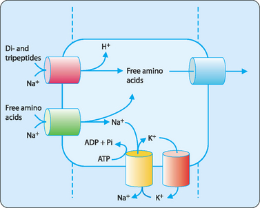
Free amino acids are taken up using a secondary active transport system very similar to that for glucose (Fig 15-2) and several transporters have been identified that will each transport a number of different amino acids dependent on their chemical nature. Di- and tripeptides are transported into enterocytes using a peptide–proton symporter after which they are degraded by intracellular peptidases to their constituent amino acids. Free amino acids are then exported to the blood by transporters in the basolateral membrane.


