Fig. 29.1 The arachidonic acid cascade of eicosanoid synthesis.
Arachidonic acid liberated from membrane phospholipids can be utilised by cyclo-oxygenases (COX-1, COX-2, COX-3) to form prostanoids (prostaglandins, prostacyclin and thromboxane) or by the 5-lipoxygenase pathway to form leukotrienes. The types and amounts of these eicosanoid products that are generated depend on the relative expression of the COX isozymes, 5-lipoxygenase and their respective downstream synthases in different cell types. After release from the cell, the eicosanoids have a multitude of actions via their selective G-protein-coupled receptors on the surface of target cells, such as bronchial, uterine and vascular smooth muscle cells, endothelial cells, platelets and leucocytes (Table 29.2). FLAP, five-lipoxygenase activating protein; 5-HPETE, 5-hydroperoxyeicosatetraenoic acid; LT, leukotriene; PG, prostaglandin; TX, thromboxane.
Arachidonic acid is mostly derived from dietary linoleic acid, which is found in vegetable oils such as sunflower oil. Linoleic acid is converted in the liver in several steps to arachidonic acid, which is then incorporated into glycerophospholipids in cell membranes. Arachidonic acid is released from membrane phospholipids by lipases such as phospholipase A2. In the COX pathways, the initial products of arachidonic acid metabolism are unstable intermediates known as cyclic endoperoxides. These are converted by cell-specific synthases and isomerases to various receptor-active prostanoids (Fig. 29.1). The products of the COX pathway therefore differ among various tissues depending upon the synthases present, generating a diverse range of actions tailored to the individual requirements of each cell type. Most cell types can form different prostanoids simultaneously and in different quantities, with the pattern of production modulated by regulatory influences on the cell.
There are three COX isoenzymes: COX-1, COX-2 and COX-3. COX-1 is constitutively expressed in the endoplasmic reticulum of most cell types. Prostanoids generated via the COX-1 enzyme are produced in small amounts by many cells in the resting state and contribute to the regulation of several homeostatic processes such as renal and gastric blood flow, gastric cytoprotection and platelet aggregation (Table 29.1). COX-2 is present in greater concentrations in the nuclear envelope than in the endoplasmic reticulum. It is found in many cells such as endothelial cells, macrophages, synovial fibroblasts, mast cells, chondrocytes and osteoblasts. Its expression in these cells is induced by cytokines and other inflammatory stimuli, so that COX-2-derived prostanoids are formed in large amounts in response to inflammation, pain and fever. However, the concept that there is only a pathological role for inducible COX-2-derived prostanoids and a ‘housekeeping’ physiological role for the constitutive COX-1 enzyme is now recognised as too simplistic. In many organs, particularly the kidney, central nervous system (CNS), cardiovascular system and reproductive system, there is no clear separation between the functions of COX-1 and COX-2. Either isoform may be involved in the production of ‘physiological’ or ‘pathological’ prostanoids (Table 29.1).
Table 29.1
Some biological roles of cyclo-oxygenase (COX) 1 and 2
| COX-1 homeostatic roles | COX-2 homeostatic roles |
| Gastrointestinal protection | Renal function |
| Platelet aggregation | CNS function |
| Blood flow regulation | Tissue repair and healing (including gastrointestinal) |
| CNS function | Reproduction |
| Uterine contraction | |
| Blood vessel dilation | |
| Pancreas | |
| Inhibition of platelet aggregation | |
| Airways | |
| COX-1 pathological roles | COX-2 pathological roles |
| (Possible involvement in inflammation) | Inflammation |
| Chronic pain | Chronic pain |
| Raised blood pressure | Fever |
| Blood vessel permeability | |
| Reproduction | |
| Alzheimer’s disease | |
| Angiogenesis, inhibition of apoptosis | |
| Tumour cell growth |
COX-3 is a splice variant of COX-1 that retains the entire COX-1 transcript and has an additional conserved intronic sequence. It is mainly expressed in the brain and spinal cord and to a lesser extent in the heart, endothelial cells and monocytes. COX-3 may have a role in pain perception, but its precise functions are uncertain.
The actions of prostaglandins and thromboxanes depend upon the circumstances and site of their formation, and whether they are formed in excessive amounts. For example, prostaglandin E2 (PGE2) is generated in low physiological amounts by COX-1 in gastric mucosa where it is important for maintaining mucosal integrity by a variety of mechanisms, including bicarbonate and mucus production and maintaining mucosal blood flow. In contrast, damage to many tissues leads to increased PGE2 synthesis via enhanced COX-2 expression. This contributes to inflammation and pain by vasodilation, increased vascular permeability and sensitisation of pain fibre nerve endings to the nociceptive action of bradykinin, serotonin and other mediators (Ch. 19). However, in the later stages of repair following tissue damage, COX-2-derived prostanoids may contribute to the processes of wound healing.
Prostanoids act via five main classes of G-protein-coupled receptors on cell surfaces (Fig. 29.1). Some of the actions of prostaglandins are shown in Table 29.2.
Table 29.2
Main biological actions of the eicosanoids
| Tissue | Effect | Eicosanoid |
| Platelets | Increased aggregation | TXA2 |
| Decreased aggregation | PGI2, PGD2 | |
| Vascular smooth muscle | Vasodilation | PGI2, PGE2, PGD2 |
| Vasoconstriction | TXA2, LTC4, LTD4 | |
| Other smooth muscle | Bronchodilation | PGE2 |
| Bronchoconstriction | LTC4, LTD4, LTE4, PGD2, PGF2, TXA2 | |
| Gastrointestinal tract (contraction/relaxation, depends on muscle type) | PGF2, PGE2, PGI2, PGD2 | |
| Uterine contraction | PGE2, PGF2α | |
| Vascular endothelium | Increased permeability | LTC4, LTD4, LTB4 |
| Potentiates histamine/bradykinin | PGE2, PGI2 | |
| Leucocytes | Chemotaxis of neutrophils, monocytes, lymphocytes | LTB4 |
| Chemotaxis of eosinophils, basophils | LTE4, PGD2 | |
| Gastrointestinal mucosa | Reduced acid secretion | PGE2, PGI2 |
| Increased mucus secretion | PGE2 | |
| Increased blood flow | PGE2, PGI2 | |
| Nervous system | Inhibition of noradrenaline release | PGD2, PGE2, PGI2 |
| Endogenous pyrogen in hypothalamus | PGE2 | |
| Sedation, sleep | PGD2 | |
| Endocrine/metabolic | Secretion of ACTH, GH, prolactin, gonadotrophins | PGE2 |
| Inhibition of lipolysis | PGE2 | |
| Kidney | Increased renal blood flow | PGE2, PGI2 |
| Antagonism of ADH | PGE2 | |
| Renin release | PGI2, PGE2, PGD2 | |
| Pain | Potentiates pain through bradykinin, serotonin | PGE2, PGD2 |
Only the main eicosanoids are shown. Inhibition of COX isoenzymes by NSAIDs reduces the synthesis only of prostanoids. Synthesis of leukotrienes is reduced by 5-lipoxygenase inhibitors (zileuton, not available in UK). Antagonists of the cysteinyl-leukotriene (LTC4, LTD4, LTE4) receptor 1 are used in asthma prophylaxis (Ch. 12).
ACTH, adrenocorticotrophic hormone (corticotropin); ADH, antidiuretic hormone (vasopressin); GH, growth hormone; LT, leukotriene; PG, prostaglandin; TX, thromboxane.
The second route for arachidonic acid metabolism is via the 5-lipoxygenase pathway to produce leukotrienes (Fig. 29.1). These are also involved in the inflammatory process by enhancing vascular permeability and smooth muscle contraction (particularly the cysteinyl-leukotrienes LTC4, LTD4 and LTE4; see also Ch. 12) and through chemotactic attraction of leucocytes (particularly LTB4) (Table 29.2).
NSAIDs
NSAIDs have three major therapeutic activities: anti-inflammatory, analgesic and antipyretic.
Mechanisms of action
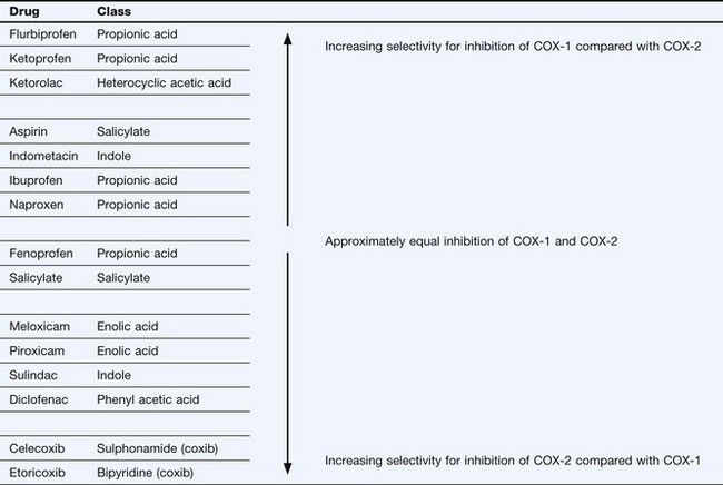
All NSAIDs share a common mode of action by inhibition of the COX isoenzymes (Table 29.3). Most NSAIDs bind reversibly to the site in the COX enzymes that accepts arachidonic acid, but the irreversible inactivation produced by aspirin (acetylsalicylic acid) involves acetylation of a serine residue in the enzyme. This mechanism of action is important in the use of aspirin as an antiplatelet drug (see below and Ch. 11).
Different NSAIDs inhibit the two main isoenzymes (COX-1 and COX-2) to varying extents. The degree of COX selectivity of each NSAID will also depend on the dosage used. The clinical responses and unwanted effect profiles of individual NSAIDs reflect the ability of the drug to inhibit the diverse biological actions of each isoenzyme (Tables 29.1 and 29.2). Highly selective inhibitors of COX-2 (coxibs) have been developed with the aim of reducing the unwanted effects associated with concomitant inhibition of COX-1 by the non-selective NSAIDs (see below). Neither the non-selective NSAIDs nor the selective COX-2 inhibitors directly affect the production of leukotrienes by 5-lipoxygenase. However, PGE2 normally inhibits leukotriene synthesis and reduced PGE2 generation as a consequence of NSAID use may increase leukotriene synthesis.
NSAIDs have additional anti-inflammatory effects that appear to be independent of COX inhibition. These effects may be mediated by actions on peroxisome proliferator-activated receptors (PPARs), particularly PPAR-γ. PPARs have key roles in modulating immune responses by suppressing transcription of pro-inflammatory genes, such as those encoding tumour necrosis factor α (TNFα), interleukin-1 (IL-1) and inducible nitric oxide synthase (iNOS) in macrophages. NSAIDs also have complex effects on lymphocytes, including inhibition of T-cell activation and increased T-cell apoptosis, which may reflect PPAR-mediated actions. NSAIDs may also modulate inflammatory gene transcription via actions on the nuclear transcription factor nuclear factor κB (NF-κB) and on other cell-signalling proteins including mitogen-activated protein (MAP) kinases. Some of these COX-independent actions only occur in vitro at NSAID concentrations higher than those required for anti-inflammatory activity or analgesia in vivo, and their clinical relevance remains to be determined.
Classification of NSAIDs
Table 29.3 shows the principal chemical types of NSAIDs with an indication of their selectivity in inhibiting COX-1 or COX-2. Many classical NSAIDs produce greater inhibition of COX-1 than of COX-2, although at clinical doses this selectivity may not be apparent and both isoenzymes will be effectively inhibited. The coxibs are highly selective inhibitors of COX-2 at clinical doses.
Actions and effects of non-selective NSAIDs
Some of the properties and actions of a selection of commonly used analgesic drugs are compared in Table 29.4.
Analgesic effect
The analgesic action of NSAIDs is in part a peripheral action at the site of pain and is most effective when the pain has an inflammatory origin (see Ch. 19). It is achieved predominantly through inhibition of COX-2-derived prostaglandins in inflamed or injured tissues. The main pain-inducing action of prostanoids is at sensory nerve endings on first-order neurons in peripheral tissues. PGE2 enhances the ability of several mediators (serotonin, substance P, bradykinin) to stimulate Aδ and C nociceptive fibres. There is also a CNS component to the analgesic action of NSAIDs which is due to inhibition of COX isoenzymes and reduction of prostaglandin production within the CNS pain pathways. COX inhibition decreases PGE2 production in the dorsal horn of the spinal cord, which inhibits neurotransmitter release and reduces the excitability of second-order neurons in the pain relay pathway. The analgesic action of NSAIDs is apparent after the first dose, but does not reach its maximal effect until about 1 week with repeated dosing.
Anti-inflammatory effect
Inhibition of vasodilation and oedema is partly related to a reduction in peripheral COX-2-generated prostaglandin synthesis. However, NSAIDs also affect several other inflammatory processes unrelated to their effects on prostaglandins. For example, they probably reduce harmful superoxide free radical generation by neutrophils. They may also uncouple G-protein-regulated processes in the cell membrane of inflammatory cells, which reduces their responsiveness to a variety of agonists released by damaged tissues. The anti-inflammatory effects of NSAIDs develop gradually over about 3 weeks.
Antipyretic effect
Fever is reduced through inhibition of hypothalamic COX-2. Circulating pyrogens such as interleukin-1 enhance PGE2 production in the hypothalamus, which depresses the response of temperature-sensitive neurons. NSAIDs do not affect normal body temperature.
Stay updated, free articles. Join our Telegram channel

Full access? Get Clinical Tree


