Non-Langerhans Cell Histiocytoses
Aaron Auerbach, MD, PhD
Key Facts
Terminology
Wide range of histiocytic disorders that are not derived from Langerhans cells
Sometimes difficult to categorize because of overlapping morphologic and clinical findings
Microscopic Pathology
Macrophages are large (15-80 µm in diameter) phagocytic cells with irregular shapes and pseudopodia
Indented nuclei, fine chromatin, abundant cytoplasm
Ancillary Tests
Macrophages are positive for CD14, CD68, CD163
Dermal/interstitial dendritic cells are positive for CD14, CD163, FXIIIA, fascin
Top Differential Diagnoses
Benign cephalic histiocytosis
Rare cutaneous condition in children characterized by skin lesions that initially present on the head and consist of histiocytes
Indeterminate cell histiocytosis
Features of macrophages, dendritic cells, and Langerhans cells, and is S100(+), CD68(+), CD1a(+)
Progressive nodular histiocytosis
Part of xanthogranuloma family of disease, but more clinically aggressive and disfiguring than others
Atypical granuloma annulare
May be either clinically atypical (associated with malignancies) or histologically atypical (more cellular, pleomorphic, and mitoses)
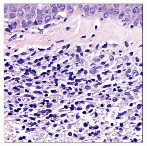 Indeterminate cell histiocytosis is a very rare histiocytosis composed of cells with histologic and immunohistochemical similarities to Langerhans cells, but lacking Birbeck granules by electron microscopy. |
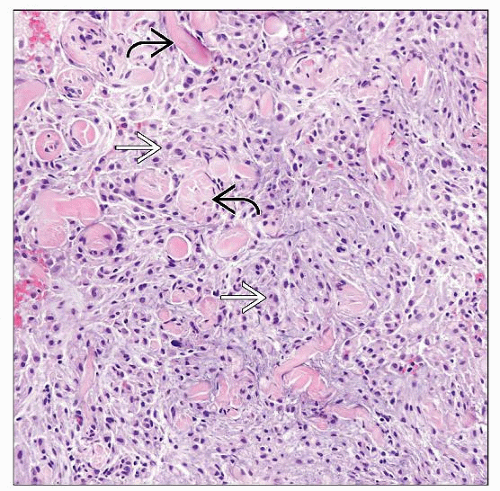 Hereditary progressive mucinous histiocytosis shows a dermal proliferation composed of epithelioid histiocytes  and mucin separating broad bundles of collagen and mucin separating broad bundles of collagen  . . |
TERMINOLOGY
Synonyms
Non-X histiocytoses
Definitions
Wide range of histiocytic disorders that are not derived from Langerhans cells
Sometimes difficult to categorize because of overlapping morphologic and clinical findings
Includes juvenile xanthogranuloma, reticulohistiocytoma, and Rosai-Dorfman disease
Histiocyte = bone marrow-derived cell belonging to monocytes/macrophage &/or dendritic cell lineage and functional variants
Includes macrophages, Langerhans cells, interstitial dendritic cells, interdigitating dendritic cells, plasmacytoid dendritic cells, microglia, Kupffer cells, and alveolar macrophages
CLINICAL ISSUES
Epidemiology
Age
Depends on type of histiocytosis
Treatment
Benign tumors that do not require treatment in most cases
Prognosis
Depends on type of histiocytosis
MICROSCOPIC PATHOLOGY
Histologic Features
Macrophages are large (15-25 µm in diameter) phagocytic cells with irregular shapes and pseudopodia
Nucleus is round, but may be indented or reniform
Nuclear membrane is indistinct, and chromatin is fine
Cytoplasm is abundant and can be granulated
Cytoplasmic vacuoles may be seen, and phagocytosed material may be present
Other features depend on the type of histiocytosis
ANCILLARY TESTS
Immunohistochemistry
Depends on type of histiocytosis
Macrophages are typically positive for CD14, CD68, and CD163
Dermal/interstitial dendritic cells are positive for FXIIIA, CD14, CD163, and fascin
Indeterminate cells are S100(+), CD1a(+), FXIIIA(+)
Electron Microscopy
Non-Langerhans cell histiocytoses do not have Birbeck granules by ultrastructural studies, unlike Langerhans cell histiocytosis (LCH)
DIFFERENTIAL DIAGNOSIS
Benign Cephalic Histiocytosis
Rare histiocytosis characterized by asymptomatic, self-healing reddish to brownish macules and papules on the head, which spread later to trunk and arms
Begins in children ≤ 3 years old
Morphology
Dermal histiocytes, fairly small with round nuclei
± vacuolated cytoplasm
Positive for CD68, CD11a, and CD11c; negative for S100 and CD1a
Patterns include papillary dermal, lichenoid, and diffuse
Usually no epidermotropism
Electron microscopy shows comma-shaped bodies, coated vesicles, and desmosome-like structures with absence of Birbeck granules
Generalized Eruptive Histiocytoma
Rare benign histiocytosis characterized by crops of hundreds of blue-red papules, affecting mainly adults, and self-healing
Symmetrically distributed on trunk and extremities
May become hyperpigmented macules when they regress
Clinical course
Usually self-limiting; may last from 1 month to over 12 years
May persist, may resolve, or may relapse
Sometimes associated with underlying tumors
Macules/papules may regress when underlying malignancy is treated
Morphology
Histiocytes in upper dermis
Sometimes small and vacuolated cytoplasm
± perivascular distribution
Usually no multinucleated giant cells
Older lesions may show fibrosis or giant cells
CD68(+), S100(-), CD1a(-)
Electron microscopy shows dense bodies with myelin, but no Birbeck granules
Indeterminate Cell Histiocytosis (ICH)
Dendritic cells in dermis with features of histiocytes and Langerhans cells but no Birbeck granules
Only few cases reported
Presentation
Numerous red/brown papules
May coalesce
Morphology
Monomorphous infiltrate of mononuclear histiocytes intermixed with clusters of lymphocytes
Rarely multinucleated cells
Often increased numbers of reactive T cells interspersed among histiocytic cells
Usually ample pale cytoplasm and clefted nuclei
Rare spindle cell variant has been described
Stroma can be myxoid
Immunohistochemistry
S100(+), CD68(+), CD1a(+), FXIIIA(±)
Sometimes look like Langerhans cells with ample cytoplasm and grooves
But lack some Langerhans markers such as Langerin and Birbeck granules
Progressive Nodular Histiocytosis
Normolipemic non-Langerhans cells histiocytosis with multiple yellow-brown papules and nodules on skin and mucous membranes; part of xanthomatous family of lesions
Nodules mostly on trunk
Rarely systemic lesions
Stay updated, free articles. Join our Telegram channel

Full access? Get Clinical Tree


