Neurophysiology
AUTONOMIC NERVOUS SYSTEM
What are the two structural divisions of the human nervous system?
- Central nervous system
- Peripheral nervous system
What are the two functional divisions of the human nervous system?
- Somatic nervous system
- Autonomic nervous system (ANS)
What is the function of the somatic nervous system?
Innervates skeletal muscle; largely under voluntary control
What is the function of the ANS?
Maintenance of homeostasis through involuntary coordination of glandular, cardiac, and smooth muscle activity throughout the body
What are the constituent parts of the ANS?
Enteric nervous system, sympathetic nervous system (SNS), and parasympathetic nervous system (PNS)
Generally speaking, what is the function of each part of the ANS?
Enteric nervous system
Coordination of various gut functions (motility, secretion, etc) (see Chap. 6)
Sympathetic nervous system
“Fight or flight” response. Coordinates the body’s response to stressors.
Parasympathetic nervous system
“Rest and digest” response. Coordinates the process of energy conservation and replenishment.
Where is the anatomic origin of SNS?
Intermediolateral cell column of the spinal cord between segments T1 to L3 (e.g., thoracolumbar)
Where is the anatomic origin of the PNS?
Nuclei of cranial nerves (CN) III, VII, IX, and X and spinal cord segments S2 to S4 (e.g., craniosacral spinal cord)
What type(s) of neurotransmitter(s) and receptors are present in the preganglionic fibers of the PNS and SNS?
PNS and SNS preganglionic neurons use ACh as the neurotransmitter and nicotinic receptors for transmission
Is the neurotransmitter/receptor combination used at the preganglionic synapse the same as that used at the neuromuscular junction (NMJ)?
No, while these receptors are nAChR (nicotinic Acetylcholine receptors) they differ from those at the NMJ. The nicotinic subunits here have a different subunit makeup allowing for differences in pharmacologic influences
Describe the axon length for the PNS and SNS in the following locations:
PNS
SNS
Preganglionic nerve axon
Long
Short
Postganglionic nerve axon
Short
Long
Anatomically, where do most preganglionic sympathetic fibers synapse?
Either at the paravertebral chain ganglion, or at collateral ganglia that follow large vessels in the abdomen (celiac, superior mesenteric, etc)
Anatomically, where do most preganglionic parasympathetic fibers synapse?
In microscopic ganglia associated with the target organs
What receptor types are primarily used at the effector organs of the PNS and SNS? What are their respective neurotransmitters?
PNS: all receptors are muscarinic and ACh is the neurotransmitter
SNS: α1, α2,, β1, or β2; the primary neurotransmitter is norepinephrine (NE)
There is an exception to the above rule regarding the SNS. What is it?
Sweat glands have muscarinic receptors. The associated postganglionic SNS neurons release ACh.
What is unique about the adrenal medulla?
It is a specialized SNS ganglion where preganglionic fibers synapse directly with the effector organ (chromaffin cells)
What does SNS stimulation of chromaffin cells induce?
Secretion of epinephrine (Epi; 80%) and NE (20%) into the circulation
What is the purpose of releasing adrenal hormones into the circulation?
These substances activate organs that receive little innervation (fat cells, hepatocytes etc) allowing them to assist in the stress response
What are the SNS receptor types in the following locations and what are their effects?
Heart
SA nodeα
β1: increased pacemaker activity
AV node
β1: increased conduction velocity
Myocardium
β1: increased contractility
Vascular smooth muscle
Skin and splanchnic circuits
α1 and α2: constriction
Skeletal muscle and pulmonary circuits
β2: dilation
Peripheral veins
Eye
Ciliary muscle
β1: relaxes muscle (for far vision)
Radial muscle
α1: muscle contraction → dilates pupil
Bladder
Detrusor muscle
β2: relaxes
Bladder sphincter
α1: constricts
Bronchioles
Bronchial muscle
β2: dilates smooth muscle
Bronchial glands
α1: inhibits secretion
β2: stimulates secretion
Gastrointestinal tract
Sphincters
α1: constricts
Secretion
α2: inhibition
Motility
α1, α2, β2: decrease
Kidney
β1: increase renin secretion
Male sex organs
α1: ejaculation
Sweat glands
Muscarinic: increase sweat production
Adipose tissue
β1, β3: increase lipolysis
For which of the above SNS locations is there no corresponding PNS innervation?
Vascular smooth muscle, kidney, sweat glands, fat cells, and liver
What is the mechanism of action for the following receptor types?
α1
Inositol 1,4,5-triphosphate (IP3) formation and increased intracellular [Ca2+]
α2
Adenylate cyclase inhibition and decreased cyclic adenosine monophosphate (cAMP)
β1
Adenylate cyclase activation and increased cAMP
β2
Adenylate cyclase activation and increased cAMP
Nicotinic
Ion channel for Na+ and K+
Muscarinic
Heart (sinoatrial [SA] node): adenylate cyclase inhibition
Smooth muscle and glands: IP3 formation and increased intracellular [Ca+]
What is the effect on the ANS of the following pharmacologic agents?
ACh
Nicotinic and muscarinic agonist
Albuterol
β2 Agonist
Atropine
Muscarinic antagonist
Butoxamine
β2 Antagonist
Carbachol
Nicotinic and muscarinic agonist
Clonidine
α2 Agonist
Curare
Nicotinic antagonist
Dobutamine
β1 Agonist
Hexamethonium
Nicotinic antagonist (ganglion only)
Isoproterenol
β 1 and β2 agonist
Metoprolol (at therapeutic doses)
β1 Antagonist
Muscarine
Muscarinic agonist
Nicotine
Nicotinic agonist
NE
α1 and β1 agonist
Phenoxybenzamine
α12 Antagonist
Phentolamine
α12 Antagonist
Phenylephrine
α1 Agonist
Prazosin
α1 Antagonist
Propranolol
β1 and β2 antagonist
Yohimbine
α2 Antagonist
What autonomic centers are located in the following areas?
Medulla
Respiratory; swallowing, coughing, and vomiting; and vasomotor
Pons
Pneumotaxic
Midbrain
Micturition
Hypothalamus
Regulation of food and liquid intake and temperature regulation
SENSORY SYSTEMS
What four qualities of sensation must be encoded for effective transmission?
- Modality
- Location
- Intensity
- Duration
What is a sensory receptor?
A specialized cell that transduces physical environmental stimuli into neural signals
What types of cells are usually sensory receptors?
Specialized epithelial and neuronal cells
What is meant by “receptive field”?
Area on the body that changes the firing rate of its sensory neuron when stimulated
What is the field called if it increases the firing rate?
Excitatory
What is the field called if it decreases the firing rate?
Inhibitory
A sensory receptor must transmit its findings back to the CNS for interpretation. How does the CNS differentiate between an AP (action potential) from a retinal cell and an AP from a somatosensory cell?
The receptors are simple transducers, each converting a different type of energy into electrical impulses. These impulses follow defined pathways into the CNS and relay on specific nuclei. These pathways define how sensory perception is recognized.
For an example of this, think about rubbing your eyes. That pressure is always perceived as flashes of light.
How are afferent neurons of the sensory systems classified?
By diameter (roman numerals I-IV) and conduction velocity (A and C)
Give the relative size of the following sensory neuron classifications:
I
Largest
II
Medium
III
Small
IV
Smallest
Give the relative conduction velocity of the following sensory neuron classifications:
Aα
Fastest
Aβ
Medium
Aδ
Medium
C
Slowest
Describe the series of events that occur in sensory transduction.
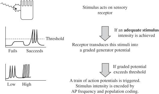
Figure 2.1 Sensory transduction sequence.
What effect does intensity of the stimulus have on the receptor potential generated?
Larger stimuli create larger graded potentials (e.g., receptor potential)
What direction does the current usually flow when sensory receptor channels open?
Positive inward flow, depolarizing the cell
What is an exception to this flow direction?
Photoreceptors: stimulation decreases inward current and hyperpolarizes the membrane
As seen in Fig. 2.1, how does the body encode intensity changes in sensory stimuli?
Primarily with frequency coding; greater intensity leads to a higher frequency. Also with population coding; a greater number of receptors will be triggered by a stimulus of increasing intensity.
What is sensory adaptation?
The dynamic change in the frequency of triggered AP’s with a static stimuli; this occurs at the level of the receptor
What types of adaptation do sensory receptors exhibit?
Tonic (slowly adapting) and phasic (rapidly adapting)
Which type of sensory adaptation detects the beginning of (onset) and the end of (offset) of a stimulus?
Phasic
What type of sensory adaptation responds consistently to prolonged stimuli?
Tonic
What type of sensory adaptation detects steady stimuli?
Tonic
What happens to action potential frequency in phasic receptors with constant stimulation?
Decreases
Label the three types of adaptation seen below:
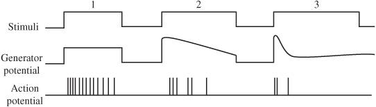
Figure 2.2 Adaptation.
- No Adaptation, also called tonic adaptation
- Slowly adapting sensory neurons
- Rapidly adapting sensory neurons, also called phasic adaptation
What is the location of the following structures?
First-order neurons
Dorsal root ganglia
Second-order neurons
Spinal cord or brain stem (depending on system)
Third-order neurons
Thalamus (relay nuclei)
What goes on beyond the relay nucleus?
Extensively overlapping cortical circuitry that interprets external stimuli and relays information throughout the cortex
Somatosensory System
What sensations are perceived by the somatosensory system?
Touch, movement, and position, as well as temperature, and pain
What two anatomical pathways does the somatosensory system use?
- Dorsal-column system
- Anterolateral system
What sensations are detected by the following systems?
Dorsal-column system
Touch, vibration, and proprioception
Anterolateral system
Temperature and pain
What path does sensory information take in the dorsal-column system?
Receptors with cell bodies in the
dorsal root ganglia receive stimulus (first order)
↓
Signal ascends to the nucleus gracilis
and nucleus cuneatus in medulla
(still first order)
↓
Signal crosses the midline and
ascends to enter contralateral
thalamus (second order)
↓
Signal ascends to the somatosensory
cortex (third order)
What path does sensory information take in the anterolateral system?
Receptors with cell bodies in the
dorsal root ganglia receive stimulus
(first order)
↓
Signal crosses the midline and enters
the anterolateral quadrant of the
spinal cord (second order)
↓
Signal ascends to contralateral thalamus
(still second order)
↓
Signal sent to somatosensory cortex
(third order)
What are the major differences between the two systems with respect to the anatomy of the ascending information?
Dorsal columns don’t cross midline until the medulla; anterolateral system crosses at the level of the peripheral nerve
Ascending information in dorsal columns carried by first-order neuron; in the anterolateral system, it is carried by the second-order neuron
What are the tracts of Lissauer?
These are found at the dorsal root where the anterolateral system enters the spinal cord. They allow for fibers to move vertically one or two segments before synapsing.
What is another name for the sensory cortex?
Sensory homunculus
How is the homunculus arranged?
Upside down (face most lateral, with feet and genitals inside the central sulcus)
Why are the face, hands, and genital regions of the homunculus so large?
They possess the highest density of nerve fibers
What are the types of mechanoreceptors that detect touch and pressure?
Meissner corpuscle
Merkel disk
Pacinian corpuscle
Ruffini corpuscle
What sensation is encoded by the following mechanoreceptors?
Meissner corpuscle
Velocity
Merkel disk
Pressure-small receptive field
Pacinian corpuscle
Vibration
Ruffini corpuscle
Pressure-large receptive field
Which types of mechanoreceptors demonstrate phasic adaptation?
Meissner and Pacinian corpuscles
Which types of mechanoreceptors demonstrate tonic adaptation?
Merkel disks and Ruffini corpuscles
What is nociception?
Detection and perception of noxious stimuli (e.g., pain)
What types of receptors detect pain?
No specialized receptors; pain is detected by free nerve endings
Any excessive application of energy (chemical, thermal, or mechanical)
Where are pain receptors located?
Skin, muscle, and viscera
What two pathways does visceral pain stimulate?
- Visceral afferent fibers ascend with sympathetic nerves
- Referred to the skin in a dermatomal fashion
Explain referred pain:
Afferent pain fibers serving the viscera enter the spinal cord at particular levels. Since the body is not used to visceral pain, it confuses those stimuli with dermatomal pain from the same spinal root.
What fibers carry fast pain signals?
Group III
What fibers carry slow pain signals?
C fibers
Opiates inhibit the release of what neurotransmitter for nociception?
Substance P
VISION
In the image below, label the numbered portions of the eye.
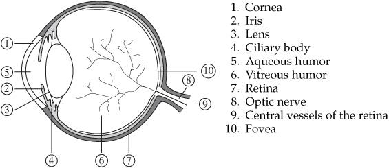
Figure 2.3 The eye.
- Cornea
- Iris
- Lens
- Ciliary body
- Aqueous humor
- Vitreous humor
- Retina
- Optic nerve
- Central vessels of the retina
- Fovea
What is the function of the lens?
Focuses light onto the retina
What is the condition called when the curvature of the lens is not uniform?
Astigmatism
What structure produces aqueous humor?
Ciliary epithelium covering the surface of the ciliary body
How does aqueous humor drain?
Via the canal of Schlemm
What conditions are described by the following?
Lens focuses light onto the retina
Emmetropia (this is normal)
Lens focuses light in front of the retina
Myopia (nearsighted)
Lens focuses light behind the retina
Hyperopia (farsighted)
What is accommodation?
Focusing of light by the lens
How does accommodation occur?
The smooth muscle of the ciliary body contracts to focus the lens
What is presbyopia?
Loss of accommodation due to stiffening of the lens
Identify the labeled cell types of the retina in the image below.
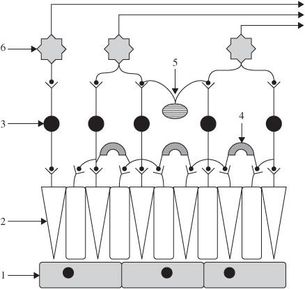
Stay updated, free articles. Join our Telegram channel

Full access? Get Clinical Tree


