FIGURE 20-1
(A) Anterior horn of spinal cord
(B) Dentate nucleus
(C) Nucleus basalis of Meynert
(D) Red nucleus
(E) Substantia nigra
2. Creutzfeldt cells, a form of reactive astrocytes with abundant cytoplasm and fragmented nuclear material, are most likely to be encountered in which of the following settings?
(A) Demyelinating disease
(B) Diffuse astrocytomas
(C) Elevated ammonia levels
(D) Radiation therapy
(E) Viral encephalitis
3. Microglial cells are best highlighted by which of the following antibodies?
(A) CD3
(B) CD48
(C) CD68
(D) CD138
(E) GFAP
4. Herring bodies in the pituitary gland are filled with neurosecretory granules containing which of the following?
(A) Adrenocorticotropic hormone
(B) Growth hormone
(C) Prolactin
(D) Thyroid stimulating hormone
(E) Vasopressin
5. All of the following are neuronal neuropathologic features of normal aging except
(A) Corpora amylacea
(B) Ferrugination
(C) Lipofuscin accumulation
(D) Marinesco bodies
(E) Neurofibrillary tangles
6. What percent of the total cardiac output circulates to the brain?
(A) 5%
(B) 15%
(C) 25%
(D) 35%
(E) 50%
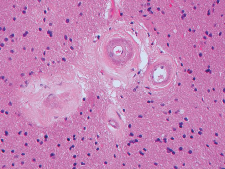
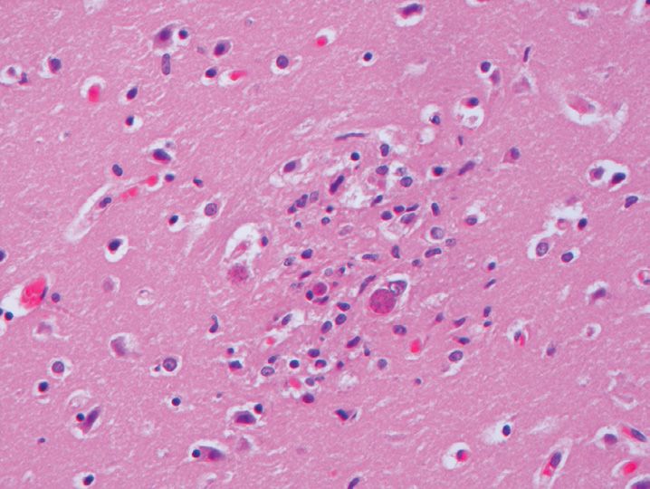
7. This section (Figure 20-2) was taken from an autopsy in a 72-year-old woman who had stagnant hypoxia for the last 24 hours of her life. A section from which part of the brain would contain neurons that are the most susceptible to anoxic damage?
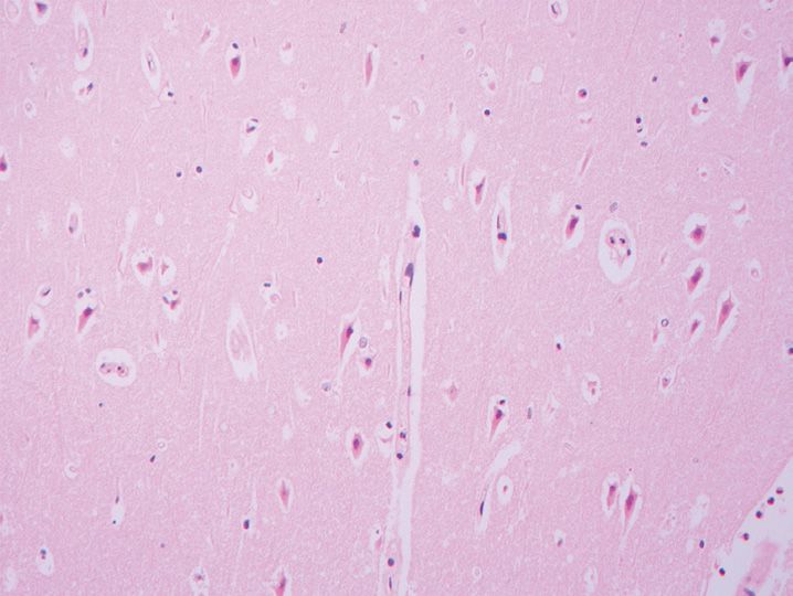
(A) Amygdala
(B) Basal ganglia
(C) Hippocampus
(D) Mamillary bodies
(E) Nucleus basalis of Meynert
8. Which of the following cells are most sensitive to ischemic damage?
(A) Astrocytes
(B) Endothelial cells
(C) Meningothelial (arachnoidal cap) cells
(D) Neurons
(E) Oligodendrocytes
9. The earliest light microscopic evidence of acute hypoxic damage is typically observed how many hours after the hypoxic event?
(A) <2 hours
(B) 3–4 hours
(C) 5–7 hours
(D) 9–11 hours
(E) 13–15 hours
10. An acute infarct involving the occipital lobe visual cortex would be most likely due to an occlusion in which artery?
(A) Anterior cerebral artery
(B) Anterior inferior cerebellar artery
(C) Middle cerebral artery
(D) Posterior cerebral artery
(E) Superior cerebellar artery
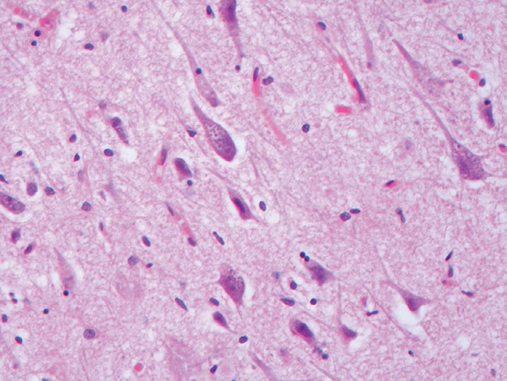
11. The most common location for an acute hypertensive hemorrhage is which of the following locations?
(A) Basal ganglia
(B) Cerebellum
(C) Frontal lobe
(D) Parietal lobe
(E) Pons
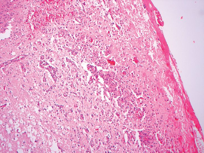
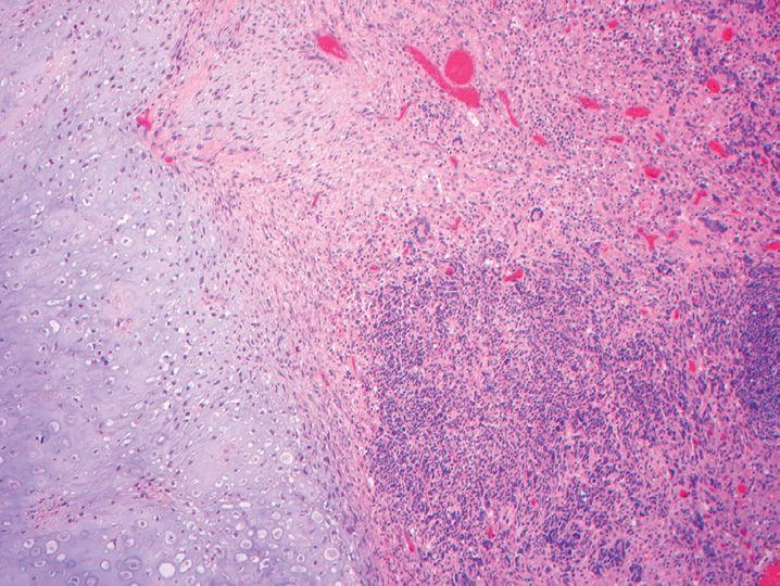
12. The lesion shown in Figure 20-3 was found at autopsy in a 69-year-old man. The lesion was diagnosed as an infarct. The best approximation of the age of this lesion would be which of the following?
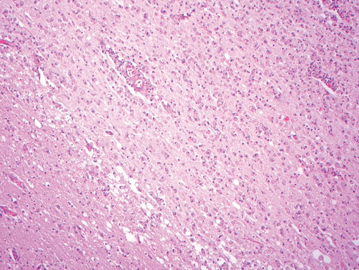
(A) 10–12 hours
(B) 3 days
(C) 1 week
(D) 3 weeks
(E) >3 months
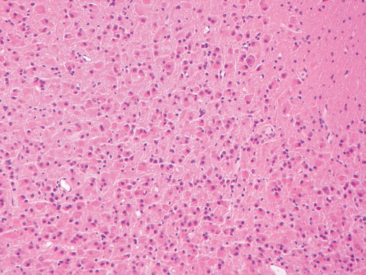
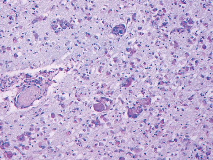
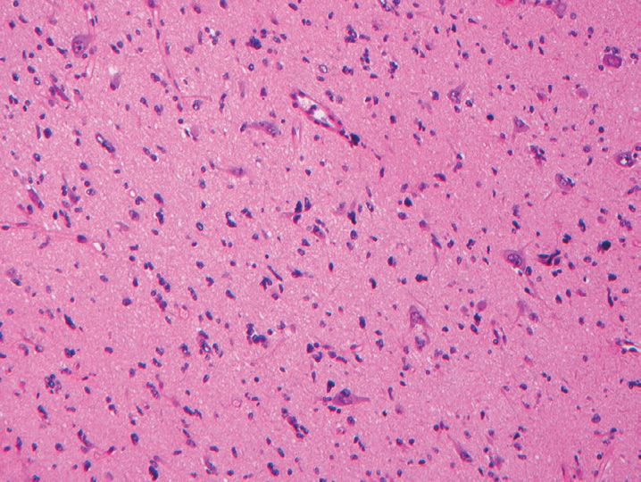
13. The abnormality illustrated (Figure 20-4) in this section from the frontal lobe in a 22-year-old man represents which of the following?
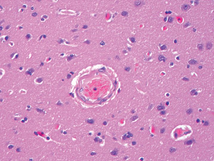
(A) Alzheimer type II astrocytes
(B) Anoxic changes
(C) Microglial cell proliferation
(D) No abnormality is present
(E) Red cell disorder
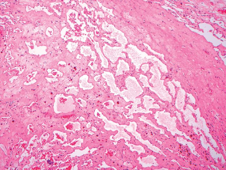
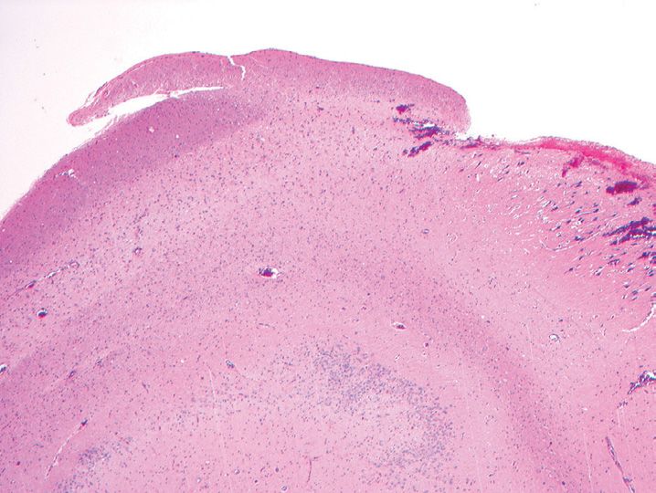
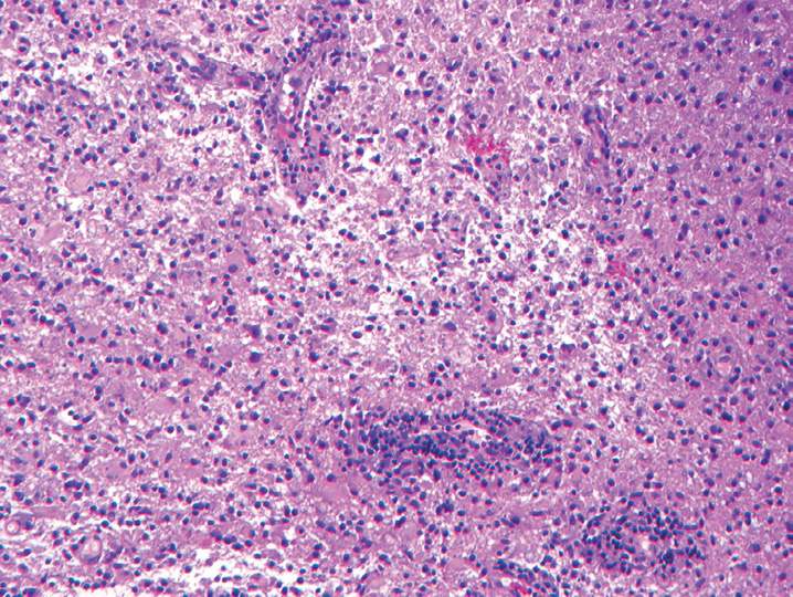
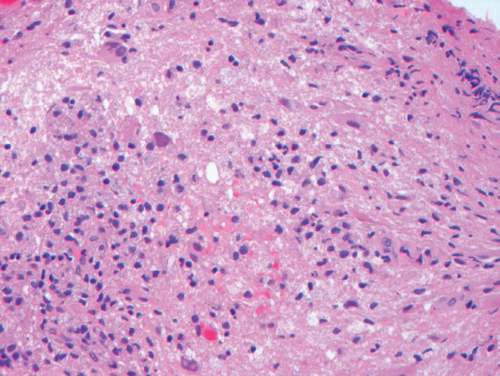
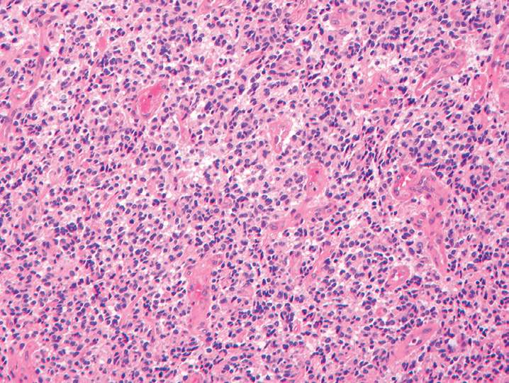
14. The most common location for the lesion shown in Figure 20-5 is which of the following arterial branch points?
(A) Anterior cerebral-anterior communicating arteries
(B) Basilar-posterior cerebral arteries
(C) Internal carotid-posterior communicating arteries
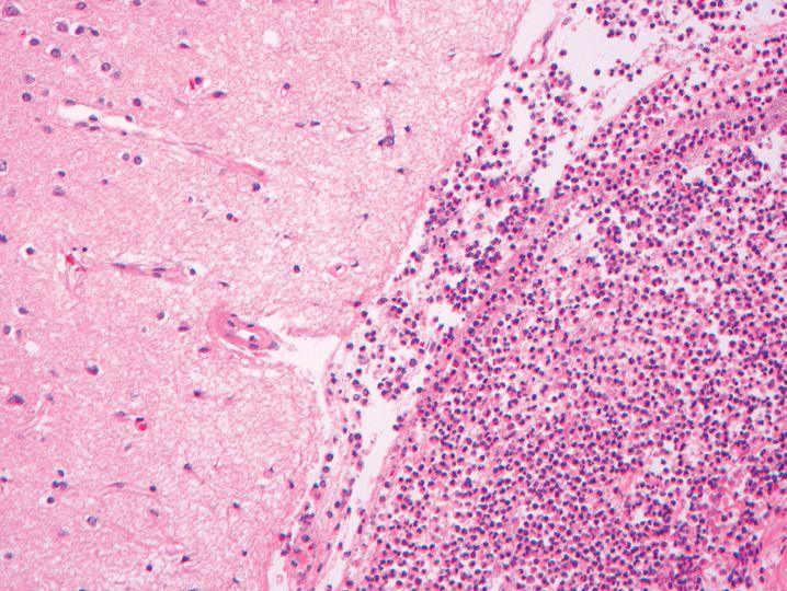
(D) M1-M2 division of the middle cerebral artery
(E) Vertebral-basilar arteries
15. The most common cause for dolichoectasia is which of the following?
(A) Amyloid
(B) Atherosclerosis
(C) Moyamoya syndrome
(D) Mycotic aneurysm
(E) Subarachnoid hemorrhage
16. All of the following lesions are associated with a circle of Willis berry aneurysm except
(A) Arterial fibromuscular dysplasia
(B) Coarctation of the aorta
(C) Hemangioblastoma
(D) Marfan disease
(E) Polycystic kidney disease
17. Most arteriovenous malformations occur within which of the following vascular distributions?
(A) Anterior cerebral artery
(B) Anterior choroidal artery
(C) Middle cerebral artery
(D) Posterior cerebral artery
(E) Superior cerebellar artery
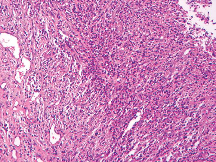
18. The arteriovenous malformation resected (Figure 20-6) here is located where?
(A) Brain stem
(B) Cannot tell
(C) Caudate or putamen
(D) Cerebellum
(E) Hippocampus
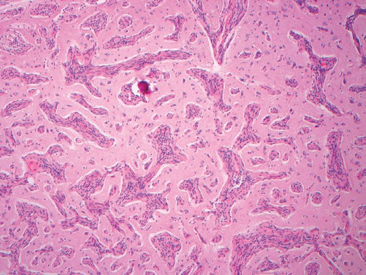
19. All of the following are true regarding the lesion illustrated in Figure 20-7 except
(A) About one-third of patients present with focal epilepsy
(B) Annual risk of hemorrhage exceeds 5%
(C) More common in men
(D) More common in young versus old adults
(E) Most are supratentorial
20. Familial forms of cavernous angioma are associated with mutations of the CCM1 gene on chromosome 7q 11-21, which encodes for which of the following?
(A) Beta-1-integrin
(B) GTPase
(C) ICAP-1
(D) KRIT1
(E) RAS
21. All of the following proteins are potentially associated with cerebral amyloid angiopathy except
(A) Abeta-amyloid peptide
(B) Beta-2-microglobulin
(C) Cystatin C
(D) Prion protein
(E) Transthyretin
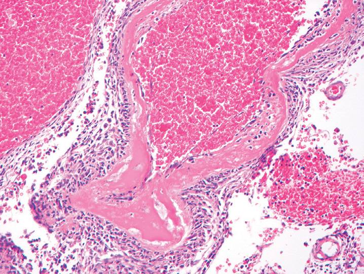
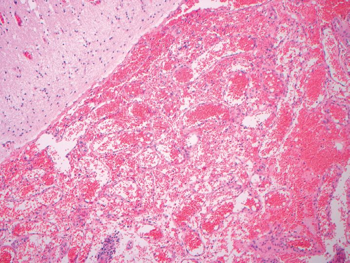
22. A 62-year-old patient presents with a large bleed. Evacuation of the clot with some adjacent brain parenchyma was performed. The blood vessels show thioflavin-S staining. The most likely location of the bleed is which of the following? (Figure 20-8)
(A) Basal ganglia
(B) Frontal lobe
(C) Occipital lobe
(D) Parietal lobe
(E) Temporal lobe
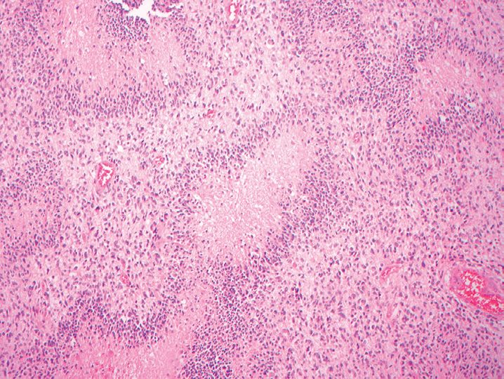
23. The vasculitic pattern of injury shown (Figure 20-9) in the brain is most likely associated with which of the following processes?
(A) Amyloid-associated vasculitis
(B) Giant cell arteritis
(C) Lymphoproliferative disorder
(D) Polyarteritis nodosa
(E) Primary angiitis of the central nervous system
24. The changes shown in Figure 20-10 are best diagnosed as which of the following?
(A) Amyloid angiopathy
(B) Atherosclerotic disease
(C) Binswanger disease
(D) CADASIL (cerebral autosomal-dominant arteriopathy with subcortical infarcts and leukoencephalopathy)
(E) MELAS (mitochondrial encephalomyopathy with lactic acidosis and stroke-like episodes)
25. All are true regarding CADASIL except
(A) Associated with seizures in most patients
(B) Associated with white matter infarcts
(C) Mean age of onset 40–50 years
(D) NOTCH 3 gene mutations
(E) Systemic disorder
26. The most common source of blood in an epidural hematoma associated with skull fracture is
(A) Bridging vein
(B) Dural sinus
(C) Middle meningeal artery
(D) Middle meningeal vein
(E) None of the above
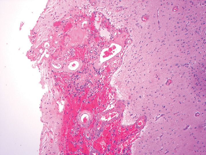
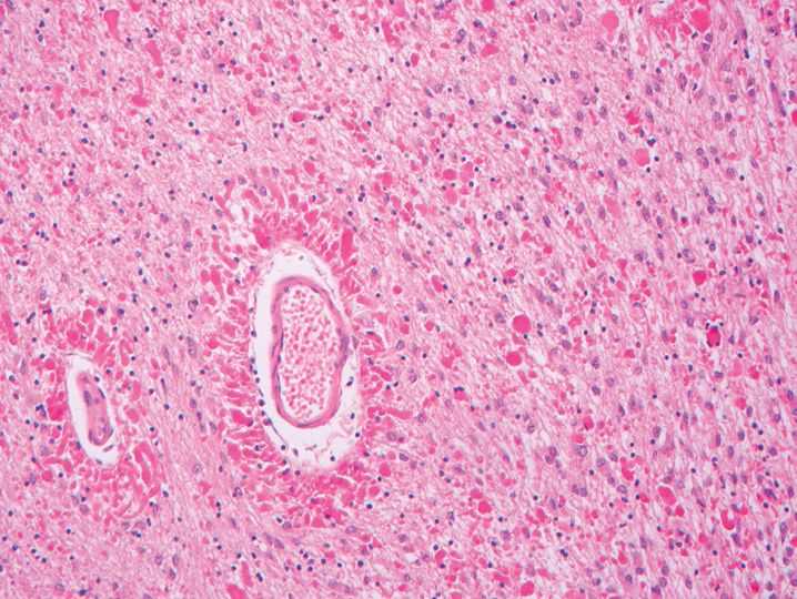
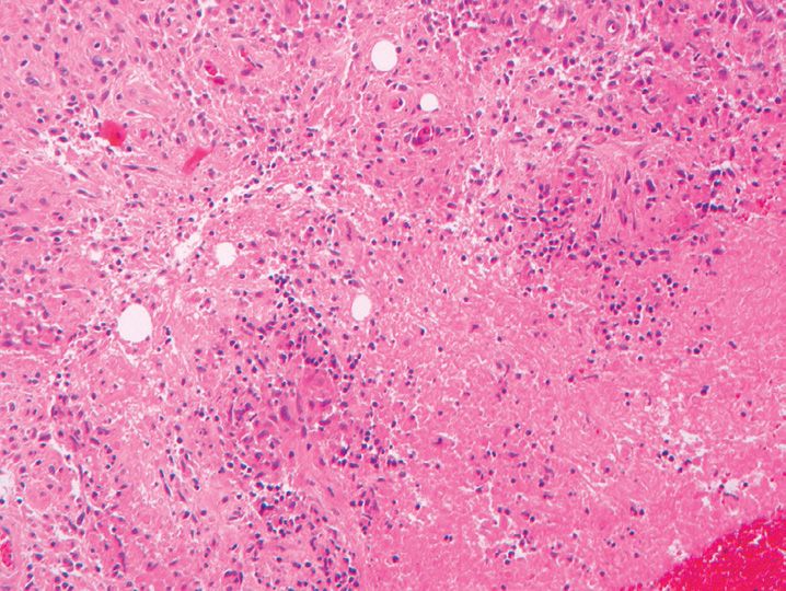
27. The most likely etiology of the lesion shown in Figure 20-11 in the temporal lobe of a 82-year-old man is which of the following?
(A) Carbon monoxide poisoning
(B) Embolism
(C) Infection
(D) Trauma
(E) Tumor
28. Which of the following stains is useful in highlighting dystrophic axons in diffuse axonal injury after about 2 hours survival time?
(A) Beta-amyloid precursor protein
(B) NeuN
(C) S-100 protein
(D) Synaptophysin
(E) Tau protein
29. All of the following are causes of microcephaly except
(A) Achondroplasia
(B) Down syndrome
(C) Maternal alcohol abuse
(D) Prenatal radiation
(E) Prenatal toxoplasmosis infection
30. All of the following are causes of secondary megalencephaly except
(A) Alexander disease
(B) Canavan disease
(C) Maternal anticonvulsant therapy
(D) Mucopolysaccharidoses
(E) Sphingolipidoses
31. Potential risk factors for the development of neural tube closure defects include all of the following except
(A) Anticonvulsant therapy
(B) Hypothermia
(C) Maternal diabetes
(D) Deletions on chromosome 22q11
(E) Vitamin deficiency
32. Which of the following is most associated with the development of holoprosencephaly?
(A) Klinefelter syndrome
(B) Monosomy 18
(C) Trisomy 13
(D) Trisomy 21
(E) Turner syndrome
33. Which of the following features is not associated with holoprosencephaly?
(A) Absent ears
(B) Cleft lip and palate
(C) Cyclopia
(D) Hypotelorism
(E) Proboscis
34. All of the following are gross manifestations of focal cortical dysplasia or malformations of cortical development except
(A) Cortical tuber
(B) Alexander disease
(C) Lissencephaly
(D) Pachygyria
(E) Polymicrogyria
35. All of the following are associated with a Dandy–Walker malformation except
(A) Agenesis of the cerebellar vermis
(B) Association with congenital heart disease
(C) Associated with maternal diabetes
(D) Cystic dilatation of the fourth ventricle
(E) Hydrocephalus
36. The cerebellar lesion shown in Figure 20-12 was excised in a 22-year-old patient with headaches. This lesion is associated with which of the following?
(A) Cowden syndrome
(B) Joubert syndrome
(C) Neurofibromatosis type 1
(D) Tuberous sclerosis
(E) von Hippel–Lindau syndrome
37. Which of the following features is most characteristic of a Chiari type I malformation?
(A) Cerebellar polymicrogyria
(B) Cerebellar tonsillar displacement below the foramen magnum
(C) Hydrocephalus
(D) Myelomeningocele
(E) Platybasia
38. The changes seen in ulegyria are usually attributable to which of the following?
(A) Germinal matrix bleed
(B) Infection
(C) Ischemic or hypoxic damage
(D) Malformation of development
(E) Trauma
39. The highest risk period for the development of periventricular leukomalacia is in infants delivered at how many weeks postconception?
(A) 26–30 weeks
(B) 30–34 weeks
(C) 34–36 weeks
(D) 40–44 weeks
(E) After the 1st month of life
40. A germinal matrix bleed that ruptures into the ventricles without expansion of the ventricles is classified radiologically as which grade?
(A) Grade I
(B) Grade II
(C) Grade III
(D) Grade IV
(E) Grade V
41. A 26-year-old woman presents with a 16-year history of medically intractable seizures. The patient undergoes a temporal lobe resection. The findings shown (Figure 20-13) in the hippocampal region suggest which of the following?
(A) Double dentate nucleus
(B) Focal cortical dysplasia
(C) Ganglioglioma
(D) Hippocampal sclerosis
(E) Trisomy 21
42. All of the following brain findings are associated with Down syndrome except
(A) An atrophic superior temporal gyrus
(B) A subependymal giant cell astrocytoma
(C) Cerebellar atrophy
(D) Foreshortening of the frontal lobe
(E) Increased risk of Alzheimer disease
43. All of the features are major pathologic criteria for the diagnosis of neurofibromatosis type 1 except
(A) Axillary freckling
(B) Optic nerve glioma
(C) Plexiform neurofibroma
(D) Six or more café-au-lait macules
(E) Two vestibular schwannomas
44. The lesion shown in Figure 20-14 is most commonly associated with which of the following?
(A) Neurofibromatosis type 1
(B) Neurofibromatosis type 2
(C) Sturge–Weber syndrome
(D) Tuberous sclerosis
(E) von Hippel–Lindau syndrome
45. All of the following are associated with neurofibromatosis type 2 except
(A) Autosomal-dominant inheritance
(B) Meningioma
(C) Mutated merlin protein
(D) Pheochromocytoma
(E) Posterior subcapsular lens opacity
46. All of the following features are associated with tuberous sclerosis except
(A) Renal angiomyolipoma
(B) Renal cell carcinoma
(C) Shagreen patch
(D) Subependymal giant cell astrocytoma
(E) Subungual fibroma
47. The VHL gene for von Hippel–Lindau disease is located on which of the following chromosomes?
(A) 1
(B) 3
(C) 11
(D) 17
(E) 22
48. The lesion illustrated in Figure 20-15 represents a section from the meninges of a 6-year-old patient with refractory seizures. The most likely diagnosis for the patient based on the pathology is which of the following?
(A) Ataxia-telangiectasia
(B) Neurofibromatosis type 1
(C) Sturge–Weber syndrome
(D) Tuberous sclerosis
(E) von Hippel–Lindau disease
49. A defect in the PTEN/MMAC1 gene on chromosome 10q23 is associated with all of the following lesions except
(A) Breast carcinoma
(B) Dysplastic gangliocytoma of the cerebellum
(C) Pituitary adenoma
(D) Trichilemmoma
(E) Thyroid nodules
50. All of the following tumors may be encountered in patients with Li–Fraumeni syndrome except
(A) Astrocytoma
(B) Choroid plexus papilloma
(C) Ganglioglioma
(D) Medulloblastoma
(E) Meningioma
51. All of the following central nervous system findings have been noted in Gorlin syndrome except
(A) Agenesis of the corpus callosum
(B) Glioblastoma
(C) Macrocephaly
(D) Medulloblastoma
(E) Meningioma
52. Which of the following disorders is X-linked and caused by a defect in the adenosine 5’-triphosphate-binding cassette transporter (ABCD1) gene, resulting in an accumulation of long-chain fatty acids?
(A) Adrenoleukodystrophy
(B) Canavan disease
(C) Krabbe disease
(D) Metachromatic leukodystrophy
(E) Pelizaeus–Merzbacher disease
53. All of the following are true regarding metachromatic leukodystrophy except
(A) Adult form presents with psychiatric changes
(B) Autosomal-dominant condition
(C) Deficiency of arylsulfatase A
(D) Preferentially involves the frontal lobes
(E) Spares the subcortical U-fibers
54. The lesion shown in Figure 20-16 is from the brain of an 8-month-old girl who presented with hyperirritability, limb stiffness, and weight loss. Imaging shows confluent and symmetrical periventricular lesions. The best diagnosis for this lesion is which of the following?
(A) Alexander disease
(B) Krabbe disease
(C) Metachromatic leukodystrophy
(D) Multiple infarcts
(E) Multiple sclerosis
55. The lesion illustrated in Figure 20-17 presented at age 2 with developmental delay, seizures, and psychomotor retardation. Which of the following is the underlying defect in this patient?
(A) Deficiency of aspartoacylase
(B) Deficiency of galactosylceramidase
(C) Mutation in the GFAP gene
(D) Mutation in the PLP1 gene
(E) Mutation in the PTEN gene
56. The highest incidence area for multiple sclerosis of the countries listed here is which of the following?
(A) Brazil
(B) Egypt
(C) India
(D) Mexico
(E) United Kingdom
57. Multiple sclerosis is associated with which of the following?
(A) HLA-B3
(B) HLA-B8
(C) HLA-D3
(D) HLA-D5
(E) HLA-DR15
58. All of the following are useful stains in evaluating the lesion shown in Figure 20-18 except
(A) CD3
(B) CD68
(C) Luxol fast blue
(D) Neurofilament
(E) Reticulin
59. All of the following are true regarding acute disseminated encephalomyelitis except
(A) Can occur after measles
(B) More frequent in children than pediatric multiple sclerosis
(C) Most develop multiphasic disease course
(D) Most present with pyramidal signs or acute hemiplegia
(E) Perivascular demyelination
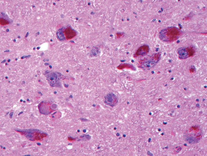
60. The lesions illustrated in Figure 20-19 represents which of the following?
(A) Granulovacuolar degeneration
(B) Hirano bodies
(C) Neurofibrillary tangles
(D) Pick bodies
(E) Senile plaques
61. All of the following stains are useful in highlighting the lesion shown in Question 60 except
(A) Alpha-synuclein
(B) Bielschowsky
(C) Bodian
(D) Gallyas
(E) Tau
62. All of the following genes are associated with early onset Alzheimer disease except
(A) Alpha-synuclein
(B) Amyloid precursor protein
(C) Apolipoprotein E
(D) Presenilin-1
(E) Presenilin-2
63. The lesion shown (Figure 20-20) in a 76-year-old woman with Alzheimer disease stains with all of the following markers except
(A) Neurofilament
(B) Tau
(C) Thioflavin-S
(D) Tubulin
(E) Ubiquitin
64. In neurofibrillary tangle predominant dementia, a high density of neurofibrillary tangles are found in all of the following locations except
(A) Amygdala
(B) Basal ganglia (caudate and putamen)
(C) CA1 region of the hippocampus
(D) Entorhinal cortex
(E) Subiculum
65. Mutations in all of the following genes are associated with familial cases of Parkinson disease except
(A) Alpha-synuclein
(B) Parkin
(C) PINK1
(D) Tau
(E) UCHL1
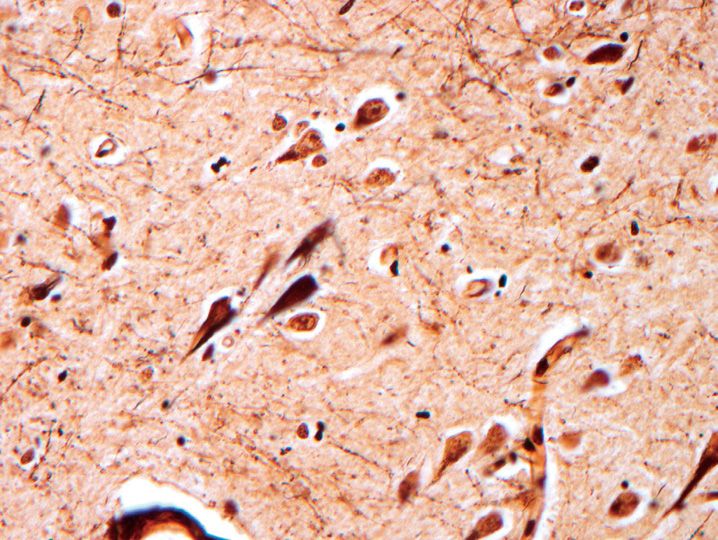
66. The most sensitive and specific marker for the eosinophilic cytoplasmic inclusions shown in Figure 20-21 is which of the following?
(A) Alpha-synuclein
(B) GFAP
(C) Neurofilament
(D) Tau
(E) Ubiquitin
67. All of the following are true regarding multiple system atrophy except
(A) Atrophy of the caudate and putamen in parkinsonian predominant type
(B) Cerebellar degeneration with Purkinje cell loss in cerebellar predominant type
(C) Male preponderance
(D) May present with akinesias, dystonia, and rigidity
(E) Neuronal cytoplasmic inclusions
68. All of the following are features of Pick disease except
(A) Argyrophilic intracytoplasmic inclusions
(B) Ballooned neurons
(C) Frontotemporal atrophy
(D) GFAP-positive Pick cells
(E) Spared involvement of the posterior superior temporal gyrus
69. Which of the following is the most common type of frontotemporal lobar degeneration?
(A) FTLD-1F
(B) FTLD-ni
(C) FTLD-tau
(D) FTLD-TDP
(E) FTLD-U
70. All of the following are true regarding frontotemporal lobar atrophy-U except
(A) Alpha-synuclein negative
(B) Association with charged multivesicular body protein mutation 2B gene
(C) Neuron loss and gliosis in frontal and temporal lobes
(D) Patients may develop progressive aphasia
(E) Tau positivity
71. A 62-year-old man is suspected of having amyotrophic lateral sclerosis) All of the following are findings expected at autopsy except
(A) Bunina bodies
(B) Lateral corticospinal tract degeneration
(C) Loss of anterior horn cells
(D) Loss of Betz cells in the primary motor cortex
(E) Loss of neurons in locus ceruleus
72. A 6-month-old boy presents with recent onset areflexia and fasciculations. Over the next few years, he develops contractures and kyphoscoliosis. The best diagnosis is which of the following?
(A) Familial amyotrophic lateral sclerosis
(B) Myotonic dystrophy
(C) Spinal muscular atrophy 1
(D) Spinal muscular atrophy 2
(E) Spinal muscular atrophy 3
73. All of the following are trinucleotide repeat disorders except
(A) Becker muscular dystrophy
(B) Friedreich ataxia
(C) Huntington disease
(D) Myotonic muscular dystrophy
(E) Spinal bulbar muscular atrophy
74. A patient with suspected Huntington disease has neuronal loss and astrocytosis involving most of the caudate and putamen with sparing of the nucleus accumbens. Which Vonsattel grade most accurately describes the findings?
(A) Grade 0
(B) Grade 1
(C) Grade 2
(D) Grade 3
(E) Grade 4
75. All of the following are features of hereditary spinocerebellar ataxia type 3 (Machado–Joseph disease) except
(A) Inferior olivary nuclei relatively preserved
(B) Loss of neurons in the putamen
(C) Neuronal loss in the cerebellar dentate nucleus
(D) Spinocerebellar tract degeneration
(E) Substantia nigra degeneration
76. A patient with Friedreich ataxia might be expected to have all of the following except
(A) Cataracts
(B) Gait ataxia
(C) Hypertrophic cardiomyopathy
(D) Scoliosis
(E) Sensorineural hearing loss
77. All of the following are features associated with neurodegeneration with brain iron accumulation (Hallervorden–Spatz disease) except
(A) Axonal spheroids
(B) Glial cytoplasmic tau positive inclusions
(C) Globus pallidus and substantia nigra neuronal loss
(D) Mutation in pantothenate kinase gene
(E) X-linked condition
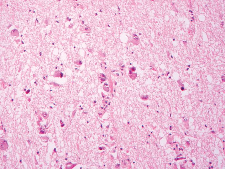
78. In a hospitalized patient, the most likely etiology for the changes shown in Figure 20-22 is which of the following?
(A) Gram-negative bacilli
(B) Listeria monocytogenes
(C) Neisseria meningitidis
(D) Nocardia asteroides
(E) Streptococcus pneumoniae
79. All of the following are conditions that predispose to the development of the changes shown in Figure 20-23 except
(A) Bacterial endocarditis
(B) Cardiac disease with left-to-right shunt
(C) Chronic pulmonary disease
(D) Dental infection
(E) Urinary tract infection
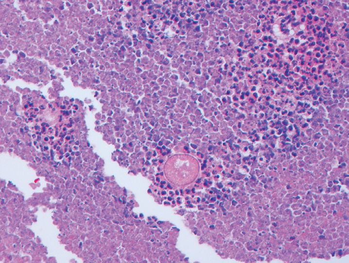
80. The most likely etiology of the parietal lobe lesion shown in Figure 20-24 is which of the following?
(A) Actinomycosis
(B) Candida albicans
(C) Group B streptococcus
(D) Mycobacterium leprae
(E) Mycobacterium tuberculosis
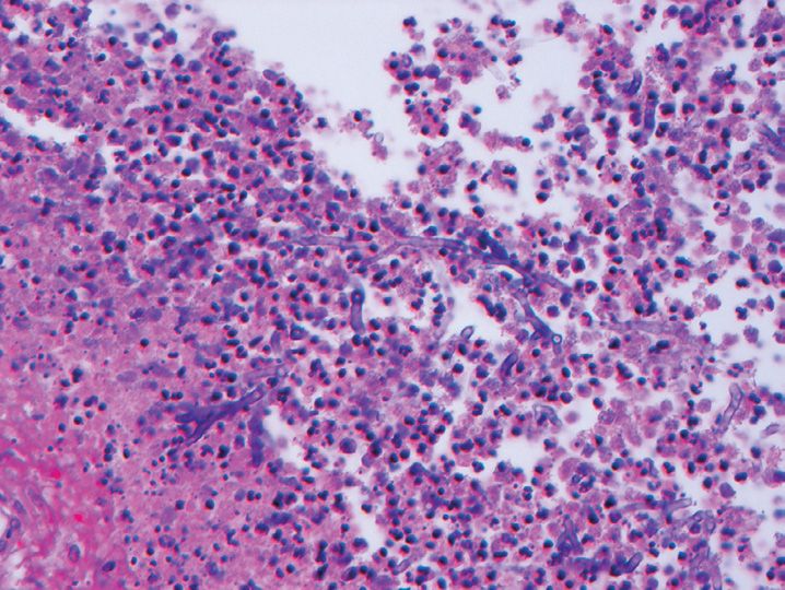
81. A 62-year-old cardiac transplant patient develops right parietal lobe abscess. A Gomori methenamine silver (GMS) stain of the lesion is shown in Figure 20-25. The most likely etiology of the abscess is which of the following?
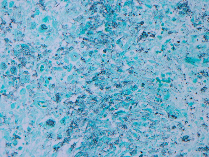
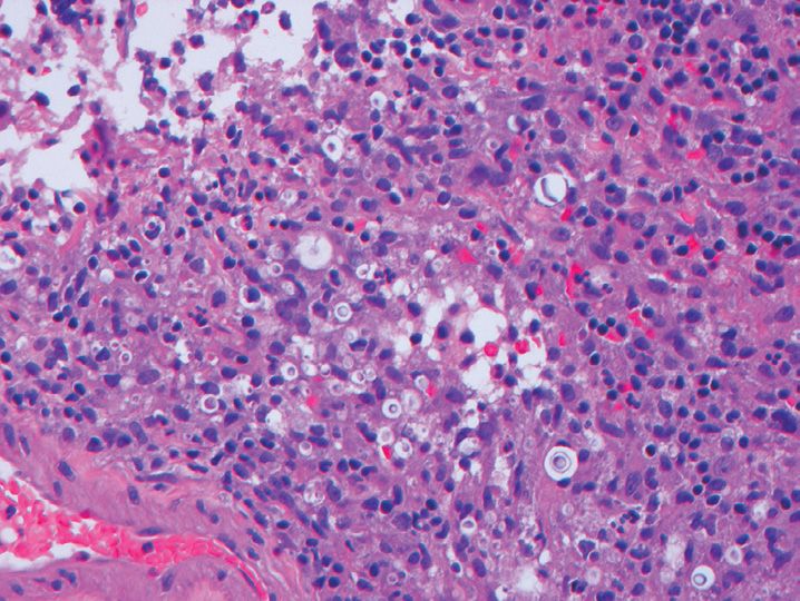
(A) Aspergillus
(B) Fusarium
(C) Mucor
(D) Mycobacterium
(E) Nocardia
82. The meningitis shown in Figure 20-26 was identified in a 38-year-old immunocompromised man. The most likely etiology is which of the following?
(A) Blastomycosis
(B) Candidiasis
(C) Cannot tell the etiology
(D) Coccidioidomycosis
(E) Cryptococcosis
83. The lesion shown in Figure 20-27 is associated with all of the following conditions except
(A) Collagen vascular disease
(B) Corticosteroid therapy
(C) Diabetes mellitus
(D) Organ transplantation
(E) Travel to Southwest United States
84. The best diagnosis for this lesion shown (Figure 20-28) in a biopsy from a 59-year-old man is which of the following?
(A) Blastomycosis
(B) Coccidioidomycosis
(C) Glioblastoma
(D) Sarcoidosis
(E) Tuberculosis
85. A patient is diagnosed with histoplasmosis meningitis. He is most likely a resident of which state?
(A) Arizona
(B) Florida
(C) Maine
(D) Ohio
(E) Texas
86. A 12-year-old girl presents with fever, headaches, and new onset seizures. She is diagnosed with herpes encephalitis, based on a cerebrospinal fluid (CSF) polymerase chain reaction (PCR) study. Where in the brain is she most likely to have radiographic abnormalities?
(A) Basal ganglia
(B) Cerebellum
(C) Mamillary bodies
(D) Pons
(E) Temporal lobes
87. The changes shown (Figure 20-29) in the optic nerve most commonly are marked by which of the following patterns of injury?
(A) Abscess
(B) Acute meningitis
(C) Empyema
(D) Inflammatory pseudotumor
(E) Microglial nodules
88. A 68-year-old man with a history of hypertension and non-Hodgkin lymphoma develops limb weakness and confusion. On imaging, he has multiple subcortical white matter lesions. One is biopsied and shown in Figure 20-30. The best diagnosis for this patient is which of the following?
(A) Cytomegalovirus encephalitis
(B) Metastatic lymphoma
(C) Multiple sclerosis
(D) Progressive multifocal leukoencephalopathy
(E) Subacute sclerosing panencephalitis
89. Tropical spastic paraparesis is caused by which of the following?
(A) Arbovirus
(B) HIV
(C) HTLV-1
(D) HTLV-2
(E) Syphilis
90. A liver transplant patient develops frontotemporal lesions on imaging. These abnormalities are due to which of the following? (Figure 20-31)
(B) Chagas disease
(C) Histoplasmosis
(D) Leishmaniasis
(E) Toxoplasmosis
91. A 10-year-old boy presents with severe frontal headaches, nausea, vomiting, and stiff neck. (He recently has been playing in a pond in his backyard.) Which of the following is the most likely cause of this presentation?
(A) Acanthamoeba
(B) Balamuthia
(C) Cysticercosis
(D) Naegleria
(E) Toxoplasmosis
92. All of the following are true regarding neuroschistosomiasis except
(A) Eggs may embolize to the brain from the portal mesenteric system
(B) May be associated with granulomatosis inflammation
(C) More common in men
(D) Most cases are asymptomatic
(E) Most commonly due to Schistosoma haematobium
93. All of the following are true regarding Creutzfeldt–Jakob disease except
(A) About 10% are familial cases
(B) Associated with a prion protein abnormality
(C) Elevated protein 14-3-3 in CSF
(D) Rapidly progressive dementia
(E) Spongiform changes with microglial nodules
94. Which of the following is least effective in decreasing the risk of transmission of prion disease?
(A) Autoclaving
(B) Bleach
(C) Formalin fixation
(D) Lye
(E) Postfixation with formic acid
95. Which ethnic group or region has a higher incidence of Neimann–Pick disease?
(A) Ashkenazi Jews
(B) Finland
(C) Mexico
(D) Northern Europe
(E) Nova Scotia
96. All of the following are sphingolipidoses that present in the neonatal period except
(A) Fabry disease
(B) Farber disease
(C) Krabbe disease
(D) Niemann–Pick disease type B
(E) Pompe disease
97. All of the following are lysosomal storage diseases that can affect the nervous system except
(A) Alexander disease
(B) Ceroid lipofuscinosis
(C) Gaucher disease
(D) Globoid cell leukodystrophy
(E) Metachromatic leukodystrophy
98. All of the following are associated with Tay–Sachs disease except
(A) Blindness
(B) Cherry red spots
(C) Hepatosplenomegaly
(D) Hexosaminidase A deficiency
(E) Membranous cytoplasmic bodies by electron microscopy
99. Angiokeratoma corporis diffusion or cutaneous telangiectasis in a bathing trunk distribution and renal dysfunction are features associated with which of the following?
(A) Fabry disease
(B) Farber lipogranulomatosis
(C) Gaucher disease
(D) Niemann–Pick disease type A
(E) Sandhoff disease
100. All of the following ultrastructural features are associated with neuronal ceroid lipofuscinoses except
(A) Curvilinear bodies
(B) Fingerprint bodies
(C) Granular osmophilic deposits
(D) Lipofuscin-like deposits
(E) Zebra bodies
101. All of the following represent mitochondrial diseases except
(A) Kearns–Sayre Syndrome
(B) Leigh syndrome
(C) MELAS
(D) Progressive external ophthalmoplegia
(E) Zellweger syndrome
102. All of the following are features of MELAS except
(A) Age of presentation typically 5th and 6th decades
(B) Dementia
(C) Elevated cerebrospinal fluid lactate
(D) Headaches
(E) Infarct-like lesions
103. All of the following are true regarding Menkes disease except
(A) Abnormal hair
(B) Alzheimer type II astrocytes
(C) Copper deficiency
(D) Failure to thrive
(E) X-linked recessive disorder
104. At autopsy, the mamillary bodies are noted to be brown in discoloration and atrophic. The most likely underlying abnormality is a deficiency in which of the following?
(A) Copper
(B) Thiamine
(C) Vitamin B12
(D) Vitamin E
(E) Zinc
105. All of the following are associated with the development of a neuronopathy except
(A) Aluminum
(B) Lead
(C) Mercury
(D) Methanol
(E) Toluene
106. All of the following are features used in grading fibrillary or diffuse astrocytomas except
(A) Cellularity
(B) Mitoses
(C) Necrosis
(D) Secondary structures of Sherer
(E) Vascular proliferation
107. Which of the following antibodies would be best at differentiating this lesion from gliosis? (Figure 20-32)
(A) EGFR
(B) GFAP
(C) IDH-1
(D) Ki-67
(E) S-100 protein
108. The best diagnosis for the frontal lobe lesion illustrated (Figure 20-33) in a 62-year-old patient is which of the following?
(A) Chordoma
(B) Gliosarcoma
(C) Malignant meningioma
(D) Metastatic chondrosarcoma
(E) Metastatic osteosarcoma
109. Which of the following abnormalities is most consistently found in this glioblastoma variant? (Figure 20-34)
(A) Chromosome 1p deletion
(B) Chromosome 19q deletion
(C) EGFR overexpression
(D) IDH-1 mutation
(E) p53 mutation
110. Which of the following antibodies is least likely to stain the neoplasm shown in Figure 20-35?
Stay updated, free articles. Join our Telegram channel

Full access? Get Clinical Tree


