Headache
Headache is the most frequently reported neurological symptom, accounting for considerable morbidity in the general population. The clinician’s challenge is to exclude a treatable underlying intra- or extracranial secondary cause (Table 15.1) and to make a definitive diagnosis. The history provides important clues and should ascertain:
- date of onset, frequency and duration of episodes
- precipitating and relieving factors
- type and location of pain
- severity
- associated features.
Table 15.1 Causes of Headache and Facial Pain
| Type | Examples |
| Primary headache syndrome | Tension headache Migraine Cluster headache |
| Headache secondary to other disorders | Raised intracranial pressure Idiopathic (‘benign’) intracranial hypertension Meningeal irritation (e.g. meningitis, SAH) Post-traumatic (head injury) Hypertension |
| Giant-cell arteritis | |
| Sinusitis Perioral disease (poor dentition, salivary gland disorders) Cervical spine disease | |
| Ocular, orbital and retro-orbital disease | |
| Ear disease | |
| Temporomandibular joint dysfunction | |
| Metabolic disturbance (e.g. hypoxia, hypercapnia, hypoglycaemia) | |
| Drugs (e.g. nitrates, vasoactive agents) | |
| Facial pain | Trigeminal neuralgia |
| Postherpetic neuralgia | |
| Atypical facial pain | |
| SAH, subarachnoid haemorrhage. | |
Primary Headache Syndromes
Tension Headache (Chronic Daily Headache)
Characteristically a continuous severe pressure is felt bilaterally over the vertex, occiput or eyes. It may be ‘band-like’ or non-specific and of variable intensity. Aetiology remains unclear but may be musculoskeletal in origin. It is most common in middle-aged women, but may occur at any age and in either sex, especially in the context of stress or depression.
The headache often occurs on a daily basis and may persist for months or even years. Standard analgesics are reported to be ineffective and continuous analgesic use may exacerbate the situation, especially when the effects of medication wear off (so-called analgesic headache or rebound headache). Aside from nausea, there are no other associated features and neurological examination is normal.
Treatment is difficult: reassurance that there is no sinister underlying cause may help in some cases, as may teaching relaxation techniques and addressing underlying stressors. The patient should be advised to avoid excessive analgesic use, but a small dose of amitriptyline taken at night may help.
Migraine
Migraine is episodic and affects approximately 10% of the population. It usually begins around puberty and continues intermittently to middle age. It is three times more common in women and there is often a family history. It may be associated with menstruation or triggered by contraceptive pill usage, physical exercise, alcohol, various specific foods (especially chocolate, cheese and red wine) or heightened emotions. There is a link with hypertension and prior head injury.
The pathophysiology remains poorly understood. Prodromal sensory phenomena (‘aura’) have been attributed to vasoconstriction within intracerebral vessels, although a wave of depolarisation spreading across the cerebral cortex may account for this early phase. Thereafter, vasodilatation of extracerebral vessels correlates with the onset of headache. A number of vasoactive peptides including calcitonin-gene-related peptide (CGRP) and serotoninergic (5-HT) pathways have been implicated in the pathogenesis.
Clinical Presentation
Classical Migraine with Aura
Characteristically migraine starts with a sense of ill health (lasting up to several hours) followed by a visual aura (e.g. shimmering lights, fortification spectra, scotomata) usually in the field opposite to the side of the succeeding headache and lasting up to 1 h. In severe cases the patient may develop a homonymous hemianopia or even complete blindness. Sensory symptoms (e.g. unilateral numbness/paraesthesia) are less commonly seen, and speech disturbance or motor weakness are very rare. Thereafter, a throbbing unilateral headache is associated with anorexia, nausea, vomiting, photophobia and withdrawal. The headache lasts several hours and even days in some patients, and is aggravated by movement (e.g. coughing, sneezing, bending). Neurological examination is usually within normal limits, and between episodes the patient remains well.
Migraine without Aura
Although the classical aura is absent, patients may feel non-specifically unwell prior to the onset of headache.
Hemiplegic and Ophthalmoplegic Migraine
Rarely focal neurological features may persist for several days. Other structural lesions (e.g. arteriovenous malformation, aneurysm) must be excluded.
Investigation
When the diagnosis is clear, investigation is not required; otherwise brain imaging should be performed.
Management
Acute Attack
- Sleeping in a quiet darkened room is effective in many patients.
- Simple analgesics (e.g. aspirin, paracetamol) and an antiemetic agent.
- 5-HT1B/D agonists (e.g. rizatriptan, sumatriptan, zolmitriptan) are effective when taken early and may abort an established attack. They are contraindicated in patients with known/suspected coronary or cerebrovascular disease or uncontrolled hypertension and must be used cautiously in those with vascular risk factors.
Clinical trials with a novel class of agent, CGRP antagonists, are currently underway and may be effective in the third of all migraine sufferers who do not respond well to ‘triptans’.
Prophylaxis
Precipitating causes should be identified and avoided. Oestrogen-containing preparations must be used with caution. Preventative treatment for migraine should be considered for patients who suffer:
- > 1 acute attack per month
- increasing frequency of headaches
- significant disability despite appropriate treatment for acute attacks.
Therapeutic options include:
- β-blockers – usually propranolol
- amitriptyline
- pizotifen
- topiramate
- others, e.g. sodium valproate, verapamil, methysergide (the latter has potentially serious fibrotic side effects and must only be used under expert supervision).
Cluster Headache
These are relatively short-lived (30–120 min) episodes of severe pain, typically centred on one eye and affecting men more than women ( 3 : 1), with an age of onset between 20 and 60 years. Attacks start without warning and are associated with red eye, eye and nose watering and vomiting. They may occur several times a day, often waking the patient from sleep. Usually cluster headaches are recurrent for several days, weeks or months before the disorder remits and the patient becomes pain-free for months or years. Alcohol is a recognised precipitant.
Sumatriptan (self-administered by subcutaneous injection) is the treatment of choice for cluster headaches – simple analgesics are rarely effective in this condition. High-flow oxygen and corticosteroids have also been reported to be efficacious in some patients. Prophylaxis with verapamil or lithium may be tried (methysergide is reserved for refractory cases and, as with migraine prophylaxis, must be used under expert supervision because of the risk of inducing fibrotic disorders).
Secondary Causes of Headache
See Table 15.1.
Raised Intracranial Pressure
Usually secondary to an intracranial tumour, haematoma or abscess, the pain is worse on waking and associated with nausea and vomiting. It improves 1–2 h after rising and is exacerbated by coughing, sneezing, straining and bending down. Visual function may be preserved despite papilloedema, but other neurological symptoms and signs related to the primary lesion are usually evident. The pain often responds to simple analgesics.
Idiopathic (‘Benign’) Intracranial Hypertension (IIH; Pseudotumour Cerebri)
IIH is commonest in young obese women with symptoms and signs of raised intracranial pressure but no mass lesion on brain imaging. Altered cerebrospinal fluid (CSF) dynamics (with impaired absorption) have been suggested to underlie the disorder. The patient may report visual disturbance, including diplopia and obscurations (abrupt onset transient visual loss secondary to changes in posture), and examination reveals bilateral papilloedema. Occasionally, bilateral sixth cranial nerve palsies are present and reflect raised intracranial pressure (‘false-localising’ sign). Pulsatile tinnitus is another recognised feature.
CT/MRI scanning of the brain is normal without hydrocephalus but lumbar puncture confirms raised CSF pressure. Intracranial venous sinus thrombosis, disorders of calcium metabolism, drugs (tetracyclines, isotretinoin, hormonal contraceptives, growth hormone and corticosteroids), systemic lupus erythematosus and hypervitaminosis A may all present with a syndrome similar to IIH, thus secondary causes must always be excluded.
Weight loss may facilitate spontaneous remission. Serial ‘therapeutic’ lumbar punctures can be used to lower CSF pressure but are unpopular with patients. In more chronic cases, medical therapy with acetazolamide, other diuretics or corticosteroids may be tried but surgical intervention (lumboperitoneal shunt or optic nerve sheath decompression) is often required to relieve symptoms and/or protect vision – prolonged raised intracranial pressure predisposes to optic atrophy.
Meningeal Irritation
Irritation of the meninges (meningism) occurring in meningitis or following subarachnoid haemorrhage characteristically produces a triad of symptoms:
- severe headache – global or occipital associated with nausea/vomiting
- photophobia
- neck stiffness.
In meningitis the headache evolves over minutes to hours whereas in subarachnoid haemorrhage it is abrupt in onset and may be followed by loss of consciousness.
Post-Concussion
Similar to tension headache but usually associated with dizziness (not vertigo) and impaired concentration, post-concussion headache persists for months and there may be a history of inadequate recovery following the head injury.
Giant-cell Arteritis
This is an important cause of headache in patients over 50 years of age (see rheumatology, p. 284).
Neuralgias
Neuralgias are intermittent, brief, severe, lancinating pains occurring along the distribution of a nerve.
Trigeminal Neuralgia
Trigeminal neuralgia predominantly affects those over 50 years of age. It reflects compression of the sensory root of the trigeminal nerve (e.g. by a tumour or aberrant vessel) or may complicate multiple sclerosis. The agonising sharp pain is confined to the distribution of the trigeminal nerve on one side, commonly the maxillary or mandibular divisions. It lasts only seconds and is usually triggered from a place on the lips, side of the face or nose, by chewing, eating, speaking, or by a cold breeze. It tends to get worse with age, and eventually a continuous background pain may develop if left untreated. Physical examination is usually normal but may reveal neurological signs in the presence of an underlying mass lesion.
Simple analgesics are generally ineffective. Usually carbamazepine provides good symptom control, but gabapentin, sodium valproate, clonazepam and tricyclic antidepressants may be tried.
Radiofrequency thermocoagulation or chemical (glycerol) ablation of the trigeminal ganglion produce benefits in some patients.
Glossopharyngeal Neuralgia
A rare disorder precipitated by swallowing, which produces pain in the pharynx or deep inside the ear.
Postherpetic Neuralgia
Patients have a history of herpes zoster infection (shingles) in the distribution of one of the branches of the trigeminal nerve (usually ophthalmic). Pain, itching and altered sensation develop along the course of the affected nerve and persist after the rash has healed. The pain may be difficult to treat, but sometimes responds to tricyclic antidepressants, carbamazepine or topically applied capsaicin.
Atypical Facial Pain
This describes episodic aching in the jaw and cheek (in a non-anatomical distribution), lasting several hours and usually occurring in young to middle-aged women who often exhibit coexistent features of anxiety or depression. It is often bilateral and may respond to antidepressants.
Epilepsy
Epilepsy results from intermittent paroxysmal electrical discharges of cerebral neurons causing stereotypical attacks of altered consciousness, motor or sensory function, behaviour or emotion. A single unprovoked episode (i.e. a seizure) is insufficient to make a diagnosis as the term should be reserved for those with a recurring tendency to seizures. Ideally, all patients with a first unexplained seizure should be rapidly assessed by a neurologist in a specialist clinic.
Up to 1% of the general population suffers from epilepsy. Each year a small number of individuals with this condition (1–2 per 100,000) die prematurely as a consequence of status epilepticus (see below), accidental injury or sudden unexplained death – the latter is assumed to be related to seizure activity with associated cardiorespiratory dysfunction.
Classification
Partial Seizures
These have a single focus of activity, which may be scar tissue related to previous trauma, a cerebrovascular accident or tumour. They are classified as:
- simple partial seizures: with no impairment of consciousness
- complex partial seizures: consciousness is impaired at some stage.
Partial seizures may progress to generalised seizures.
Generalised Seizures
Generalised seizures are typified by widespread activity affecting both cerebral hemispheres and include:
- childhood absence seizures (petit mal) and atypical absence seizures
- myoclonic epilepsy
- tonic–clonic (grand mal) seizures
- tonic (spasm), clonic (jerking), atonic or akinetic seizures.
Aetiology
Most epilepsy is idiopathic. There may be a family history suggesting genetic susceptibility, particularly with petit mal seizures. Seizures may be secondary to cerebral disorders, metabolic dysfunction and drug ingestion (Table 15.2).
Table 15.2 Causes of Epilepsy
| Type | Cause |
| Idiopathic | Often unknown, but likely significant inherited component |
| Secondary to other disorders | |
| Neonatal | Birth trauma, including intracranial haemorrhage |
| Hypoxia | |
| Childhood | Congenital anomalies |
| Tuberous sclerosis | |
| Metabolic storage disorders | |
| Adulthood | Head injury |
| Drug* and/or alcohol intoxication or withdrawal | |
| Elderly | Cerebrovascular disease |
| Degenerative disorders (e.g. Alzheimer’s disease, Huntington’s disease) | |
| All/most ages | Metabolic disturbance (e.g. hypoglycaemia, hypocalcaemia, hyponatraemia) |
| Cerebral infection (e.g. meningitis, encephalitis, abscess) | |
| Cerebral tumour or arteriovenous malformation | |
| Inflammation (e.g. vasculitis, SLE, rarely demyelination) | |
| *A wide variety of drugs have been reported to provoke seizures – a list is available at www.epilepsy.com. | |
Provocation of Seizures
A variety of factors can provoke seizures in patients not usually prone to epilepsy (e.g. drug overdose, hypoglycaemia). In those with known epilepsy, seizures may be provoked by sleep deprivation, stress, alcohol and, occasionally, stimuli such as television or strobe lighting. In some women seizures may increase in frequency around the time of menstruation.
Differential Diagnosis
- syncope: e.g. vasovagal faints, postmicturition and cough syncope
- cerebrovascular disease: e.g. transient ischaemic attacks, critical carotid artery stenosis, vertebrobasilar ischaemia
- vestibular disorders
- low cardiac output states
- metabolic: e.g. hypoglycaemia
- postural hypotension
- narcolepsy
- psychiatric disorders: e.g. conversion hysteria
Clinical Features
Epilepsy in Childhood
Absence Seizures (‘Petit Mal’)
This usually presents between 4 and 10 years and is more common in girls. It is characterised by brief (10–15 s) moments of absence without warning (e.g. the child stops talking and stares blankly) followed by immediate recovery. It rarely continues beyond puberty, although 5–10% of children will develop adult seizures.
Febrile Convulsions
These are seizures occurring in the context of fever, usually in young children under 5. The majority are ‘one-off’ events although up to 5% go on to develop epilepsy. They are usually generalised and brief but occasionally longer lasting or focal in nature.
Infantile Spasms
These are brief spasms (typically ‘shock-like’ with flexion of the arms, head and neck and drawing up of the knees) associated with progressive learning difficulties. Aetiology includes perinatal asphyxia, metabolic disorders, encephalitis and cerebral malformations.
Juvenile Myoclonic Epilepsy
This form of primary generalised epilepsy with typical onset in teenagers is characterised by relatively infrequent generalised seizures, daytime absences and myoclonus.
Epilepsy in Adulthood
Primary Generalised Epilepsy (Tonic–Clonic Seizures/Grand Mal)
Seizures may be preceded by a prodrome/aura in which the patient reports dizziness, irritability or other non-specific symptoms. This is followed by loss of consciousness and the tonic phase (characterised by generalised muscle spasms), which usually lasts up to 30 s. Cyanosis may occur. The clonic phase, characterised by sharp repetitive muscular jerks in all limbs, follows. Tongue biting, salivation and involuntary micturition may occur. Consciousness remains impaired typically for around 30 min, with drowsiness and confusion lasting several hours.
Temporal Lobe Epilepsy
Patients typically experience an aura which may include a sense of fear or déjà-vu, hallucinations (visual, olfactory or gustatory) or a rising sensation in the epigastrium. Confusion and anxiety may develop and some patients exhibit automatism (organised stereotyped movements, e.g. chewing, lip-smacking).
Jacksonian (Focal) Epilepsy
Epileptic activity originates in one part of the motor cortex. Each seizure begins in one body part and may proceed to involve that side of the body and then the whole body. Temporary paresis of the originally affected limb may persist after the attack (Todd’s paralysis). Sensory epilepsy is a parallel condition originating in the sensory cortex.
Investigation
The object is to detect treatable underlying brain disease and identify provoking factors.
A full history and clinical examination should identify other causes of loss of consciousness. Biochemical evidence of excess alcohol, hypoglycaemia, hyponatraemia or hypocalcaemia should be sought.
An EEG should help to confirm the diagnosis, but both false positive (in 1% of the normal population) and false negative results occur. EEG diagnosis can be enhanced by prolonged recording especially after sleep deprivation.
CT or MRI scanning is performed in most adult patients presenting with a seizure to identify structural lesions. Imaging is of particular value in late-onset epilepsy, partial seizures and in patients with generalised epilepsy where the EEG discloses a focal abnormality.
Management
A single fit rarely requires treatment but an underlying cause should be sought. Most neurologists would begin treatment with prophylactic anticonvulsants after a second episode. However, it may be prudent to treat after a first seizure when neuroimaging reveals a structural lesion or when there is no reversible precipitant.
The choice of pharmacological therapy is determined by the type of epilepsy, for example:
- Partial seizures (with or without secondary generalisation): carbamazepine, lamotrigine, oxcarbazepine and sodium valproate are the drugs of choice; second-line agents include clobazam, gabapentin, levetiracetam, pregabalin and topiramate.
- Generalised tonic–clonic: carbamazepine, lamotrigine and sodium valproate are first-line agents; clobazam, levetiracetam, phenytoin and topiramate are useful second-line options.
- Absence: ethosuximide or sodium valproate are the drugs of choice for classical absence seizures; clonazepam and lamotrigine are alternatives.
- Myoclonic: sodium valproate is the drug of choice for most cases; clonazepam, levetiracetam and topiramate may be tried as second-line agents.
In addition, age, sex, child-bearing potential, comorbidity and concomitant medication should be taken into account. Seizure control with minimal adverse effects can be achieved using a single anticonvulsant in  75% of patients. The addition of a second drug produces satisfactory control in a further subgroup.
Refractory epilepsy (inadequate control on multiple agents) may reflect:
- poor compliance
- pseudoseizures or non-epileptic attacks (either alone or in combination with genuine seizures)
- an underlying structural brain lesion
- excess alcohol or illicit drug usage.
Status Epilepticus
This is defined as recurring or continuous seizures, in which the patient does not regain consciousness between attacks. It is a medical emergency as hypoxia/anoxia can lead to permanent brain damage or even death. Key principles of management include:
- basic life support/resuscitation
- seizure control
- identification and correction of predisposing cause.
The choice of agent used to terminate seizure activity depends on the stage/duration, but may include:
- Intravenous lorazepam (4 mg) (clonazepam and diazepam are alternatives) – dose may be repeated after 10 min if seizures recur/continue.
- Intravenous phenytoin (15 mg/kg, maximum rate of 50 mg/min), fosphenytoin (prodrug of phenytoin), both of which require ECG monitoring, or phenobarbitone (10 mg/kg, maximum rate of 100 mg/min) should be used when there is established status.
- Intravenous thiopentone (bolus followed by infusion), combined with ventilation/neuromuscular blockade, is required when seizures continue beyond 30–60 min. Midazolam and propofol have also been used in this setting. EEG monitoring is required to confirm termination of seizure activity.
Patients should be nursed in a high dependency/intensive care setting. Regular anticonvulsant therapy should be reinstituted as soon as possible in those with known epilepsy.
Surgical Treatment of Epilepsy
This should be considered in patients with intractable epilepsy if a focus for seizure onset can be identified using MRI and electrophysiology mapping.
Epilepsy and Pregnancy
Uncontrolled seizures in pregnancy present a serious risk to both mother and fetus. Anticonvulsant drugs must be continued especially if there is a history of recent seizure activity. In women with no recent (2– 3 years) history of seizures, a trial off therapy before pregnancy should be considered.
Women with epilepsy who wish to become pregnant should receive pre-pregnancy counselling about the risk of congenital abnormality and the individual pros and cons of continuing treatment. Wherever possible, the lowest dosage of a single agent should be used. Screening for neural tube defects is especially indicated in women taking sodium valproate or carbamazepine, and folic acid supplementation is essential both preconception and throughout the pregnancy.
For mothers taking carbamazepine, phenobarbitone or phenytoin (enzyme inducing agents), vitamin K should be prescribed before delivery and for the newborn.
NB Carbamazepine, phenytoin and phenobarbitone all induce hepatic microsomal enzymes and increase metabolism of oestrogens and progestagens, making oral contraception unreliable.
Epilepsy and Driving
In the UK all patients with a history of seizures must report their condition to the Driver and Vehicle Licensing Agency (DVLA). Currently, for an isolated seizure, a 6-month ‘off driving period’ is stipulated providing the individual has undergone assessment by an appropriate specialist and no relevant abnormality has been identified on investigation. A 12-month ‘off driving period’ applies if there is deemed to be the potential for further seizures. Patients with a history of epilepsy must be seizure free for 1 year before being allowed to drive. More stringent regulations apply to licences for heavy goods or passenger-carrying vehicles (www.dft.gov.uk/dvla/drivers.aspx). Patients with sleep-related epilepsy may drive if they have an established pattern of seizures that have occurred only in relation to sleep during the previous 3 years.
Epilepsy and Employment
There are certain statutory employment restrictions for individuals with epilepsy, including in relation to the emergency and armed services, pilots and train drivers.
Epilepsy and Lifestyle
There are no ‘absolute rules’, but it is sensible to avoid heights/ladders, operating heavy machinery, working underground and activities such as unsupervised swimming until seizure control is established. Fires should be guarded and children should not be left in the bath unattended.
Prognosis in Epilepsy
The long-term prognosis of epilepsy is good, with most patients attaining a 5-year remission and many stopping treatment in due course. The decision to discontinue anticonvulsant therapy is determined by: type of epilepsy; duration of remission; potential deleterious effects of seizure recurrence (driving and employment) and side effects of treatment.
Stroke
Stroke is characterised by rapidly developing symptoms and/or signs of loss of central nervous system function. It is distinguishable from a transient ischaemic attack (see below) by virtue of symptoms persisting for more than 24 h. Stroke has an annual incidence of 1–2 per 1000 population, is the third most common cause of death in industrialised countries and is a major cause of morbidity in those who survive.
Aetiology and Pathophysiology
Approximately 85% of cases are ischaemic (thrombosis or embolism) in origin, 10% are caused by intracerebral haemorrhage and 5% by subarachnoid haemorrhage.
Thrombosis is due to one or more of the following:
- abnormal blood vessel wall
- polycythaemia
- altered blood flow.
Emboli may arise from vascular or cardiac sources, including:
- degenerative vascular disease
- atrial fibrillation
- cardiac valvular disease
- post-myocardial infarction.
Degenerative arterial disease is the most common cause of stroke. Risk factors include family history of premature vascular disease, smoking, hypertension, hyperlipidaemia, diabetes mellitus, excess alcohol ingestion and certain oral contraceptive preparations.
A small number of cases have a non-vascular lesion (tumour, subdural haematoma, migraine, intracranial infection, metabolic disturbance).
In the absence of a collateral blood supply, the brain territory supplied by an occluded artery undergoes infarction. Potentially salvagable surrounding areas of brain which lie within the so-called ischaemic penumbra remain viable for a period of time and may recover function if their blood supply is restored.
Both cytotoxic (accumulation of water in damaged neurones and glial cells) and vasogenic (extracellular fluid accumulation secondary to disruption of the blood–brain barrier) oedema may complicate infarction.
Clinical Features
Symptoms and signs are dictated by the vascular territory that is rendered ischaemic. As a general rule, the carotids supply the anterior and middle cerebral arteries (anterior circulation), while the vertebrobasilar system feeds the posterior cerebral arteries which, together with branches supplying the cerebellum and the brainstem, constitute the posterior circulation.
Total Anterior Circulation Infarct
- hemiplegia (corticospinal tract)
- homonymous hemianopia (optic tract/radiation)
- cortical deficits, e.g. dysphasia (dominant hemisphere), visuospatial loss (non-dominant hemisphere)
Partial Anterior Circulation Infarct
- two of the above or cortical deficit alone
Lacunar Infarct
- pure motor or sensory stroke or ataxic hemiparesis
Multiple/Serial Lacunar Infarcts
These may produce cumulative neurological deficits including:
- cognitive impairment (multi-infarct dementia)
- gait disturbance/apraxia.
Posterior Circulation Infarct
- brainstem dysfunction, e.g. vertigo, diplopia, altered conscious level
- homonymous hemianopia
Spinal Cord Infarct
See below (spinal disorders, p. 192).
Diagnosis and Initial Investigation
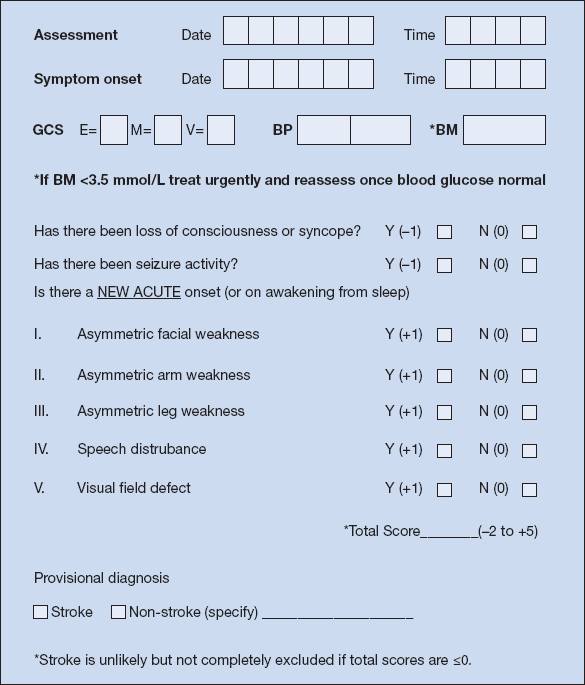
The introduction and promotion of ‘diagnostic aids’ such as ‘FAST’ (Box 15.1) and ‘ROSIER’ (Fig. 15.1) have facilitated earlier assessment and treatment, including thrombolysis in selected cases.
 Box 15.1 The Face, Arm and Speech Test (‘FAST’)
| Facial weakness | Ask the person to smile or show their teeth. | |
| Look for an unequal smile or grimace – has their mouth or eye drooped or is there obvious facial asymmetry? | ||
| Arm weakness | Lift the patient’s arms together and ask them to hold the position for 5 s after you have let go. | |
| Does one arm drift down or fall rapidly? | ||
| Speech problems | Check for difficulties with speech. | |
| Can the person speak clearly and understand what you say? Is there any slurring of speech or difficulty finding words/naming common objects? | ||
| Time to call emergency services | ||
| Adapted from Harbison et al., Stroke 2003; 34: 71–76. Reproduced with permission from Wolters Kluwer Health. | ||
Figure 15.1 The Recognition of Stroke In the Emergency Room scale (‘ROSIER’). BM = blood glucose; BP = blood pressure (mmHg); GCS = Glasgow Coma Scale; E = eye; M = motor; V = verbal component. Reproduced with permission from Elsevier. Nor et al., Lancet Neurology 2005; 4: 727–734.
Although the diagnosis of stroke is primarily clinical, CT scanning is required to reliably distinguish cerebral infarction from haemorrhage, and early scanning is therefore recommended in the majority of patients (on CT a low-density area appears within a few hours of a cerebral infarct, whereas a high-density area appears immediately after a bleed; brainstem, cerebellar and small cortical infarcts may not be visible). MRI is more sensitive for detecting ischaemic stroke, but takes longer to perform and requires greater cooperation from the patient. Immediate CT/MRI scanning is especially important if any of the following are present:
- indication for thrombolysis or early anticoagulation treatment
- on anticoagulant therapy or known bleeding diathesis
- reduced level of consciousness or unexplained progressive/fluctuating symptoms
- papilloedema, neck stiffness or fever
- headache at onset of symptoms.
Investigations to Establish the Aetiology
- full blood count (± ESR), coagulation (± thrombophilia) screen, urea and electrolytes, glucose/HbA1c, fasting lipids
- ECG
- chest X-ray
- echocardiography
- duplex imaging of the extracranial carotid and vertebral arteries
Less common causes of stroke should be considered, particularly in young patients and those without risk factors (serum protein electrophoresis, autoantibody screen, protein C, S and antithrombin III levels, sickle test, blood cultures, urine for homocystinuria). Angiography may be helpful in detecting cerebral vasculitis.
Early Management
Wherever possible, patients should be admitted as soon as possible to a specialist stroke unit, as research demonstrates significant improvements in outcome when patients are managed by a multidisciplinary specialist team. Thrombolysis with recombinant tissue plasminogen activator (alteplase) should be considered and commenced within 3 h of symptom onset for patients whose CT or MRI scan excludes cerebral haemorrhage. The efficacy of thrombolysis is highly time dependent and the decision to thrombolyse should be taken by a specialist stroke physician/neurologist.
For patients with haemorrhagic stroke, any predisposing coagulopathy (including warfarin therapy) should be immediately corrected. Neurosurgical intervention is only rarely indicated in acute stroke, e.g. for symptomatic hydrocephalus.
Once admitted to the stroke unit, further assessment/management includes:
- administration of aspirin 300 mg unless contraindicated; this is typically continued for 2 weeks after symptom onset, following which long-term antithrombotic therapy is instituted (see below)
- swallowing screen and institution of alternative feeding strategies if required
- malnutrition screen and nutritional support as required
- early mobilisation wherever possible
- specialist physiotherapy, occupational therapy and speech therapy input.
Prevention and Treatment of Complications
- Antithrombotic therapy: long-term aspirin (or clopidogrel) is indicated in most patients for secondary prevention. Anticoagulants should be considered for those with atrial fibrillation or other cardiac sources of emboli.
- Hypertension: in the longer term, good blood pressure control is crucial to reducing the risk of further cerebrovascular events. Unless there is evidence of hypertensive encephalopathy, hypertensive cardiac or renal failure, aortic dissection or pre-eclampsia/eclampsia, most physicians do not treat high blood pressure in the early stages following stroke for fear of lowering cerebral blood flow and exacerbating ischaemia.
- Chest infection: aspiration is common and treatment with antibiotics and physiotherapy should be started early.
- Deep venous thrombosis and pulmonary embolism: use graduated compression stockings and consider low molecular weight heparin prophylaxis.
- Pressure sores: avoidance requires careful positioning, regular turning and use of appropriate pressure-relieving mattresses.
- Urinary infections/septicaemia: especially in those requiring catheterisation.
- Seizures: (5% patients) may require treatment with anticonvulsants.
- Hyponatraemia: may complicate intravenous fluid use or reflect syndrome of inappropriate antidiuretic hormone secretion.
Venous Infarction
Thrombosis of the intracranial venous sinuses produces clinical syndromes which are distinct from those of arterial infarction. Superior sagittal sinus thrombosis may present with headache, papilloedema and features suggestive of raised intracranial pressure, together with seizures and bilateral neurological deficits. It may arise during extreme dehydration, in the puerperium and in those taking oral contraceptive preparations.
Treatment is aimed at the underlying cause, with antibiotics for any predisposing infection; heparinisation should be considered for non-infective cases.
Transient Ischaemic Attack (TIA)
TIA describes sudden onset of focal neurological dysfunction of presumed vascular origin that, by definition, resolves within 24 h (usually much sooner). It is a predictor of progression to completed stroke. Non-focal features such as syncope, confusion or dizziness are not diagnostic.
Aetiology
The most common cause is thromboembolism from atherosclerotic neck vessels. A cardiac source of emboli (e.g. atrial fibrillation) may be present or, rarely, cerebral vasculitis, hypercoagulable states or arterial dissection.
Non-vascular conditions that may mimic TIA include seizures, migraine, intracranial tumour and vascular malformation, subdural haematoma, multiple sclerosis, vestibular dysfunction, hypoglycaemia and psychogenic disorders.
Clinical Features
Symptoms and signs depend on the arterial territory involved:
- Carotid artery TIAs affect the cortex inducing ipsilateral monocular visual loss (amaurosis fugax) or contralateral weakness or sensory disturbance. Involvement of the dominant hemisphere may produce dysphasia.
- Vertebrobasilar TIAs affect the brainstem causing dizziness, ataxia, vertigo, dysarthria, diplopia with unilateral or bilateral weakness and numbness in the limbs. Bilateral sudden visual loss may occur.
At presentation, neurological signs have often fully resolved. Cholesterol emboli may be seen on fundoscopy in patients with amaurosis fugax. A detailed history, risk factor assessment and a full physical examination should be performed, checking for hypertension, cutaneous stigmata of hyperlipidaemia, atrial fibrillation, cardiac murmurs and carotid bruits.
Subclavian Steal Syndrome
Rarely, stenosis in the proximal subclavian artery is associated with retrograde flow down the vertebral artery when the arm is exercised. There may be an audible bruit in the root of the neck and reduced blood pressure in the affected arm.
Diagnosis and Investigation
Recent recommendations, such as the UK National Clinical Guideline for Stroke, have emphasised the importance of early clinical assessment in patients suspected of suffering a TIA. As outlined above for stroke, effective early intervention in TIA is dependent on patients seeking medical help as soon as they develop symptoms/signs.
Assessment of those at Highest Risk of Stroke Following a TIA
Epidemiological studies have shown that the highest risk of progression to stroke is seen in older people (> 60 years) and those with hypertension, diabetes mellitus, longer duration of symptoms, speech problems or motor weakness.
A scoring system such as ‘ABCD2’ may identify patients who need immediate specialist assessment (within 24 h) and those requiring assessment within 1 week (Table 15.3). A score of ≥ 4 is deemed ‘high risk’ (> 4% risk of stroke over the next week) and requires immediate referral to a specialist unit. For patients with a score of < 4, aspirin should be commenced immediately and the patient referred for specialist review as soon as possible.
Table 15.3 The ‘ABCD2’ Score for Assessment of risk of Stroke Following Transient Ischaemic Attack (TIA)
| Age | 60 years | = 1 point |
| Blood pressure elevation | Systolic ≥ 140 mmHg or diastolic ≥ 90 mmHg | = 1 point |
| Clinical features | Unilateral weakness | = 2 points |
| Speech impairment without weakness | = 1 point | |
| Duration of TIA | 60 min | = 2 points |
| 10–59 min | = 1 point | |
| Diabetes | = 1 point | |
| Adapted from Johnston et al., Lancet 2007; 369: 283–292. Reproduced with permission from Elsevier. | ||
NB Scoring systems such as ‘ABCD2’ may fail to identify some ‘high risk’ patients (e.g. those suffering recurrent events (‘crescendo TIA’ = ≥ 2 TIAs in a week) or receiving treatment with anticoagulation) who merit urgent evaluation. In addition, their relevance to patients who present late is unclear.
Management
Acute Management
- Exclude hypoglycaemia: this is an important mimic of stroke/TIA – correct the blood sugar and reassess the patient.
- Confirm no ongoing/residual neurological deficit.
- Check full blood count (± ESR), electrolytes/renal function, glucose/HbA1c, fasting lipids, ECG.
- Commence aspirin (300 mg/day, unless contraindicated).
- Establish ‘high’ or ‘low risk’ – i.e. do they need specialist review within 24 h or within 1 week?
Specialist Management
- Confirmation of the diagnosis.
- Assessment/treatment of risk factors (e.g. hypertension, diabetes mellitus, dyslipidaemia, smoking, atrial fibrillation) and institution of secondary prevention measures/advice.
- Early pharmacological therapy.
- Timely referral for brain and carotid imaging.
Brain imaging is recommended when the diagnosis remains unclear, when the vascular territory affected is uncertain and the patient is a potential candidate for carotid endarterectomy, and to exclude intracerebral haemorrhage.
Diffusion-weighted MRI is the imaging modality of choice for patients with suspected TIA. It should be performed within 24 h of onset of symptoms if the patient is deemed at ‘high risk’ (‘ABCD2’ score ≥ 4) of subsequent stroke, and within 1 week in other cases. Patients with anterior circulation events who are deemed potential candidates for carotid endarterectomy should be referred for carotid imaging within 1 week.
Medical Treatment
Aspirin reduces the risk of stroke, myocardial infarction and vascular death in patients with a history of TIA. Clopidogrel is an alternative in those who are aspirin intolerant. In the absence of risk factors for cardioembolism there are no conclusive data to support the routine use of oral anticoagulants.
Surgical Treatment
The decision to perform carotid endarterectomy depends on several factors, including the severity of the stenosis and the extent of comorbidities. In centres with low surgical morbidity/mortality, endarterectomy is of proven benefit in patients with a severe stenosis (70–99% according to the European Carotid Surgery Trial (ECST) criteria) and should be undertaken within 2 weeks of symptom onset.
Extracerebral Haemorrhage
Extradural Haematoma
This results from traumatic damage to the middle meningeal artery as it passes upwards on the inside of the temporal bone. Momentary loss of consciousness is followed by apparent recovery, but if left untreated progressive neurological dysfunction with deteriorating consciousness and even death ensues. A high index of clinical suspicion is required to facilitate early neurosurgical intervention.
Subdural Haematoma
This occurs most frequently in the elderly, especially in those with a coagulopathy. It often follows trauma, which may seem relatively minor at the time. A small venous haemorrhage occurs and the clot slowly enlarges in size, absorbing fluid osmotically from the CSF.
Symptoms may develop over a period of weeks to months. Headache, confusion and progressive loss of conscious level occur with fluctuation of consciousness. Focal neurology and/or signs of raised intracranial pressure may be evident. If undiagnosed, patients with chronic subdural haematoma may present with features of dementia. Following radiological confirmation, evacuation of the haematoma may permit full recovery, irrespective of the patient’s age. Subdural haematomas are not infrequently bilateral.
Subarachnoid Haemorrhage
Subarachnoid haemorrhage presents with the abrupt onset of severe generalised headache, loss of consciousness, vomiting or seizures. Meningeal irritation causes neck stiffness and photophobia. Focal neurological signs may point to the site of the lesion or may reflect raised intracranial pressure (false localising signs), coexistent intracerebral haemorrhage or vasospasm (due to the irritant effect of blood). It may result from:
- rupture of an aneurysm of the circle of Willis
- arteriovenous malformation
- trauma
- mycotic aneurysm
- pituitary apoplexy.
NB In the case of an aneurysm, some patients give a history of previous similar but milder episodes, possibly caused by smaller leaks.
Unenhanced CT scanning shows subarachnoid blood in over 90% of cases and reveals haematoma, the site of a leaking aneurysm and associated hydrocephalus. If intracranial blood is not seen, but there is a reasonable clinical index of suspicion, then lumbar puncture should be performed to examine CSF for uniform bloodstaining (i.e. frank blood that fails to clear in subsequent bottles) and xanthochromia (the CSF supernatant becomes straw-coloured due to the presence of haemoglobin breakdown products). Lumbar puncture can be safely performed once imaging has excluded a mass lesion, providing there is no bleeding diathesis.
Management
- resuscitation
- analgesia/bedrest
- Nimodipine (to reduce vasospasm)
- cerebral angiography followed by surgical intervention (e.g. clipping of the aneurysm)
The timing of investigation and surgery in patients with severe subarachnoid haemorrhage and impaired consciousness remains a matter of debate.
Aneurysmal subarachnoid haemorrhage carries a very high mortality (up to 30–40% of patients within the first few days). There is a significant risk of rebleeding, with the second bleed often more severe than the first. Hydrocephalus occurs in 20% of patients and may require ventricular drainage. Delayed ischaemic brain damage caused by cerebral vasospasm presents with deteriorating conscious level or focal neurological signs.
Dementia
Dementia is a major public health problem, with substantial social and financial implications in ageing populations. It is characterised by significant impairment in two or more domains of cognition, one of which must be memory (the others being abstract thought, language, praxis, personality, social behaviour or visuospatial skills). Typically, there is progressive, global impairment of intellectual function in the context of normal consciousness.
Dementia must be distinguished from delerium (i.e. an acute confusional state in which patients may exhibit lack of clarity of thought (and hence speech), memory impairment (especially with respect to new material/recent events), mood change, altered sleep (with disturbed sleep–wake cycle) and possible hallucinations) and psychiatric disorders (e.g. depression or schizophrenia) which may cause ‘pseudodementia’.
Causes (Table 15.4)
Many diseases including infective, inflammatory, metabolic/endocrine and neurodegenerative disorders may predispose to cognitive impairment which may be reversible – hence the need to ascertain the underlying cause wherever possible.
Table 15.4 Causes of Dementia
| Type | Examples |
| Inherited | Familial Alzheimer’s disease |
| Huntington’s disease | |
| Some forms of spinocerebellar ataxia | |
| Wilson’s disease | |
| Infective | AIDS-related |
| Herpes simplex encephalitis | |
| Progressive multifocal leucoencephalopathy | |
| Subacute sclerosing panencephalitis | |
| Syphilis | |
| Inflammatory | Multiple sclerosis |
| Sarcoidosis | |
| Systemic lupus erythematosus | |
| Vasculitis | |
| Metabolic/endocrine | Myxoedema Vitamin B12 deficiency Chronic renal (uraemic) or hepatic (encephalopathic) failure |
| Drug/toxin-induced | Alcohol Carbon monoxide Lead |
| Vascular | Multi-infarct |
| Trauma | Chronic subdural haematoma |
| Severe/recurrent head injury (e.g. ‘punch-drunk’ syndrome) | |
| Neoplasia | Frontal lobe tumours |
| Multiple metastases | |
| Paraneoplastic | |
| Posterior fossa tumour with associated hydrocephalus | |
| Degenerative | Alzheimer’s disease |
| Pick’s disease | |
| Prion diseases | |
| Parkinson’s disease and other akinetic-rigid syndromes | |
| Other | Normal-pressure hydrocephalus |
| Pseudodementia in depressive illness | |
| AIDS, acquired immune deficiency syndrome. | |
Clinical Assessment
The history should include information from a knowledgeable relative or friend. A full physical examination and cognitive assessment should be performed.
Investigation
This is aimed at establishing the diagnosis and excluding treatable causes. The following should be considered:
- full blood count and ESR
- creatinine and electrolytes, glucose, liver, bone and thyroid function tests, vitamin B12
- chest X-ray (for bronchial carcinoma)
- CT/MRI scan of the head.
In other patients additional investigations may be required, including:
- serological tests for syphilis and HIV (human immunodeficiency virus)
- functional imaging (e.g. with PET) may be useful in cases where diagnostic uncertainty persists
- EEG (useful if Creutzfeld–Jacob disease (CJD) or epileptic amnesia suspected)
- CSF analysis
- genetic testing (in patients with a relevant family history or appropriate clinical phenotype)
- tissue biopsy (very rarely indicated).
Alzheimer’s Disease
Alzheimer’s disease (AD) is the most common form of dementia in all age groups, with increasing prevalence with advancing age.
Aetiopathogenesis
AD is a neurodegenerative disorder. The so-called amyloid hypothesis proposes that altered metabolism of the transmembrane amyloid precursor protein (APP; genetic locus 21q21–22) results in the production of amyloid β-peptides (Aβ). Studies of patients with Down syndrome (trisomy 21) also support a pathogenic role for amyloid, where a ‘gene dosage effect’ has been implicated in early onset AD in this setting. Understanding of the pathogenesis of AD has been aided by the study of rare familial (autosomal dominant) cases, arising as a consequence of mutations in three different genes: APP, presenilin-1 and presenilin-2, all of which alter Aβ production. However, even when taken together, mutations in these genes account for < 1% of all AD. Susceptibility to AD is also conferred by genetic variation at other loci, the most important to date being the ε4 allele of the lipid transport protein apolipoprotein E (ApoE). Genome-wide association studies have identified other potential susceptibility genes, several of which may influence Aβ metabolism.
Two characteristic pathological features are found in the brain of patients with Alzheimer’s disease:
Stay updated, free articles. Join our Telegram channel

Full access? Get Clinical Tree


