Chapter 21 Neurology and the Neuromuscular System
Acetaminophen
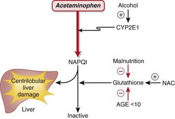
MOA (Mechanism of Action) (Figure 21-1)
 Acetaminophen is neither a narcotic nor a nonsteroidal antiinflammatory drug (NSAID). It is in a drug class of its own called aniline analgesics.
 Acetaminophen has analgesic and antipyretic effects similar to those of aspirin and NSAIDs and most likely exerts its effects through the inhibition of cyclooxygenase (COX).
 COX catalyzes the formation of prostaglandins (PGs) and other mediators that are important in the processing and signaling of pain and control of the thermoregulatory center in the brain.
 In contrast to NSAIDs and aspirin, acetaminophen is not an antiplatelet agent, nor does it possess antiinflammatory properties; therefore there are differences in the mechanism of action compared with aspirin or NSAIDs:
 Tissue selectivity: Acetaminophen demonstrates variable COX inhibition in different tissues. Of primary importance, it inhibits prostaglandin E2 (PGE2) production in the central nervous system (CNS), which is probably the primary mediator of its analgesic and antipyretic properties.
 COX site binding: COX possesses two different catalytic sites: a COX site and a peroxidase site. Acetylsalicylic acid (ASA) and NSAIDs inhibit the COX site, whereas acetaminophen inhibits the peroxidase site.
 Inhibition by hydroperoxide: Hydroperoxide is produced by macrophages, which are important inflammatory cells; the hydroperoxide at the sites of inflammation displaces acetaminophen from the peroxidase site and thus dramatically limits the potential antiinflammatory action of acetaminophen. Furthermore, platelet 12-lipoxygenase produces hydroperoxide in platelets, which is likely the explanation for lack of antiplatelet effect by acetaminophen.
Important Notes
 Therapeutic doses of acetaminophen have no effect on the cardiovascular and respiratory systems or platelet function and do not produce gastric irritation, erosion, or bleeding.
Overdose
 Acetaminophen exposure is the most commonly reported drug exposure reported to U.S. poison control centers.
 The therapeutic index is about 10: the toxic dose is 10 times greater than the therapeutic dose (15 mg/kg versus 150 mg/kg). For example, if you weigh 100 kg, then an upper limit of a therapeutic dose would theoretically be 1500 mg and a toxic dose would be 15,000 mg. To put this in perspective, tablets and suppositories are usually in the range of 325 to 650 mg each. A U.S. Food and Drug Administration (FDA) panel of experts in 2009 recommended that the maximum single dose of acetaminophen in adults be lowered from 1000 to 650 mg.
 Inadvertent co-ingestion of acetaminophen with other medications that also contain acetaminophen (such as cold remedies) is a cause of accidental overdose. Overdose can also occur with repeated subtoxic doses that, when combined over time, become toxic.
 Because of hepatic metabolism, the liver is the predominant organ that is initially injured, and fatal hepatic necrosis can occur in severe and untreated overdoses. In less severe conditions, significant liver injury and dysfunction can occur, resulting in an increase in transaminases (aspartate transaminase [AST], alanine transaminase [ALT]) and abnormalities in coagulation because of abnormal production of hepatic coagulation proteins.
 The mechanism of injury is as follows:
 Acetaminophen is metabolized in the liver via CYP2E1 to N-acetyl-p-benzoquinone imine (NAPQI). NAPQI is highly reactive, electrophilic, and toxic.
 When GSH stores become depleted, then NAPQI accumulates in the liver and starts to react with hepatocytes and proteins, causing permanent cellular damage. Injury is most prevalent where CYP2E1 activity is highest, which is in a centrilobular distribution.
 Treatment is specifically targeted at replenishing GSH stores. This is accomplished through the administration of a GSH precursor called N-acetylcysteine (NAC).
 NAC has been proposed to function as an antidote to acetaminophen via two additional mechanisms in addition to replenishment of GSH:
 NAC can substitute directly for GSH because it has antioxidant properties and directly reduces NAPQI.
Evidence
Analgesia
Acetaminophen versus Placebo for Treatment of Osteoarthritis
 A Cochrane review in 2005 (seven studies) demonstrated that acetaminophen was superior to placebo in five of the seven randomized controlled trials (RCTs). A pooled analysis demonstrated a statistically significant but minimal difference that is of questionable clinical significance. The relative percent improvement in pain score from baseline was 5%, with an absolute change of 4 points on a 0-to-100 scale.
Acetaminophen versus Nonsteroidal Antiinflammatory Drugs for Treatment of Osteoarthritis
 The same Cochrane review in 2005 (10 studies) demonstrated that acetaminophen was less effective overall than NSAIDs in terms of pain reduction, global assessments, and improvements in functional status. Patients taking traditional NSAIDS were more likely to experience an adverse GI event (relative risk [RR] 1.47). However, the median trial duration was only 6 weeks, which is too short to adequately assess adverse outcomes.
Acetaminophen plus Codeine versus Placebo
 A Cochrane review in 2008 (26 studies, N = 2295 patients) of postoperative patients demonstrated significant differences for obtaining at least 50% pain relief over 4 to 6 hours, with a number needed to treat (NNT) of 2.2 for high doses (800 to 1000 mg acetaminophen plus 60 mg codeine) and smaller effect sizes for medium and smaller doses (as low as 325 mg acetaminophen with 30 mg codeine).
Acetaminophen Plus Codeine versus Acetaminophen Alone
 A Cochrane review in 2008 (14 studies, N = 926 patients) of postoperative patients demonstrated that addition of codeine increased the proportion of participants achieving at least 50% pain relief over 4 to 6 hours by 10% to 15% and reduced the proportion of patients needing rescue medication by about 15%.
Opioids
MOA (Mechanism of Action)
 The pain pathways in the body are very complex and only briefly summarized here. In short, opioids reduce the signaling and processing of pain pathways through a variety of receptor types, receptor locations, and complex interactions.
 Exogenous opioids also bind multiple opioid receptors (Table 21-1). Opioid receptors are present in both the brain and the spinal cord, specifically:
 The descending pathways in the spinal cord decrease the pain processing that occurs in the spinal cord.
 The opioid receptors are G protein coupled to the inhibitory G protein (Gi), but the downstream effects of each receptor are dependent on the cell type where it is located.
TABLE 21-1 Main Classes of Opioid Receptors
| Receptor | Action |
|---|---|
| Mu (µ) | |
| Kappa (κ) | |
| Delta (δ) |
Pharmacokinetics (Table 21-2)
 For equivalency, note that:
 Fentanyl and sufentanil doses are measured in micrograms and are therefore much more potent than all other narcotics listed; remember that potency and efficacy are not the same.
 Routes of administration:
 Neuraxial (injected into the epidural or intrathecal spaces) administration by anesthesiologists is common as an adjunct to spinal or epidural anesthesia.
 Important metabolites:
 Most opioids are metabolized by CYP3A4 and renally eliminated (except where the following information states otherwise).
 Morphine → morphine-3-glucuronide (90%), morphine-6-glucuronide (10%).
• The glucuronides (being water soluble) are eliminated by the kidneys; renal disease can prolong and increase the effects of morphine.
• The glucuronides are water soluble; thus they do not readily cross the blood-brain barrier, but with high concentrations of drug, brain levels will increase.
• Morphine-6-glucuronide is more potent and longer acting than the parent compound morphine. Its accumulation can result in an increased and prolonged opioid effect
 Remifentanil is an ester and is metabolized within minutes by pseudocholinesterase. Its metabolites are essentially inactive.
Contraindications
 Decreased level of consciousness: There is an increased risk of further decreased level of consciousness and resultant respiratory compromise.
Side Effects
 Respiratory depression: All narcotics are powerful respiratory depressants. They can cause hypoventilation, hypoxia, and apnea. The effect is dose dependent. Low-efficacy opioids (codeine) are less likely to result in profound respiratory depression.
 Nausea and vomiting: Some patients are exquisitely sensitive. This is a very common side effect. The mechanism involves stimulation of the chemoreceptor trigger zone, the area of the brain responsible for vomiting.
 Pruritus (itching): This effect is not mediated by histamine (the most common mediator for itchy skin) but is rather a centrally (in the brain) mediated effect. Patients will often scratch their nose about 45 seconds after receiving intravenous fentanyl. For some patients the itching can be very uncomfortable and distressing and could mandate changing to a different opioid or stopping opioids completely.
 Constipation: This effect can be extremely severe. Codeine even in low doses can produce profound constipation. Mu receptors in the bowel are responsible for this side effect.
 Truncal rigidity: The thorax and abdomen can exhibit increased muscular tone. This effect is more common in patients administered high doses of the synthetic opioid fentanyl and related opioids. It usually does not occur with typical doses of morphine or codeine.
Important Notes
 The cardinal signs of opioid withdrawal include rhinorrhea, lacrimation, yawning, chills, piloerection (goose bumps), hyperventilation, hyperthermia, mydriasis, muscular aches, vomiting, diarrhea, anxiety, and hostility.
 A new antagonist called methylnaltrexone is a methylated version of naltrexone. It is special because it does not cross the blood-brain barrier. Therefore it has the ability to antagonize all the peripherally mediated side effects of narcotics (mostly constipation) without reducing the analgesic effects (which are mediated within the CNS). The most common indication for methylnaltrexone is treatment of constipation.
 Loperamide is an antidiarrheal opioid. It is absorbed very poorly from the gastrointestinal tract and therefore, when given orally, acts on mu receptors in the stomach, intestine, and colon only, reducing motility.
 Methadone is most commonly used to control withdrawal symptoms in patients who are recovering from narcotic addiction (usually heroin) because it has a long half-life. Furthermore, methadone is also an N-methyl-d-aspartate (NMDA) receptor antagonist, and this property has been implicated in a role of preventing tolerance to methadone. NMDA antagonists are also analgesics. Methadone also has monoamine oxidase inhibitor (MAOI) properties. MAOIs are antidepressants.
 Tramadol is a very weak mu agonist whose mechanism of action is predominantly based on blockade of serotonin reuptake. It does not cause respiratory depression. It can cause seizures, as can meperidine, which is in the same chemical class.
 The partial agonists all have kappa receptor activity and only limited mu receptor activity. This is significant because the mu receptor mediates analgesia, so partial agonists are less effective analgesics than full agonists.
 Opioid antagonists can precipitate severe withdrawal in patients who are physically dependent on opioids. They should be administered carefully.
 Tolerance: Opioid receptor down-regulation occurs with chronic administration of opioids. The result is that higher and higher doses are required to achieve the same effect. Neuromodulation is another term for tolerance.
 Although respiratory depression is a side effect of opioids, the same mechanism is responsible for reducing the ventilatory drive in patients who are short of breath; therefore opioids can reduce the unpleasant sensation of breathlessness and can be effective at relieving discomfort related to breathing in selected patients.
Evidence
Opioids versus Nonsteroidal Antiinflammatory Drugs for Treatment of Renal Colic
 A systematic review in 2004 (20 trials, 1613 participants) found that both NSAIDs and opioids led to clinically important reductions in patient-reported pain scores. Pooled analysis of six trials showed a greater reduction in pain scores for patients treated with NSAIDs than with opioids. Patients treated with NSAIDs were significantly less likely to require rescue analgesia (RR 0.75). Most trials showed a higher incidence of adverse events in patients treated with opioids. Compared with patients treated with opioids, those treated with NSAIDs had significantly less vomiting (0.35).
Opioids for Treatment of Chronic Back Pain
 A systematic review in 2007 examined multiple questions pertaining to opioid treatment of chronic low back pain. 11 studies showed that there was significant variation in how opioids are prescribed for chronic low back pain. A meta-analysis of the four studies assessing the efficacy of opioids compared with placebo or a nonopioid control did not show reduced pain with opioids. A meta-analysis of the five studies directly comparing the efficacy of different opioids demonstrated a nonsignificant reduction in pain from baseline.
 With respect to risk of addiction, the prevalence of lifetime substance use disorders ranged from 36% to 56%; the prevalence of current substance use disorders was as high as 43%; and aberrant medication-taking behaviors (“drug seeking”) ranged from 5% to 24%. The authors found that the study was limited by retrieval and publication biases and poor study quality. No trial evaluating the efficacy of opioids was longer than 16 weeks.
FYI
 The term opioid refers broadly to all compounds related to opium, although only morphine and codeine are actually produced by the opium poppy, Papaver somniferum.
 The name morphine is from Morpheus, the Greek god of dreams. Morphine was first isolated in 1803 by Sertürner.
 The word narcotic is derived from the Greek word meaning stupor. The term narcotic is often used in a legal context to refer to a variety of illegal substances; the term in medical vocabulary refers only to opioids.
 Etorphine is an opioid with about 2000 times the potency of morphine. It is used as a large animal immobilizer (for veterinary use). One drop on the skin of a human would be fatal because of the profound respiratory depression. Carfentanil, another veterinary opioid, is 10,000 times more potent than morphine (and 10 times more potent than sufentanil, the most potent opioid for humans). Nonhuman mammals do not experience the same degree of respiratory depression as humans do, and so these drugs are suitable as immobilizing agents in large animals.
α2 Agonists
MOA (Mechanism of Action)
 Glaucoma is characterized by increased intraocular pressure (IOP). Strategies to reduce intraocular pressure include reducing the production and secretion of aqueous humor and facilitating its drainage.
 The exact mechanism by which α2 agonists reduce IOP has not been established, but is likely multifactorial, employing several strategies:
 Through a secondary mechanism, brimonidine may also have a neuroprotective role, mitigating the damage to the optic nerve caused by the elevated IOP.
 The antihypertensive effect of α2 agonists is a result of inhibition of presynaptic release of vasoconstrictors such as norepinephrine. Recall that the α2 receptor is an autoreceptor (see Chapter 3). An autoreceptor is a receptor that when stimulated by an agonist, reduces release of transmitter into the synaptic cleft.
Pharmacokinetics
 Brimonidine and apraclonidine are administered topically to the eye, and only a small amount reaches the systemic circulation.
Contraindications
 Concomitant MAOIs: MAOIs prevent the breakdown of norepinephrine, and this could lead to elevated blood pressure.
Side Effects
Oral Use
 Sedation: Stimulation of central α2 receptors has an inhibitory effect on neurotransmitter release. This is the desired effect when used for sedation.
 Bradycardia, likely caused by inhibition of catecholamine release, can be significant in some individuals.
Important Notes
 The ability of clonidine to modulate neurotransmitter release has also led to its increasing use in attention-deficit/hyperactivity disorder (ADHD).
Evidence
In Primary Open Angle Glaucoma and Ocular Hypertension
 A 2007 Cochrane review of all medical interventions for glaucoma and ocular hypertension found three trials comparing brimonidine to timolol. There were no differences in visual field progression (glaucoma) or visual field defects (ocular hypertension) within 1 year. Timolol was better tolerated than brimonidine, as measured by the incidence of dropouts from drug-related adverse events (OR 0.21). There were no trials comparing brimonidine with placebo.
Inhaled Anesthetics
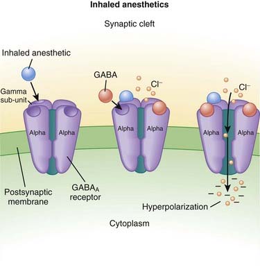
MOA (Mechanism of Action) (Figure 21-2)
 The primary site of action in causing CNS depression is most likely the γ-aminobutyric acid A (GABAA) receptor. Inhaled anesthetics activate these receptors.
 The GABAA receptor is linked to a chloride channel; this is important because one way to turn off a neuron is to hyperpolarize it so that it cannot be depolarized enough to trigger an action potential. Opening a chloride channel will allow the negatively charged chloride ion to enter the cell and will reduce the electrical charge inside the cell (thereby hyperpolarizing it) and effectively render the neuron unresponsive to incoming stimuli that would otherwise depolarize the cell.
Pharmacokinetics
 As their name suggests, these drugs are administered only via inhalation. They are supplied in liquid form and then vaporized using very precise vaporizers that are part of the anesthetic machine, the anesthetic oxygen and air mixture is combined with calculated doses of the inhaled anesthetic.
 Very small amounts of the drug are metabolized. For example, desflurane undergoes <0.02% metabolism, which is clinically insignificant. Older drugs had higher rates of metabolism, with halothane as high as 40%.
 Inhaled anesthetics move through the body by dissolving in the blood and distributing into tissues; the important tissue interfaces include the following:
 Blood ∂ fat (and other vessel-poor tissues)
• When inhaled anesthetics are administered, the drug must enter the lungs, then the blood, then the brain.
• For inhaled anesthetics to be eliminated, drug must exit the brain and other tissues, be carried to the lungs, and then exhaled.
• Vessel-poor tissues are slow to take up and release drug. Therefore vessel-poor tissues can act as a sink and slowly absorb drug at the early parts of an anesthetic procedure (lowering the drug levels) but then at the end of a long anesthetic procedure can release drug, prolonging elimination of the drug.
 The solubility of an inhaled anesthetic affects the speed at which it is taken up by the body and exerts its action (speed of onset). The key point is that the partial pressure is the measure of the drug’s active form. If a drug has a high solubility, then a lot of drug needs to be absorbed into blood and tissues before the partial pressure starts to rise; this impedes the onset of action of the drug. If the solubility is low, then the partial pressure will rise quickly. Conversely, eliminating the drug follows the same rules, and high solubility correlates with slower elimination because more drug had to be dissolved into the body initially to achieve the desired partial pressure.
• Solubility is described for the blood and is called the blood/gas coefficient. The smaller the number, the less soluble the drug is in blood and therefore the faster it can change its partial pressure (because only a small amount of drug actually needs to be dissolved into the blood). The agents are ranked from fastest to slowest (lowest to highest solubility coefficient) as follows:
Side Effects
 Respiratory depression: The respiratory drive in response to carbon dioxide is blunted, and blood CO2 levels rise. The tidal volume is reduced, but the respiratory rate is actually increased (the reverse occurs with opioid respiratory depression).
 Hepatic damage: Very rare cases (1 in 30,000) have possibly been associated with halothane, but clearly defined cause and effect have not been established
Important Notes
 The potency of inhaled anesthetics is measured by the minimum anesthetic concentration (MAC), which is strictly defined as the dose of inhaled anesthetic required to prevent movement in 50% of the population in response to a surgical stimulus when no other drugs are administered. For example, the MAC of desflurane is 6%. Note that this definition is not the same as the dose required to keep someone asleep. Drugs such as opioids decrease the MAC requirements of a drug and are called MAC-sparing agents.
 Inhaled anesthetics cause hypotension. The primary mechanism is through vasodilation, although older agents (e.g., halothane) produced cardiac depression (decreased contractility and decreased heart rate), which caused hypotension.
 In addition to being vascular smooth muscle relaxants, inhaled anesthetics also relax bronchial smooth muscle and through this mechanism can help to relieve bronchospasm in rare cases of life-threatening refractory status asthmaticus.
 MH is a rare condition that is precipitated by inhaled anesthetics or succinylcholine (a paralyzing agent). It is caused by a channelopathy, which is a mutation in an ion channel in muscle. It is a life-threatening condition that occurs as a result of pathologically high levels of skeletal muscle contraction from increased intracellular calcium that occurs in the absence of neuromuscular stimulation. Because of the muscular contraction, the following effects occur:
 MH is treated with dantrolene, a drug that reduces intracellular calcium through binding the ryanodine receptor, which mediates calcium release from the sarcoplasmic reticulum.
Advanced
 Nitrous oxide has a MAC value of 104%. Therefore, it is not potent enough to be a solo anesthetic agent, because delivering a mixture of 100% nitrous oxide would mean that no oxygen could be delivered and the patient would die of asphyxiation. The greatest concentration that is administered is about 60% to 70%—a MAC value of 0.6 or 0.7. For this reason, nitrous is used only as an adjuvant drug for general anesthesia and is added to one of the other volatile anesthetics.
FYI
 Ether was the first inhaled anesthetic. Most inhaled anesthetics (not including nitrous oxide or xenon) are fluorinated (fluorine is a halogen, thus the name “halogenated”) derivatives of ether:
 Earlier inhaled anesthetics such as chloroform and cyclopropane were flammable. They were mixed with oxygen, and the risk of explosion and fires resulted in their replacement.
Intravenous Anesthetics
MOA (Mechanism of Action)
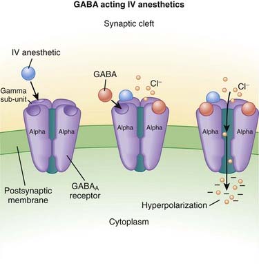
γ-Aminobutyric Acid (GABA) (Figure 21-3)
 GABA is the major inhibitory neurotransmitter in the CNS. GABA binds to three different types of receptors: GABAA, GABAB, and GABAC.
 Binding of GABA to GABAA receptors leads to the opening of the chloride (Cl−) channel, facilitating Cl− influx and cellular hyperpolarization (making the inside more negative).
 Thiopental, propofol, and etomidate all bind to GABAA receptors and increase the influx of Cl− into the neuron, resulting in decreased neuronal activity.
Pharmacokinetics
 Intravenous anesthetics work within one arm to brain circulation time; that is, if the drug is administered through an intravenous line in the arm, it is the time required to circulate back to the heart and then up to the brain (usually less than 1 minute).
 Compartmental distribution is a very important factor for intravenous anesthetics. When the drug is administered into the blood, it rapidly is taken up by the brain (inducing its effect), but very quickly the drug is also taken up by the vessel-rich organs (liver, muscle, kidney, heart), and this uptake quickly reduces the levels in the blood and therefore the brain in a matter of minutes. Therefore when the drug is given as a single bolus, the termination of action of the drug is primarily a result of redistribution of the drug and not metabolism or elimination of the drug. When the drug is given as an infusion, metabolism and elimination become important because all compartments would be equilibrated.
Side Effects
 Respiratory depression: The respiratory center is depressed with GABA agonists. It is typical for a patient who is administered a dose large enough to induce unconsciousness to develop apnea (to completely stop breathing). For this reason, administration of intravenous anesthetics (regardless of dose) must always be performed in the presence of healthcare workers who are skilled in respiratory resuscitation and who have respiratory resuscitation equipment immediately available.
 Hypotension: Intravenous anesthetics, especially propofol, are myocardial depressants. They decrease contractility and often also reduce sympathetic nervous system activity.
 Addiction: All drugs in this category both are psychologically addicting and induce physiologic dependence, tolerance, and withdrawal.
Important Notes
 Dosage: Some general concepts should be applied when choosing the dose of intravenous anesthetic. Administering a larger-than-required dose virtually guarantees significant hypotension:
 Elderly patients require lower doses compared with nonelderly adults, and children require higher doses (on a milligram-per-kilogram scale) compared with adults.
 Chronic alcohol consumers require higher doses because alcohol also works on GABA receptors and results in GABA agonist tolerance owing to receptor down-regulation. In fact, consumption of one alcoholic drink per day is enough to change responsiveness to GABA drugs.
 Ketamine:
 In contrast to GABA-mimetic drugs, ketamine:
• Is dysphoric: It produces very unpleasant sensations, hallucinations, and bad dreams. A benzodiazepine can be quite effective in reducing the dysphoria and is commonly coadministered with ketamine
 Causes sympathetic nervous system activation (resulting in increased heart rate and blood pressure) and therefore is the least likely to induce hypotension. However, it is still a direct myocardial depressant (reduces contractility), and in states in which the sympathetic nervous system is already highly activated (e.g., cardiogenic shock), ketamine retains potential for inducing hypotension.
 Causes bronchodilation (via sympathetic nervous system activation) and is therefore useful for intubation of patients with severe bronchospasm (asthmatic patients).
 Propofol:
 Is a strong myocardial depressant and should be given very cautiously, if at all, in patients with a weak heart (low systolic function).
 Is lipophilic and does not dissolve in water; therefore a lipid-containing, milk-colored emulsion is the vehicle in which it is administered. The emulsion stings veins when it is injected. It is often mixed with lidocaine, a local anesthetic, to reduce the stinging. A newer formulation called fospropofol is water soluble.
Advanced
 Etomidate and propofol preferentially act at GABAA receptors that contain β1 and β2 subunits (not to be confused with the autonomic receptors with similar names). The β2 subunit is probably the more important for mediating hypnotic and muscle-relaxing actions.
 Barbiturates also facilitate the actions of GABA at multiple sites in the CNS, but in contrast to benzodiazepines they appear to increase the duration of the GABA-gated chloride channel openings. At high concentrations the barbiturates may also be GABA-mimetic, directly activating chloride channels.
FYI
 Dextromethorphan, an NMDA antagonist, is a commonly used cough suppressant and is found in many over-the-counter cough remedies.
 The flavor-enhancing chemical MSG is monosodium glutamate; it has the potential to increase neuronal activation via increased glutamate. Seizures can be induced in animal models with MSG.
Local Anesthetics
MOA (Mechanism of Action)
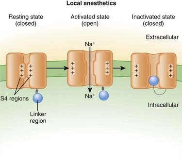
 Neuronal transmission requires that an action potential be propagated from one end of a neuron to the other. Voltage-gated sodium channels open up as the wave of depolarization travels from one end of the neuron to the other. The opening of these ion channels permits sodium to enter the cell, causing depolarization, and is the primary method by which the wave of depolarization occurs.
 Local anesthetics bind these voltage-gated sodium channels. The ion channels can exist in three states: resting, activated (open), and inactivated. The local anesthetic binds them in the inactivated state and prevents them from transitioning to the open state; thus the ion channel remains closed and unresponsive to incoming depolarizing currents (Figure 21-4).
 The local anesthetic works on the intracellular side of the ion channel. Therefore the drug must first cross the lipid cell membrane before it can exert its action.
 Neurons that are covered in myelin are more difficult to block because the myelin impedes entry of the drug into the cell.
 Larger-diameter nerves are more difficult to block than are smaller-diameter nerves. They require higher doses and are blocked for shorter durations.
 There are three major classes of nerve function: sensory, motor, and autonomic. All types of nerves are susceptible to blockade by local anesthetic, but to varying degrees because of myelination and diameter:
 Motor nerves are large and myelinated; they are the hardest to block (and first to recover from blockade).
 Voltage-gated sodium channels are the primary ion channels in phase 0 of the cardiac action potential. Therefore, local anesthetics are also classified as antiarrhythmics, specifically type 1b. This mechanism of action is discussed in more detail in the discussion of Na+ channel blockers in Chapter 11 and is also responsible for cardiac toxicity of local anesthetics.
Pharmacokinetics
 Local anesthetics are most commonly injected, but other routes of administration include topical application: oral sprays, creams, and vaporized forms (for airways).
 There are important factors that dictate the potency, speed of onset, and duration of a local anesthetic:
 Lipid solubility: Increased lipid solubility results in more drug being able to cross the cell membrane and bind the ion channel. The property of increased lipid solubility influences:
 pH: Local anesthetics are weak bases. Therefore when the environment is acidic, the following reaction occurs: B + H+ → BH+ (where B = base and represents the local anesthetic). From this equation, it can be seen that local anesthetics will be ionized (BH+) and therefore hydrophilic (not lipid soluble) in an acid environment. Therefore, acidic tissues (such as an abscess or any infection) are very difficult to anesthetize with local anesthetic. An acid pH will decrease potency, speed of onset, and duration.
 Ester local anesthetics are predominantly metabolized via ester hydrolysis by pseudocholinesterase. Ester hydrolysis is a fast reaction, and therefore ester local anesthetics have a shorter duration of action.
 Amide local anesthetics are metabolized (N-dealkylation and hydroxylation) by microsomal P-450 enzymes in the liver (Table 21-3).
TABLE 21-3 Local Anesthetic Durations
| Agent | Duration Plain (minutes) | Duration with Epinephrine (minutes) |
|---|---|---|
| 2-Chloroprocaine | 20-30 | 30-45 |
| Procaine | 15-30 | 30 |
| Lidocaine | 30-60 | 120 |
| Mepivacaine | 45-90 | 120 |
| Prilocaine | 30-90 | 120 |
| Bupivacaine | 120-240 | 180-240 |


