Neurofibroma
Key Facts
Terminology
Neurofibroma (NF)
Etiology/Pathogenesis
Proliferation of perineural fibroblasts, Schwann cells, and neurites
Clinical Issues
One of most common neural tumors
Usually adults in 3rd or 4th decade of life
Location
Posterior mediastinum
Symptoms
Chest pain
Dyspnea
Cough
Neurofibromatosis
Some patients may be asymptomatic
Treatment
Complete surgical resection
Prognosis
Good
Top Differential Diagnoses
Schwannoma
Neurofibrosarcoma
Ganglioneuroma
Solitary fibrous tumor
Leiomyoma
Diagnostic Checklist
Spindle cell proliferation without mitotic activity
Loose fibrocollagenous or myxoid stroma
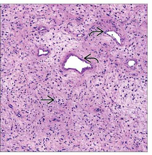 Mediastinal neurofibroma shows a classic spindle cell proliferation  embedded in a rather loose fibroconnective tissue admixed with dilated vascular structures embedded in a rather loose fibroconnective tissue admixed with dilated vascular structures  . . |
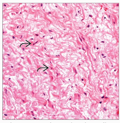 High-power view of a mediastinal neurofibroma shows a spindle cell proliferation  admixed with fibrocollagenous tissue. Note the absence of cellular pleomorphism. admixed with fibrocollagenous tissue. Note the absence of cellular pleomorphism. |
TERMINOLOGY
Abbreviations
Neurofibroma (NF)
Definitions
Benign neural neoplasm
ETIOLOGY/PATHOGENESIS
Pathogenesis
Proliferation of perineural fibroblasts, Schwann cells, and neurites
CLINICAL ISSUES
Epidemiology
Incidence
One of most common neural tumors
Age
Usually adults in 3rd or 4th decade of life
Gender
No apparent gender predilection
Site
Posterior mediastinum
Presentation
Chest pain
Dyspnea
Cough
Neurofibromatosis (von Recklinghausen disease)
Some patients may be asymptomatic
Treatment
Surgical approaches
Complete surgical resection
Prognosis
Good
MACROSCOPIC FEATURES
General Features
Well circumscribed but not encapsulated
Light tan in color
Sections to Be Submitted
In cases of patients with history of neurofibromatosis, careful sampling to rule out sarcoma
Size
Variable size
May be up to or > 10 cm in diameter
MICROSCOPIC PATHOLOGY
Histologic Features
Spindle cell proliferation
Wavy nuclei and pointed ends
Loose fibroconnective tissue
Absent mitotic activity
In some cases, stroma may be myxoid
Occasional Wagner-Meissner bodies may be seen
Pacini corpuscles may be seen
Mucin-producing glands may be seen focally
Melanin pigment may be present
DIFFERENTIAL DIAGNOSIS


