Nervous System, Skeletal Muscle, and Eye
Elisabeth J. Rushing
Mariarita Santi-Vicini
▪ Questions and Answers
1. This lesion was removed from a 40-year-old man who presented with headaches and ataxia (Fig. 13.1). What is the most likely diagnosis in this case?
a. Subependymal giant cell astrocytoma
b. Pilocytic astrocytoma
c. Arteriovascular malformation
d. Hemangioblastoma
e. Angioblastic glioma
View Answer
1. d. Hemangioblastoma is a hypercellular, vascular tumor containing foamy interstitial cells, which represent the actual neoplastic element. Hemangioblastomas occur most commonly in the cerebellum and are often associated with von Hippel-Lindau syndrome. Subependymal giant cell astrocytoma is an intraventricular tumor (lateral ventricles) resembling gemistocytic astrocytoma that is usually seen in the context of tuberous sclerosis. Pilocytic astrocytomas are biphasic neoplasms composed of alternating areas of compact spindle cells and more loosely textured zones and often accompanied by Rosenthal fibers.
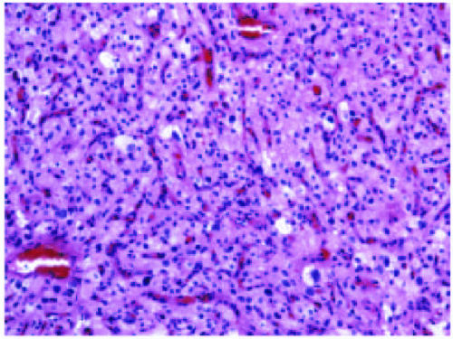 Figure 13.1 |
2. Microscopic examination of this hippocampus (Fig. 13.2) from a 68-year-old patient who suffered a cardiac arrest during surgery and survived on a respirator for 2 weeks would most likely show the following:
a. Neurofibrillary tangles in the dentate gyrus
b. Red, pyknotic neurons (acute neuronal injury) in CA1
c. Granulovacuolar degeneration
d. Hirano bodies
e. Red, pyknotic neurons (acute neuronal injury) in CA2
View Answer
2. b. Acute hypoxic/ischemic injury is associated (if the patient survives at least six hours) with shrunken eosinophilic neurons. Certain populations of neurons are selectively vulnerable, especially large neurons in CA1 sector of the hippocampus, the Purkinje cells of the cerebellum, and neurons of layers 3 through 5 of the cerebral cortex. Neurofibrillary tangles, granulovacuolar degeneration, and Hirano bodies are neurodegenerative changes. CA2 is the so-called “resistant zone” of the hippocampus that is less vulnerable to hypoxia/ ischemia.
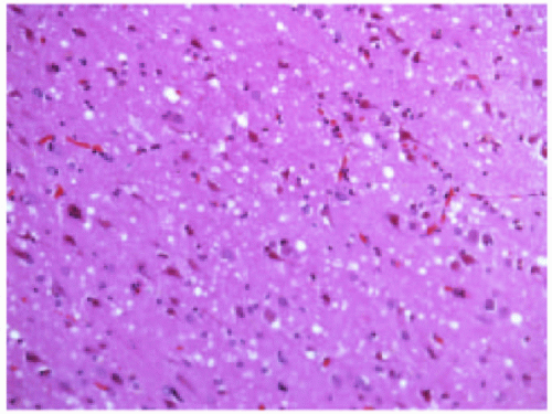 Figure 13.2 |
3. Which one of the following statements does NOT apply to the abnormality depicted in this gross dissection of the circle of Willis (Fig. 13.3)?
a. Associated processes include polycystic kidney disease and coarctation of the aorta.
b. Common at the middle cerebral trifurcation.
c. A familial inheritance pattern has been noted in fewer than 2% of intracranial aneurysms.
d. This condition is common in infants and children.
e. Hemorrhage and secondary infarcts are potential sequelae.
View Answer
3. d. Berry aneurysms involving the circle of Willis are uncommon in children. The photomicrograph shows the basilar aspect of the brain with a berry aneurysm at the trifurcation. The risk of rupture, which produces hemorrhage into the subarachnoid space at the base of the brain, is greatest for females in the fifth decade.
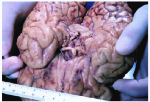 Figure 13.3 |
4. What is the most likely diagnosis based on this photomicrograph (Fig. 13.4)?
a. Multisystem atrophy
b. Unverricht epilepsy
c. Adult polyglucosan disease
d. North American blastomycosis
e. Parkinson disease
View Answer
4. e. Lewy bodies are inclusion bodies found in Parkinson disease and diffuse Lewy body disease. In pure Parkinson disease, Lewy bodies are located predominantly in the pigmented neurons in the substantia nigra (as depicted in the photomicrograph) and locus ceruleus, whereas in diffuse Lewy body disease, they are also found in the cerebral cortex.
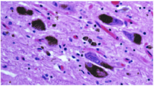 Figure 13.4 |
5. The differential diagnosis in a 7-year-old child who presented with gait ataxia and this finding (Fig. 13.5) at autopsy should include which of the following entities?
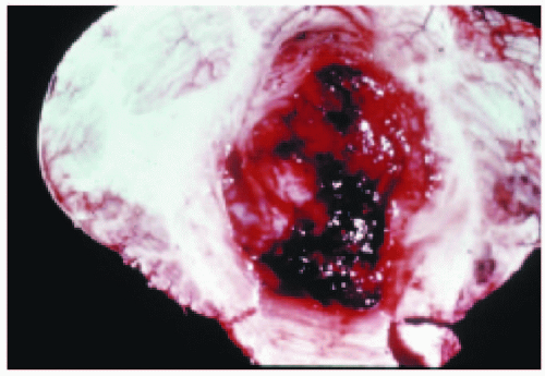 Figure 13.5 |
a. Glioblastoma multiforme
b. Medulloblastoma
c. Metastasis
d. Hemangioblastoma
View Answer
5. b. Medulloblastoma and ependymoma are the second and third most common primary central nervous system (CNS) neoplasms in children. Histopathologically, medulloblastomas are densely cellular “small blue cell” tumors that typically contain frequent mitoses and apoptotic nuclei, and sometimes frank necrosis. Ependymomas are glial neoplasms that are usually recognized by the presence of perivascular pseudorosettes. Overall, low-grade astrocytomas are the most common. In very young children, medulloblastoma is more common than astrocytic neoplasms. Glioblastoma multiforme and metastatic disease are extremely uncommon in children and are much more likely to occur in the over-60 population.
6. A 14-year-old boy with this gross finding (Fig. 13.6) most likely presented with the following clinical manifestation:
a. New-onset seizures
b. Hemiballismus
c. Parinaud syndrome
d. Cortical blindness
e. Opsoclonus
View Answer
6. c. The gross photo shows a midsagittal section of brain with a large mass in the region of the posterior third ventricle. The most common entity in this location in children is a pineal germinoma. Parinaud syndrome is associated with dorsal midbrain (pineal region) lesions and is characterized by vertical gaze palsy, convergenceretraction nystagmus, and light-near dissociation. Seizures are typically related to cortical lesions, and hemiballismus occurs with lesions of the subthalamic nucleus. Cortical blindness is seen with bilateral impairment of the posterior occipital lobes, especially those lesions that involve the calcarine fissures bilaterally.
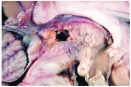 Figure 13.6 |
7. Which of the following immunohistochemical stains is the most useful for demonstrating axonal spheroids associated with diffuse axonal injury?
a. Vimentin
b. Epithelial membrane antigen
c. Glial fibrillary acidic protein
d. CD68
e. β-amyloid precursor protein
View Answer
7. e. Damage to axons, also termed diffuse axonal injury (DAI) or traumatic axonal injury (TAI), is an almost universal finding in cases of mild, moderate, and severe head trauma. Although not specific for trauma, β-amyloid precursor protein (β-APP) is a useful stain for highlighting axonal spheroids.
8. This is a coronal section from a 52-year-old man (Fig. 13.7). Which statement is FALSE?
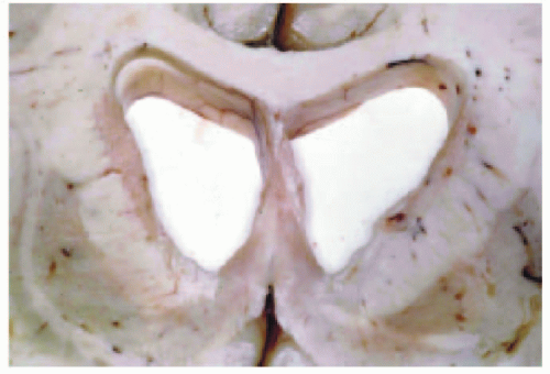 Figure 13.7 |
a. This disease selectively causes atrophy of the caudate nucleus but spares the putamen.
b. Neuronal loss preferentially affects spiny neurons.
c. CAG trinucleotide repeats characterize this disease.
d. Later generations are affected at an earlier age and often more severely.
e. Clinically, patients usually present with echolalia.
View Answer
8. e. Huntington disease is a rare autosomal dominant inherited disease with a mutation in the IT15 gene, located on chromosome 4, which alters the huntingtin protein. There are increased CAG tandem repeat sequences in the gene. The gross photo shows a coronal section of brain with bilateral effacement of the caudate nucleus. Choreiform movement and not echolalia characterize the disease. Echolalia is more common in autism.
9. Which statement applies to the condition associated with this image (Fig. 13.8)?
a. Filaments within the inclusions contain alpha-synuclein.
b. The nucleus basalis of Meynert is spared.
c. Extensive involvement of the putamen is associated with a type of dementia.
d. Immunohistochemical studies have shown that inclusions in this disease contain significant amounts of gamma amino butyric acid (GABA).
e. Patients typically present with seizures.
View Answer
9. a. In cases of idiopathic Parkinson disease, the brain shows pallor of the substantia nigra and locus ceruleus accompanied by neuronal loss, extracellular pigment, and gliosis. The remaining neurons may contain Lewy bodies, which are eosinophilic cytoplasmic structures. Ultrastructurally, Lewy bodies are composed of filaments that form the densely packed core. These filaments are made of alpha-synuclein, a lipid-binding protein that can be demonstrated by immunohistochemical methods (image b).
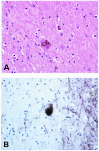 Figure 13.8 |
10. Which of the following is most likely associated with lobar hemorrhage in the elderly?
a. Metastatic prostate adenocarcinoma
b. Ruptured arteriovenous malformation
c. Cerebral amyloid angiopathy
d. Mycotic aneurysm
e. Capillary telangiectasia bleed
View Answer
10. c. Cerebral amyloid angiopathy is the most common cause of lobar hemorrhage in the elderly. The amyloidogenic proteins, nearly the same as those found in Alzheimer disease, are deposited in meningeal and cerebral vessels, thus weakening their walls and predisposing to hemorrhage.
11. A 57-year-old man was found deceased in his lakeside cabin in northern Maine where he was ice fishing. At autopsy, a coronal section of brain showed bilateral hemorrhagic lesions in the globus pallidus (Fig. 13.9). Which of the following is the most likely cause of death?
a. Blunt trauma to the head
b. Anticoagulant overdose
c. Massive ischemic cerebrovascular accident in the distribution of the middle cerebral artery
d. Cerebral aspergillosis
e. Carbon monoxide poisoning
View Answer
11. e. Carbon monoxide poisoning typically affects both globus pallidi and chronic lesions are cavitary.
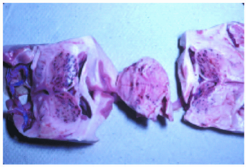 Figure 13.9 |
12. A 45-year-old missionary returned from Africa suffering from fever and mental confusion. He deteriorated rapidly and died. An autopsy was performed. Which statement does NOT apply to the entity likely diagnosed by the pathologist?
a. These changes are the result of infection with Plasmodium ovale.
b. Ring hemorrhages in the brain are a major feature.
c. People with sickle cell trait are less likely to die from infection.
d. Initial infection often causes enlargement of the spleen.
e. Sequestration of infected red blood cells in the blood-brain barrier endothelium is central to the pathologic progression.
View Answer
12. a. Cerebral malaria is not caused by P. ovale, but by Plasmodium falciparum, which infects red blood cells, causing them to clump together (rosetting) and stick to endothelial cells (sequestration). Accordingly, cerebral blood vessels are plugged with parasitized red blood cells containing dots of hemozoin pigment, which leads to stasis and tissue hypoxia. This sets the stage for ring hemorrhages and small collections of inflammatory cells known as Dürck granulomas.
13. A 28-year-woman presented with unilateral visual impairment that progressed over the course of several days. Six months later, she developed ataxia and cranial nerve signs. Which one of the following applies to the pathologic findings (Fig. 13.10)?
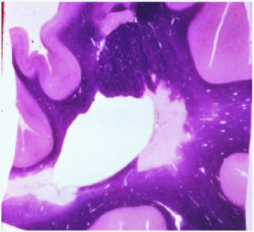
b. Lesions are commonly found adjacent to the lateral ventricles.
c. Microscopically, lesions are often macrophage-rich.
d. The Marburg variant is a fulminating form that may be confused with a neoplasm.
e. All of the above
View Answer
13. e. All of these statements apply to the diagnosis of multiple sclerosis. The photomicrograph shows a sharply demarcated focus of demyelination (myelin pallor) adjacent to the anterior horn of the lateral ventricle in a section stained for myelin.
14. Which statement applies to the condition depicted in the photomicrograph (Fig. 13.11)?
a. The pathologic changes are a result of Heubner arteritis.
b. The disease is caused by various species of Ixodes tick.
c. Proteinase K-resistant PrPsc can be detected by immunostaining or Western blotting.
d. It is associated with lactic acidosis and stroke-like episodes.
e. The disease is caused by deficiency of glutaryl-CoA dehydrogenase.
View Answer
14. c. Creutzfeldt-Jacob disease is a form of spongiform encephalopathy thought to be caused by prions that manifest clinically by rapidly progressive dementia. The spongiform change is particularly prominent in the cerebral cortex or gray matter of the brain. Accumulation of the abnormal beta-pleated sheet isoform, termed PrPsc for PrP (protease resistant) and (scrapie) is considered responsible for the pathogenesis of this disease.
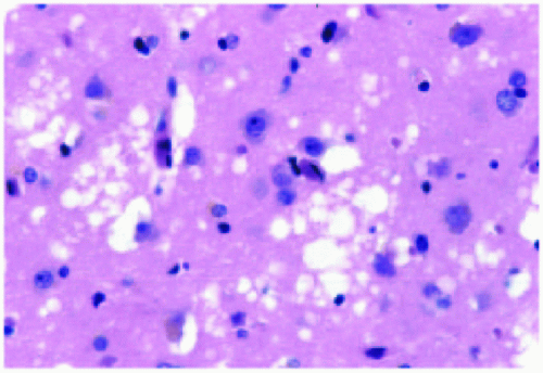 Figure 13.11 |
15. Which of the following genetic alterations is most likely to be associated with this tumor that arose in a 68-year-old man who presented with sudden-onset right hemiparesis (Fig. 13.12)?
a. Inactivation of p53 and overexpression of PDGF-A
b. Amplification of epidermal growth factor receptor (EGRF)
c. Mutation of the copper-zinc superoxide dismutase gene
d. Mutation in the Notch-3 gene
e. The A/ND5 mutation in mtDNA is pathogenic
View Answer
15. b. Glioblastoma multiforme is an extremely heterogeneous tumor characterized by varying degrees of increased cellularity, pleomorphism, mitoses, endothelial proliferation, and necrosis. Pseudopalisading necrosis shown in the photomicrograph is a common feature. Amplification of epidermal growth factor receptor (EGFR) typically indicates a primary (de novo) glioblastoma multiforme, whereas its absence suggests a secondary glioblastoma.
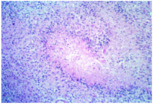 Figure 13.12 |
16. This 38-year-old woman presented with headaches of increasing severity. Which statement does NOT apply to the entity depicted in this gross photograph (Fig. 13.13)?
a. It is the most commonly diagnosed primary brain tumor of adults.
b. Sporadic meningiomas have been linked to chromosome 22 in the region of the NF2 gene.
c. The tumor expresses estrogen and progesterone receptors.
d. Clear cell and chordoid are WHO grade 2 variants.
e. Occasionally present as extracranial tumors.
View Answer
16. a. The gross photo shows a dural-based meningioma and not glioblastoma multiforme, the most common primary brain tumor in adults.
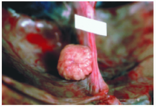 Figure 13.13 |
17. What is the approximate weight of the brain at the time of birth of a full-term infant?
a. 120 grams
b. 240 grams
c. 800 grams
d. 1450 grams
e. 360 grams
18. Which statement is NOT true about lenticular opacities of the eye?
a. Risk factors include steroids, diabetes, Wilson disease, and aging.
b. Accumulation of urochrome alters the perception of blue color.
c. A hypermature form is known as Morgagnian opacities.
d. Typically develop in eyes with shallow anterior chambers.
e. Congenital cataracts may show autosomal dominant inheritance.
View Answer
18. d. Cataracts represent lenticular opacities. Primary closed angle glaucoma develops in eyes with shallow anterior chambers.
19. Which one of the following entities is linked to highoutput cardiac failure?
a. Telangiectasia
b. Berry aneurysm
c. Aneurysm of the great vein of Galen
d. Arteriovenous malformation
e. None of the above
View Answer
19. c. Vein of Galen aneurysm is a rare congenital malformation of blood vessels that results in high-output congestive heart failure or may present with developmental delay, hydrocephalus, and seizures. The vein of Galen drains the anterior and central regions of the brain into the sinuses of the posterior cerebral fossa.
20. Which herniation category results in pupillary dilation and impairment of ocular motility on the side of the lesion?
a. Subfalcine
b. Transtentorial
c. Insular
d. Tonsillar
e. None of the above
View Answer
20. b. Transtentorial or mesial temporal herniation occurs when the medial aspect of the temporal lobe is compressed against the tentorium cerebelli. When this occurs, displacement of the temporal lobe compromises the third cranial nerve, resulting in pupillary dilatation and impaired ocular movements.
21. Epidural hematoma is most commonly associated with which one of the following lesions?
a. Rupture of the bridging veins
Stay updated, free articles. Join our Telegram channel

Full access? Get Clinical Tree


