Myelolipoma
Cyril Fisher, MD, DSc, FRCPath
Key Facts
Terminology
Tumor-like lesion resembling bone marrow composed of hemopoietic tissues, fat, and sometimes bone
Most cases involve adrenal gland
Rarely other sites
Clinical Issues
Older adults, usually after 4th decade
Microscopic Pathology
Circumscribed, nonencapsulated
Rim of adrenal cortex
Fat without atypia or lipoblasts
Hemopoietic elements
All 3 cell lines represented
Diagnostic Checklist
Typically circumscribed adrenal mass
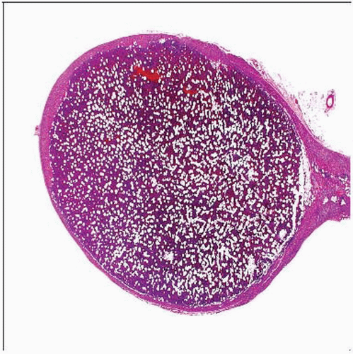 Hematoxylin & eosin shows scanning magnification appearance of myelolipoma. Adrenal glandular tissue is stretched around a circumscribed, nonencapsulated lesion that displays fatty and cellular areas. |
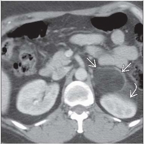 CT shows a predominantly fatty mass  replacing and expanding the left adrenal gland. The left kidney replacing and expanding the left adrenal gland. The left kidney  is displaced. These imaging appearances are characteristic of an adrenal myelolipoma. is displaced. These imaging appearances are characteristic of an adrenal myelolipoma. |
TERMINOLOGY
Definitions
Tumor-like lesion resembling bone marrow composed of hemopoietic tissues, fat, and sometimes bone
Mostly in adrenal gland
Rarely other sites
ETIOLOGY/PATHOGENESIS
Associated Conditions
Cortical adenoma, pheochromocytoma
Adrenal cortical hyperplasia
Possible effect on hemopoietic stem cell rests
Most are idiopathic
CLINICAL ISSUES
Epidemiology
Incidence
Rare
Age
Older adults, usually after 4th decade
Gender
M = F
Site
Most common in adrenal gland
Occasional cases in extraadrenal locations
Stay updated, free articles. Join our Telegram channel

Full access? Get Clinical Tree


