Mucoepidermoid Carcinoma
Key Facts
Terminology
Epithelial neoplasm composed of epidermoid and clear cells admixed with mucus-secreting cells (mucocytes)
Clinical Issues
Prognosis
Depends on grading of tumor
For low-grade tumors, prognosis is very good
For high-grade tumors, additional medical treatment may be necessary
Symptoms
Cough
Shortness of breath
Hemoptysis
Macroscopic Features
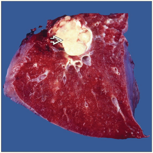
MEC usually an endobronchial tumor
Microscopic Pathology
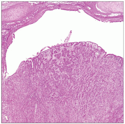
Epidermoid component without keratinization
Clear cells (intermediate cells)
Mucous-producing cells (mucocytes)
Lack of nuclear atypia or high mitotic activity
Top Differential Diagnoses
Lymphoepithelioma-like carcinoma
Adenosquamous carcinoma
Squamous cell carcinoma
Diagnostic Checklist
MEC generally does not show areas of keratinization
Epidermoid cells admixed with mucous cells
TERMINOLOGY
Abbreviations
Mucoepidermoid carcinoma (MEC)
Synonyms
Mucoepidermoid tumor
Definitions
Epithelial neoplasm composed of epidermoid cells admixed with mucus-secreting cells (mucocytes)
ETIOLOGY/PATHOGENESIS
Incidence
Tumor more common in adults but has been reported in children
CLINICAL ISSUES
Presentation
Cough
Shortness of breath
Hemoptysis
Treatment
Surgical approaches
Complete surgical resection, usually lobectomy
Prognosis
Prognosis depends on grading of neoplasm
Low-grade tumors, completely resected: Very good
High-grade tumors may require additional medical treatment in addition to surgical resection
MACROSCOPIC FEATURES
General Features
Tumor more common in central location than in periphery of lung
MEC is usually an endobronchial tumor
Size
1-5 cm in greatest dimension
MICROSCOPIC PATHOLOGY
Histologic Features
Epidermoid component without keratinization
Clear cells (intermediate cells)
Mucous-producing cells (mucocytes)
Lack of nuclear atypia or high mitotic activity
Predominant Pattern/Injury Type
Solid
Cystic
Predominant Cell/Compartment Type
Epithelial
DIFFERENTIAL DIAGNOSIS
Lymphoepithelioma-like Carcinoma
Usually shows increased mitotic activity and cellular atypia
Characteristically has inflammatory stroma composed of lymphocytes and plasma cells
Does not show the presence of mucocytes or low-grade areas of MEC
Adenosquamous Carcinoma
Squamous Cell Carcinoma
In a small biopsy, it may be difficult or impossible to separate both of these entities
Lack of keratinization and presence of mucocytes admixed with epidermoid cells will favor MEC
Presence of squamous cell carcinoma in situ would favor squamous cell carcinoma
Mucous Gland Adenoma
Does not invade beyond cartilaginous bronchial tissue
DIAGNOSTIC CHECKLIST
Clinically Relevant Pathologic Features
Gross appearance
Central tumor obstructing bronchial lumen
Pathologic Interpretation Pearls
MEC generally does not show areas of keratinization
Epidermoid cell admixed with mucous cells
GRADING
Low-Grade MEC
Low-grade tumor characterized by epidermoid component with mucus-producing cells without increased mitotic activity, nuclear pleomorphism, necrosis, or hemorrhage
High-Grade MEC
High-grade tumors characterized by epidermoid component with areas of nuclear pleomorphism, mitotic activity, necrosis, or hemorrhage
SELECTED REFERENCES
1. Vadasz P et al: Mucoepidermoid bronchial tumors: a review of 34 operated cases. Eur J Cardiothorac Surg. 17(5):566-9, 2000
2. Moran CA: Primary salivary gland-type tumors of the lung. Semin Diagn Pathol. 12(2):106-22, 1995
3. Yousem SA et al: Mucoepidermoid tumors of the lung. Cancer. 60(6):1346-52, 1987


