Mixed Tumor
Key Facts
Terminology
Synonym: Pleomorphic adenoma (PA)
Biphasic neoplasm with epithelial/myoepithelial and mesenchymal differentiation
Clinical Issues
Symptoms
Cough
Dyspnea
Chest pain
Hemoptysis
Prognosis
Excellent for benign tumors
Malignant mixed tumors may need additional therapy
Macroscopic Features
Endobronchial tumor
1-5 cm in greatest dimension
Ancillary Tests
GFAP
CK-PAN
S100
Actin-sm
Top Differential Diagnoses
Non-small cell carcinoma
Epithelial/myoepithelial carcinoma
Mucoepidermoid carcinoma
Carcinosarcoma
Pulmonary hamartoma
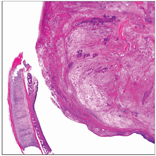 Panoramic view of a benign mixed tumor. Note the central location, as the tumor is growing just beneath the bronchial mucosa. |
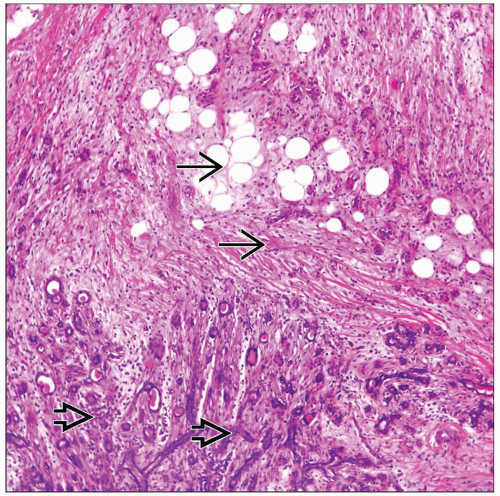 Benign mixed tumor is seen with a mixture of epithelial elements  and mesenchymal elements and mesenchymal elements  . . |
TERMINOLOGY
Abbreviations
Mixed tumor (MT)
Synonyms
Pleomorphic adenoma (PA)
Definitions
Biphasic neoplasm with epithelial/myoepithelial and mesenchymal differentiation
CLINICAL ISSUES
Presentation
Cough
Incidental finding
Hemoptysis
Treatment
Surgical approaches
Lobectomy
Prognosis
Excellent for benign tumor
Malignant mixed tumors may need additional therapy
MACROSCOPIC FEATURES
General Features
Endobronchial tumor
Size
1-5 cm in greatest dimension
MICROSCOPIC PATHOLOGY
Histologic Features
Presence of chondromyxoid background with focal cartilage admixed with epithelial component
Predominant Pattern/Injury Type
Biphasic
Predominant Cell/Compartment Type
Epithelial, biphasic, or mixed
DIFFERENTIAL DIAGNOSIS
Non-Small Cell Carcinoma
In small biopsy with only well-differentiated epithelial component sampled
In resected specimens, diagnosis should not pose a problem
Epi-myoepithelial Carcinoma
Both tumors show myoepithelial component
Epithelial myoepithelial carcinoma does not show presence of cartilage or other heterologous elements
Glandular component of inner layer of epithelial cells and outer layer of myoepithelial cells is characteristic of epithelial-myoepithelial carcinoma
Mucoepidermoid Carcinoma
Issue in small biopsy with only epithelial component sampled
In resected specimens, diagnosis is not a problem
Mucoepidermoid carcinomas show presence of mucus-producing and intermediate cells
Carcinosarcoma
Carcinosarcomas will show presence of malignant mesenchymal and malignant epithelial components
Pulmonary Hamartoma
Most hamartomas will show prominent cartilaginous component and invaginations of epithelium
Hamartomas do not show presence of myoepithelial cellular proliferation
DIAGNOSTIC CHECKLIST
Clinically Relevant Pathologic Features
Tissue distribution
Pathologic Interpretation Pearls
Presence of epithelial/myoepithelial cells admixed with chondromyxoid or cartilaginous areas
GRADING
Benign Mixed Tumors
Components are mature elements without atypical histological features
Malignant Mixed Tumor (Ex-Pleomorphic Adenoma)
Malignant mixed tumors will display conventional malignant components either in epithelial or mesenchymal component
SELECTED REFERENCES
1. Kamiyoshihara M et al: Pleomorphic adenoma of the main bronchus in an adult treated using a wedge bronchiectomy. Gen Thorac Cardiovasc Surg. 57(1):43-5, 2009
2. Fitchett J et al: A rare case of primary pleomorphic adenoma in main bronchus. Ann Thorac Surg. 86(3):1025-6, 2008
3. Méjean-Lebreton F et al: [Benign salivary gland-type tumors of the bronchus: expression of high molecular weight cytokeratins.] Ann Pathol. 26(1):30-4, 2006
Stay updated, free articles. Join our Telegram channel

Full access? Get Clinical Tree


