Metanephric Adenoma
Satish K. Tickoo, MD
Victor E. Reuter, MD
Key Facts
Terminology
Benign neoplasm composed of small primitive cells resembling early metanephric tubular differentiation
Part of spectrum of neoplasms that includes metanephric adenofibroma and metanephric stromal tumor
Etiology/Pathogenesis
Microsatellite allelotyping has shown potential tumor suppressor gene on chromosome 2p13 in 56% of informative cases
Clinical Issues
Age range 11 months to 83 years (reported mean: 41 years)
Female preponderance (M:F = 1:2)
About 12% with symptoms related to polycythemia
Benign course with no reported metastasis
Microscopic Pathology
Cellular tumor composed of crowded small acini of primitive blue cells in paucicellular intervening stroma
Papillary component, including glomeruloid appearance, fairly common
Tumor cells have minimal cytoplasm with uniform, round to ovoid nuclei, slightly larger than size of lymphocyte
Ancillary Tests
Often positive for WT1 (diffuse nuclear), AMACR (cytoplasmic, granular), and CD57
TERMINOLOGY
Abbreviations
Metanephric adenoma (MA)
Synonyms
Embryonal adenoma, nephrogenic nephroma
Definitions
Benign neoplasm composed of small primitive cells resembling early metanephric tubular differentiation
Part of spectrum of neoplasms that includes metanephric adenofibroma and metanephric stromal tumor
ETIOLOGY/PATHOGENESIS
Molecular Abnormalities
Microsatellite allelotyping has shown potential tumor suppressor gene on chromosome 2p13 in 56% of informative cases
No allelic changes in Wilms tumor (WT) gene region at chromosome 11p13 or in papillary renal cell tumor (PRCC) gene region at chromosome 17q21.32
Prior reported trisomies 7 and 17 are possibly related to the study of solid PRCC misdiagnosed as MA
CLINICAL ISSUES
Epidemiology
Age
11 months to 83 years (mean: 41 years)
Gender
Female predominance (M:F = 1:2)
Presentation
> 50% of cases detected incidentally
Most symptomatic cases show abdominal or flank pain, hematuria, and palpable mass
About 12% with symptoms related to polycythemia
Prognosis
Benign course with no reported metastasis in tumors with typical morphology
Single case of lymph node metastasis reported in child was probably a Wilms tumor because of reported atypical high mitotic activity
IMAGE FINDINGS
General Features
Calcifications seen in up to 43% of cases
MACROSCOPIC FEATURES
General Features
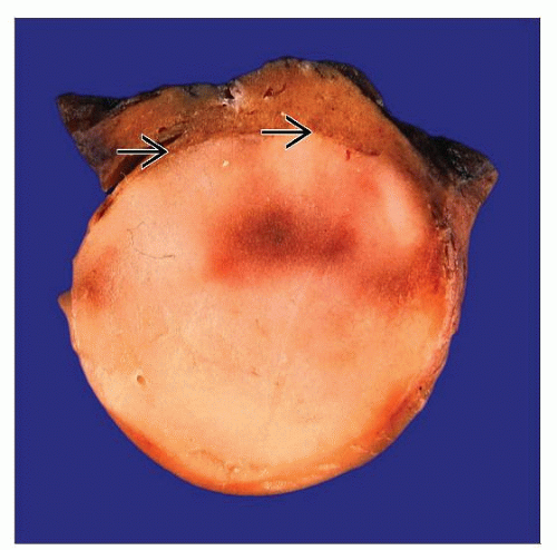
Typically unilateral, solitary, well circumscribed, and well delineated
Majority are unencapsulated, but discontinuous or continuous capsule present in some
Cut surface solid tan-pink, gray to yellow, and partially cystic in 12% of cases
Hemorrhage and necrosis are common
Gross calcifications in up to 20%, including rare instances of entirely calcified tumor
Size
Few mm to 20 cm; reported mean: 5.5 cm
MICROSCOPIC PATHOLOGY
Histologic Features
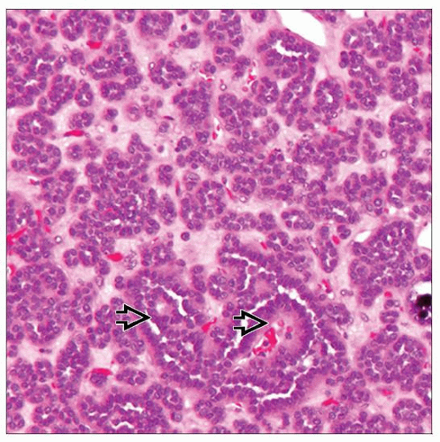
Cellular tumor composed of crowded small acini of primitive blue cells in paucicellular intervening stroma, and often with elongated tubules
Papillary component, including glomeruloid structures, fairly common
Cells have minimal cytoplasm with relatively uniform, round to ovoid nuclei bearing occasional folds
Nucleoli are generally inconspicuous, and mitosis is rare to absent
Chromatin distribution is uniform
Rare cysts and blastemal-like sheet-like patterns
Many tumors show regressive features, including hyalinization, calcifications often psammomatous, necrosis and hemorrhage, and dystrophic ossification
Predominant Pattern/Injury Type
Neoplastic
ANCILLARY TESTS
Immunohistochemistry
Often positive for WT1 (diffuse nuclear), AMACR (cytoplasmic, granular), pax-2, and CD57
Usually negative for CK7 except in branching large tubules and papillary areas
Electron Microscopy
Clusters of cells occasionally forming microlumen and surrounded by smooth, basal lamina matrix
Junctional complexes at apical end of luminal lining cells with florid microvilli
DIFFERENTIAL DIAGNOSIS
Papillary Renal Cell Carcinoma
PRCC type 1, solid variant and tubular predominant, is closest differential diagnosis
Stay updated, free articles. Join our Telegram channel

Full access? Get Clinical Tree


